


Temporal- and sex-specific changes in proteasome-dependent and independent polyubiquitination in the amygdala during fear memory formation
- Brieann Brown1,
- Natalie Preveza2,
- Gaurav Jones1,
- Meagan Turner2,
- W. Keith Ray3 and
- Timothy J. Jarome1,2
- 1School of Neuroscience, Virginia Polytechnic Institute and State University, Blacksburg, Virginia 24061, USA
- 2School of Animal Sciences, Virginia Polytechnic Institute and State University, Blacksburg, Virginia 24061, USA
- 3Department of Biochemistry, Virginia Polytechnic Institute and State University, Blacksburg, Virginia 24061, USA
- Corresponding author: tjjarome{at}vt.edu
Abstract
Strong evidence has emerged over the last two decades implicating proteasome-dependent and independent protein polyubiquitination in the memory consolidation process. Recently, it was shown that multiple forms of polyubiquitination, including proteasome-dependent K48 and proteasome-independent M1 polyubiquitination, regulate fear memory formation in a sex-dependent manner in the amygdala. However, prior work focused on single time points during the postlearning period, leaving questions about whether these are true sex differences in polyubiquitin modifications that persist throughout the extended consolidation process. Here, using unbiased polyubiquitin-type specific proteomics, we identified the protein targets of K48 and M1 polyubiquitination in the amygdala 2 and 4 h after contextual fear conditioning. Notably, we found that while the sex differences in the targeting of proteins with these polyubiquitin modifications persist for several hours after fear conditioning, the temporal dynamics of these changes vary across males and females. Further, while target protein pathways vary significantly across sexes at every time point examined, there are several notable overlaps between males and females. Together, these data provide the first comprehensive analysis of sex differences in proteasome-dependent and independent protein polyubiquitination in fear memory formation and significantly advance our understanding of the potential sex-specific roles of diverse polyubiquitin modifications in the memory consolidation process.
The process of transferring sensory information into a stable long-term memory is complex, requiring the coordinated actions of multiple brain regions, which have significant changes in gene transcription and de novo protein translation (Kandel 2012; Bisaz et al. 2014; Kandel et al. 2014; Josselyn and Tonegawa 2020). These molecular changes occur in the memory engram, a specific population of cells responsible for the storage of the long-term memory (Josselyn and Tonegawa 2020; Roy et al. 2022), and in the case of contextual fear memories are found in the amygdala and hippocampus, among other brain regions (Phillips and LeDoux 1992; Keeley et al. 2006; Parsons et al. 2006; Shin et al. 2006; Ou and Gean 2007; Hoeffer et al. 2011; Mahan et al. 2012; Morey et al. 2012). Initial sensory information is unstable and goes through the process of “consolidation” where changes in gene transcription, protein translation, and protein degradation are of critical importance to stabilize the memory and make it insensitive to disruption (McGaugh 2000).
Though it is well documented that fear memory consolidation results in transcriptional changes, a strong body of literature emerging over the last two decades has also indicated the need for protein degradation via the ubiquitin-proteasome system (UPS) (Lopez-Salon et al. 2001; Lee et al. 2008; Reis et al. 2013; Rosenberg et al. 2016a,b; Cullen et al. 2017). In the UPS, the protein ubiquitin will bind to target substrates and form polyubiquitin chains, of which those formed with linkage at lysine-48 (K48) result in degradation of the substrate by the catalytic core of the 26S proteasome complex (Jarome and Helmstetter 2013; Musaus et al. 2020). Early studies revealed that the protein degradation function of the UPS was critically involved in the memory consolidation process in the amygdala, with this work being done exclusively in males and focused primarily on pharmacological manipulations of the proteasome (Jarome et al. 2011, 2013, 2016; Rodriguez-Ortiz et al. 2011). Recently, our group has shown that both males and females require UPS-mediated protein degradation in the amygdala for contextual fear memory consolidation. Surprisingly, using an unbiased proteomic approach, we previously found that despite this shared need for protein degradation, the number and types of proteins targeted by K48 polyubiquitination in the amygdala following contextual fear conditioning varied significantly by sex (Devulapalli et al. 2021; Farrell et al. 2021). This suggests that males and females may be using protein degradation in different ways to form the same memory. However, previous studies that identified the temporal dynamics of the protein degradation process during fear memory consolidation were based exclusively on male animals, leaving questions about whether females follow a similar time course for this process in the amygdala. As our previous proteomic work was conducted at a static time point (1 h after fear conditioning), it is not clear if the sex differences we observed in the targets of K48 polyubiquitination are temporally regulated during the memory consolidation process, despite that prior work with broad western blotting approaches suggested that increases in degradation-specific polyubiquitination persist for up to 4 h after fear conditioning (Jarome et al. 2011, 2013). Further, to date, no study has systematically and unbiasedly attempted to identify the pathways targeted by K48 polyubiquitination across the consolidation period leaving a large gap in our understanding of the molecular dynamics of memory formation and the role of protein degradation in this process.
Complicating this further is that while most prior studies focused exclusively on protein degradation, ubiquitin has numerous other forms that are independent of the proteasome (Musaus et al. 2020). Recently, our group found that despite a shared need for proteasome-independent M1 polyubiquitination in the amygdala during the consolidation of contextual fear memory, males and females targeted different numbers and types of proteins with this mark in the amygdala during the consolidation of a contextual fear memory. However, similar to protein degradation, our prior work on M1 polyubiquitination was done at a single time point following behavioral training (Musaus et al. 2021). Similar to K48 polyubiquitination, to date, no study has examined the pathways targeted by proteasome-independent M1 polyubiquitination across the consolidation period and if these pathways overlap or exist independently of those targeted by K48 polyubiquitination.
To address this gap in the field, our current study aimed to test whether sex differences in degradation-dependent (K48) and degradation-independent (M1) protein polyubiquitination targets in the amygdala persisted through later phases of the consolidation process and identified the cellular pathways affected by these modifications. As prior work (Jarome et al. 2011, 2013) suggests that the ubiquitin-dependent memory consolidation period in rats persists for 4 h but is complete by 6 h after learning, we expanded upon our previous early consolidation time point (1 h) to include both a mid (2 h) and late (4 h) time point. Those data presented here present novel insights into sex differences in the use of proteasome-dependent and independent protein polyubiquitination during the memory consolidation process.
Results
K48 polyubiquitin targets in the amygdala are sex- and temporal-specific following contextual fear conditioning
Previously (Farrell et al. 2021), we demonstrated sex-specific K48 polyubiquitin targets in the amygdala 1 h after contextual fear conditioning. However, though our previous time point was chosen based on prior evidence that this was the peak of increased K48 polyubiquitin levels following fear conditioning (Jarome et al. 2011; Jarome and Helmstetter 2013; Orsi et al. 2019; Devulapalli et al. 2021), it remains unknown whether these sex differences exist throughout the entire consolidation period or if they are a result of a difference in timing between males and females. Additionally, as consolidation is a temporally graded process that lasts <6 h (Jarome and Helmstetter 2013), we wanted to identify how K48 polyubiquitin targets change across this memory consolidation period.
To test this, we trained both male and female rats with our contextual fear conditioning procedure and collected BLA tissue 2 and 4 h later as these times represent a mid and late point in the consolidation window (Fig. 1A). Following BLA collection, we utilized a K48 TUBE to enrich proteins containing K48 polyubiquitin chains and then conducted protein identification via LC/MS (n = 5–6 per group). Performance during fear conditioning for both sexes and time points is shown in Figure 1B. We found a significant effect of time (F(4,72) = 36.82, P < 0.0001) but did not see an effect of group (F(1,18) = 0.5912, P = 0.4519) or sex (F(1,18) = 3.199, P = 0.0905), and there were no significant interactions (all P > 0.05), indicating that males and females perform similarly, consistent with our previous studies (Devulapalli et al. 2021; Farrell et al. 2021). After identification of K48 polyubiquitin targets from our proteomic approach, we separated significantly altered proteins by sex and change in abundance (increased or decreased from naive controls) at our two post-fear conditioning time points. Based on our previous findings, we determined that proteins that had a positive log2 value (increased abundance) were those about to be degraded by the proteasome while those that had a negative log2 value (decreased abundance) were those that had already undergone degradation via the proteasome (Farrell et al. 2021).
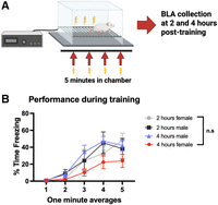
Experimental design. (A) Contextual fear conditioning training schematic. Rats were placed in the fear conditioning apparatus for 5 min, receiving a total of four unsignaled footshocks. After completing training, the basolateral amygdala (BLA) was collected at 2 and 4 h and compared with experimental naive animals that did not undergo contextual fear conditioning. (B) Performance during training did not reveal significant differences across groups. n = 5–6.
At the 2 h time point, we identified 31 proteins with a negative log2 value and 0 with a positive log2 value in male rats, with 37 targets with a negative log2 value and five with a positive log2 value being present in the female amygdala (Fig. 2A). Of interest, out of these K48 polyubiquitin targets, only two proteins overlapped between the male and female samples: CFL1 (protein cofilin 1; actin cytoskeleton dynamics) and P15212 (hexosaminidase B protein, lysosomal enzyme) with both having a negative log2 value across the sexes.
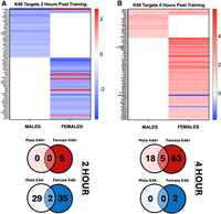
Sex-specific K48 polyubiquitin targets in the amygdala at 2 and 4 h after contextual fear conditioning. (A,B) Protein extracts from BLA samples were purified with K48 polyubiquitin TUBE and analyzed by liquid chromatography mass spectrometry and compared to naive age- and sex-matched controls. K48 polyubiquitin targets in males and females 2 h (A) and 4 h (B) after contextual fear conditioning (n = 5–6 per group). Data are displayed in heat maps with targeted proteins on the left and a colorimetric log2 fold change indicator on the right with sex being displayed on the bottom. Below the heat maps, data are summarized as Venn diagrams separated by either a significant positive (K48+) or negative (K48−) log2 value when compared to naive controls by sex. Data analysis conducted using FragPipe-Analyst with a DE log2 fold change >1 and P-value <0.05 being considered significant.
Next, we wanted to investigate if these sex-specific differences persist in the later phase of consolidation, so we looked at K48 polyubiquitination in the amygdala 4 h post-training. For males we found zero targets with a negative log2 value and 23 proteins with a positive log2 value, while in females we found two negative log2 value and 68 positive log2 value targets (Fig. 2B). Interestingly, at 4 h, we found five proteins with a positive log2 value that overlapped between sexes: ALDOC (aldolase fructose-bisphosphate C, glycolysis), EEF1a1 (eukaryotic translation elongation factor 1 α 1, delivers aminoacyl tRNAs to ribosome), ENO1 (enolase 1, glycolysis), KRT79 (keratin 79, epithelial keratin), and NSF (N-ethylmaleimide-sensitive factor, vesicle fusion and trafficking). Specific P-values and log fold changes for the 2 and 4 h time points are displayed in Tables 1 and 2.
K48 polyubiquitin targets in the male and female amygdala 2 h after contextual fear conditioning
K48 polyubiquitin targets in the male and female amygdala 4 h after contextual fear conditioning
Next, we compared our current data with what we found in our previous work that focused on 1 h post-training (Farrell et al. 2021) to better understand the sex- and temporal-specific patterns of protein degradation targets in the amygdala following fear conditioning. Interestingly, we see that males have a peak in targets at 1 h followed by a continued decrease in overall K48 polyubiquitin targets across time, while females have a biphasic pattern with peaks in targets at both 1 and 4 h post-training (Fig. 3A). Additionally, though the pattern of overall targets across time varies by sex, when we separate the targets out by whether they have a positive or negative log2 value, we see similar patterns between males and females (Fig. 3B). Taken together, these data indicate that males and females differ in the temporal dynamics of K48 polyubiquitination across the consolidation process and target largely distinct proteins.
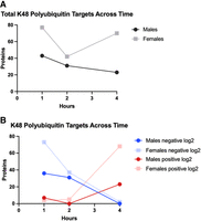
K48 polyubiquitination targets change in a sex- and time-dependent manner in the amygdala following contextual fear conditioning. (A) Utilizing previously published proteomic data for 1 h after fear conditioning (Farrell et al. 2021) publicly available with PRIDE accession number PXD027544, we compared the total number of significantly altered K48 polyubiquitin targets across the memory consolidation window to establish a temporal pattern by sex. (B) We then separated the significantly changed targets by positive and negative log2 fold changes to establish a further understanding of the temporal dynamics.
Next, as we saw very little overlap in K48 polyubiquitin targets between sexes, we wanted to use QIAGEN'S Ingenuity Pathway Analysis (IPA) to see what pathways each sex targets during memory consolidation (Fig. 4). For males at our 2 h time point, we found pathways related to translation (eukaryotic translation initiation, elongation, and termination pathways) and cellular junctions (remodeling of epithelial adherens junctions and gap junction signaling) to be highly implicated (Fig. 4A). For females at the 2 h time point, we also see translation associated pathways (eukaryotic translation initiation, elongation, and termination pathways) targeted along with ribosomal pathways (ribosomal quality control signaling pathway and major pathway of rRNA processing in the nucleus and cytosol), demonstrating a potential role of K48 polyubiquitination in de novo protein translation during consolidation (Fig. 4B). Interestingly, though there is an overlap in targeted pathways between males and females, the specific proteins in these pathways vary by sex. For the 4 h time point, in males, we observed a shift in the K48 polyubiquitination targeted pathways from a translational focus to synaptogenesis and plasticity associated pathways (clathrin-mediated endocytosis signaling, synaptogenesis signaling, activation of AMPA receptors and synaptic plasticity, and actin cytoskeleton signaling) (Fig. 4C). In females, we also see a shift away from translationally implicated pathways to those associated with synaptic receptor dynamics (translocation of GLUT4 to the plasma membrane, assembly and cell surface presentation of NMDA receptors, and activation of NMDA receptors and postsynaptic events) (Fig. 4D). Together, this pathway analysis demonstrates that though there are some similarities in the types of pathways targeted by K48 polyubiquitination at 2 and 4 h postcontextual fear conditioning, males and females still demonstrate sex-specific targets.
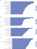
Sex-specific cellular pathways targeted by K48 polyubiquitination in the amygdala at 2 and 4 h after contextual fear conditioning. Utilizing QIAGEN IPA pathway analysis software, we analyzed our K48 polyubiquitination targets to determine cellular pathways targeted by this modification. Targeted pathways are displayed on the left with −log (P-value) being displayed on the top. Threshold displayed in orange is set to a P-value of <0.05, which equated to a −log (P-value) of 1.3. Pathways targeted at 2 h by males (A) and females (B) and pathways targeted at 4 h by males (C) and females (D) are displayed.
M1 polyubiquitin targets in the amygdala are sex- and temporal-specific following contextual fear conditioning
We next wanted to test if proteasome-independent forms of polyubiquitination also varied between sexes in a temporally specific manner. To accomplish this, we utilized the same 2 and 4 h amygdala samples from our K48 polyubiquitination experiment, and this time used a M1 polyubiquitin TUBE to enrich our samples for proteins with this specific ubiquitin chain before conducting our unbiased proteomic analysis. At our 2 h time point, in males we saw seven proteins with a negative log2 value and two with a positive log2, with females having 67 negative log2 value and 24 positive log2 value targets (Fig. 5A). Additionally, there was no overlap in target proteins between sexes.
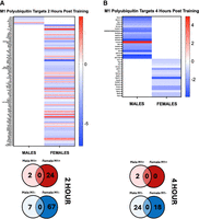
Sex-specific M1 polyubiquitin targets in the amygdala at 2 and 4 h after contextual fear conditioning. Utilizing the same BLA samples collected from Figure 1, protein extracts were purified with M1 polyubiquitin TUBE and analyzed by liquid chromatography mass spectrometry and compared to naive age- and sex-matched controls. M1 polyubiquitin targets in males and females 2 h (A) and 4 h (B) after contextual fear conditioning (n = 5–6 per group). Data are displayed as heat maps with targeted proteins on the left and a colorimetric log fold change indicator on the right with sex being displayed on the bottom. Below the heat maps, data are summarized as Venn diagrams separated by either a significant positive (M1+) or negative (M1−) log2 value when compared to naive controls by sex. Data analysis conducted using FragPipe-Analyst with a DE log2 fold change >1 and P-value <0.05 being considered significant.
At 4 h post-fear conditioning, we saw 24 negative log2 value and two positive log2 value M1 polyubiquitin targets in males and 18 negative log2 value and 0 positive log2 value targets in females. Similar to our 2 h time point, there were no overlapping M1 polyubiquitin targets between the sexes. Also, unlike K48 polyubiquitination, M1 polyubiquitin had multiple protein targets that were not present in the naive condition or became absent following fear conditioning. These proteins are denoted in Tables 3 and 4 and the Venn diagrams of Figure 5 but not shown in the heat map as log2 values could not be calculated due to the change to/from zero.
M1 polyubiquitin targets in the male and female amygdala 2 h after contextual fear conditioning
M1 polyubiquitin targets in the male and female amygdala 4 h after contextual fear conditioning
Next, we examined how M1 polyubiquitin usage varied across the consolidation phase and by sex by comparing with our previously published 1 h post-fear conditioning data (Musaus et al. 2021). For total M1 polyubiquitin targets, males had a biphasic pattern with higher target levels at 1 and 4 h. Conversely, females peaked in targets at 2 h (Fig. 6A). Additionally, when separating the targets out by positive or negative log2 value, males show a large peak in positive log2 values at 1 h followed by a drastic decline and plateau at 2 and 4 h. Females show a modest increase in positive log2 values from 1–2 h before declining significantly at the 4 h time point. For negative log2 values, neither males nor females have targets at 1 h. Females have a steep increase in negative log2 value targets at the 2 h time point before a large decrease at 4 h, while males have a gradual increase in negative log2 value targets across time. These findings indicate a sex-specific dimorphism between proteasome-dependent and proteasome-independent polyubiquitination following fear conditioning and highlight that the temporal patterns of polyubiquitination vary based on sex and linkage site. Additionally, we found no overlap between K48 and M1 polyubiquitin targets for both sexes at any time point (data not shown). This finding further indicates that while both K48 and M1 polyubiquitination are necessary for memory formation, they target entirely different proteins and do not result in mixed polyubiquitin chain linkages.
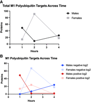
M1 polyubiquitination targets change in a sex- and time-dependent manner in the amygdala following contextual fear conditioning. (A) Utilizing previously published proteomic data for 1 h after fear conditioning (Musaus et al. 2021) publicly available with PRIDE accession number PXD025483, we compared the total number of significantly altered M1 polyubiquitin targets across the memory consolidation window to establish a temporal pattern by sex. (B) We then separated the significantly changed targets by positive and negative log2 fold changes to establish a further understanding of the temporal dynamics.
We next completed an IPA pathway analysis for M1 polyubiquitin targets at the 2 and 4 h time points (Fig. 7). In males, at 2 h, we saw a focus on metabolic pathways (sucrose degradation V, glycolysis I, and glucose metabolism) suggesting that M1 polyubiquitination may play a role in addressing increased energy needs during consolidation (Fig. 7A). In females, at the 2 h time point, the main pathways were translational (eukaryotic translational initiation, elongation, and termination, rRNA processing and modification, and ribosomal quality control) along with those associated with lysosomal processes (chaperon-mediated autophagy, late endosomal microautophagy, and aggrephagy) (Fig. 7B). Interestingly, at 4 h, the pathways targeted by males by M1 polyubiquitination changed to those associated with mitochondria (mitochondrial protein import, degradation, dysfunction, oxidative phosphorylation, cristae formation, and mitochondrial uncoupling) (Fig. 7C); however, the targeting of these pathways still suggests a role for M1 polyubiquitin in managing increased energy demands during memory consolidation. In females, the pathways implicated at 4 h were largely the same as those at 2 h (Fig. 7D). Together, this pathway analysis demonstrates that for M1 polyubiquitination, males and females demonstrate largely sex-specific targets.
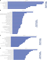
Sex-specific cellular pathways targeted by M1 polyubiquitination in the amygdala at 2 and 4 h after contextual fear conditioning. Utilizing QIAGEN IPA pathway analysis software, we analyzed our M1 polyubiquitination targets to determine cellular pathways targeted by this modification. Targeted pathways are displayed on the left with −log (P-value) being displayed on the top. Threshold displayed in orange is set to a P-value of <0.05, which equated to a −log (P-value) of 1.3. Pathways targeted at 2 h by males (A) and females (B) and pathways targeted at 4 h by males (C) and females (D) are displayed.
Discussion
We previously reported sex differences in the protein targets of proteasome-dependent (K48) and proteasome-independent (M1) polyubiquitination in the amygdala following fear conditioning (Farrell et al. 2021; Musaus et al. 2021). However, as our prior work only used a single postlearning time point, it remained unknown if these were true sex differences or reflected differences in the temporal dynamics of the memory consolidation process. Here, we found that sex differences in K48 and M1 polyubiquitin protein targets persisted beyond the early postlearning phase, continuing for several hours after fear conditioning. Importantly, our extended time course using unbiased proteomics provides the first insight into the many important functions of proteasome-dependent and proteasome-independent polyubiquitin signaling during long-term memory formation and how this varies across the sexes. Together, these data significantly advance our understanding of the potential sex-specific roles of diverse polyubiquitin modifications in the memory consolidation process.
Our current work expands on previous findings on polyubiquitination in memory consolidation by examining changes in multiple polyubiquitin targets across the mid to late consolidation phase, based on prior work suggesting that this process lasts <6 h (Jarome et al. 2011). Excitingly, we have uncovered sustained sex differences in the K48 and M1 polyubiquitin targets at both 2 and 4 h after fear conditioning. Interestingly, at 2 h, K48 polyubiquitination had only three overlapping targets between the sexes: cofilin), RACK1 (receptor for activated C kinase 1), and P15252 (hexosaminidase B protein). CFL1 is responsible for the breakdown of actin filaments and has been connected to synaptic plasticity with deficiency being tied to memory impairment in disorders such as Alzheimer's disease (Bamburg and Bernstein 2016; Ben Zablah et al. 2020; Wang et al. 2020). This could indicate that in both males and females, protein degradation is involved in actin cytoskeleton remodeling. RACK1 is an important scaffolding protein for protein kinase C (PKC). Previous research in both the hippocampus and prefrontal cortex has indicated a connection between the RACK1–PKC pathway and memory (Liu et al. 2016) and that deficiency can lead to memory impairment and has implications in Alzheimer's disease (Liu et al. 2011; Zhu et al. 2016; He et al. 2024). Our findings add to this literature by suggesting that RACK1 degradation may be involved in memory consolidation. P15252 is a lysosomal enzyme, and as learning does require de novo protein translation and degradation, cellular waste may be generated, resulting in a need for greater lysosomal enzymes to help with the cleanup process that must also be later removed. Importantly, though a small subset of overlapping protein targets exists at the 2 h time point, we see a large difference in the overall target number between the sexes. This indicates that though males and females both utilize K48 polyubiquitination during the consolidation period, how they do so differs. Despite this, both sexes primarily show negative log2 values for K48 polyubiquitination at 2 h after fear conditioning, indicating that significant degradation of targets has already occurred by the mid-phase of consolidation.
In contrast, at 4 h, we see both sexes predominantly with positive log2 values, suggesting a potential biphasic regulation of protein degradation during memory consolidation. As K48 targets are generally degraded within a short period after being marked (Kiss et al. 2025), this indicates a likely second period of mass degradation occurring during the later phase of consolidation, which perhaps reflects a cellular “cleanup” step after or around the time of increased protein translation. Similar to our 2 h time point, the targets vary significantly between sexes; however, we identified five overlapping proteins in males and females: ALDOC (adylose C), ENO1 (α enolase enzyme), EEF1A1 (elongation factor 1 α), KRT79 (keratin 79), and NSF (N-ethylmaleimide-sensitive factor). Both ALDOC and ENO1 are associated with glycolysis, and their increased targeting for degradation may be indicative of a greater need in both sexes for additional energy as both translation and degradation via the proteasome require ATP. Notably, 4 h was previously reported to be the peak time for increased proteasome activity during the memory consolidation process in the amygdala (Jarome et al. 2013; Farrell et al. 2023). EEF1A1 is associated with protein translation and is responsible for delivering aminoacyl tRNAs to the ribosome. EEF1A1 being a target for degradation at this later time point may indicate that both males and females are nearing the end of the protein synthesis phase of the consolidation process, which is consistent with prior research indicating that translation-dependent phase of memory storage is likely complete by 6 h (Nader et al. 2000). NSF is highly implicated in synaptic plasticity due to its function of disassembling the SNARE complex to allow for neurotransmitter release and ties to vesicle fusion, including those responsible for bringing additional neurotransmitter receptors to the cell surface (Tolar and Pallanck 1998; Littleton et al. 2001; Yang et al. 2024). Degradation of NSF could suggest potential synaptic remodeling during the later consolidation phase, which is a previously hypothesized role of protein degradation for which limited data existed to support (Lee et al. 2008; Kaang et al. 2009; Jarome and Helmstetter 2013). Together, these data provide evidence that our previously reported sex differences in the target substrates of K48 polyubiquitination are not due to temporary differences and persist throughout the entire consolidation process.
Previously, we found that males had more M1 polyubiquitin targets than females in the amygdala 1 h after fear conditioning and that all targets had a positive log2 value (increased M1 polyubiquitination) (Musaus et al. 2021). Interestingly, at 2 h post-fear conditioning, we observed very few targets in males, most of which were losing this M1 polyubiquitin mark, with a far greater number in females. Conversely, at 4 h after fear conditioning, males and females had a similar number of significant targets, nearly all of which were losing the M1 polyubiquitin mark. Importantly, there was no overlap in M1 polyubiquitin targets between sexes at any time point. This finding emphasizes sexually dimorphic utilization of M1 polyubiquitination in the amygdala during fear memory formation.
As noted above, our data revealed unexpected sex-specific temporal differences in K48 and M1 polyubiquitination across the consolidation period, highlighting how these modifications function independently to drive fear memory formation. Notably, for K48 polyubiquitination in females, we see a peak in targets at both 1 and 4 h, while males have the most targets at 1 h, which steadily decreases across time. Together, the total number of targets suggest a biphasic pattern in females and a monophasic pattern in males. Despite this, males and females both have greater number of negative log2 values early but more positive log2 values late in the consolidation process. This highlights that though the targets of K48 polyubiquitin vary greatly between males and females, the timing of proteasome degradation may be similar. Conversely, for M1 polyubiquitination in females, we see a single peak in total targets at 2 h, while males show peaks at 1 and 4 h. This suggests that males, not females, have a biphasic pattern of changes in M1 polyubiquitination during the consolidation process. Further, whereas males show a peak of positive log2 values at 1 h that plateaus and declines to nearly no targets for the remainder of the consolidation process, females show a peak in positive log2 values at 2 h. These findings once again demonstrate the sexual dimorphism of M1 polyubiquitination following fear conditioning and that the dynamics of K48 and M1 polyubiquitination vary greatly during the memory consolidation process.
In terms of pathways, at 2 h females are primarily using K48 polyubiquitination to target proteins related to translation, including translation initiation, elongation, and termination, ribosomal quality control signaling, and rRNA processing pathways. Within these pathways, we also see targets of both proteasome 20S and 26S subunits, as well as various ribosomal proteins. Interestingly, though the proteasome subunits could be related to increased proteasome activity during consolidation, previous evidence in yeast and rodents has shown a role for some proteasome subunits in transcriptional regulation independent of the protein degradation process (Auld et al. 2006; Jarome et al. 2021; Farrell et al. 2022, 2024). As increased transcription is a necessary mechanism for memory formation (Kim and Kaang 2017), these subunits may be performing this alternative function. Similar to females, in males, K48 polyubiquitin targets at 2 h were also involved in translation initiation, elongation, and termination, though the only overlapping protein between sexes is RACK1. This indicates that though both males and females are targeting translation pathways, the implicated proteins vary widely between sexes. In males, significant pathways also include remodeling of epithelial adherens junctions and gap junction signaling pathways, highlighting the targeting of various actin and tubulin proteins and of special interest protein phosphatase 3 catalytic subunit. Gap junction signaling has been implicated in neuronal firing synchronicity and spatial memory formation in hippocampal neurons (Allen et al. 2011; Dossi et al. 2024; Todd et al. 2025). Epithelial adherens junctions are important in blood–brain barrier maintenance and axon guidance, and some studies have shown impairment of these junctions is present in Alzheimer's models and leads to memory impairments in behavioral tasks (Udo et al. 2025). Further, previous research has suggested that protein phosphatase 3 catalytic subunit is important for limiting memory strength due to its ability to limit phosphorylation of CaMKII, a known regulator of proteasome function and fear memory formation in the amygdala (Colbran 2004; Hassan et al. 2024). At 4 h, female K48 polyubiquitin target pathways are primarily related to receptor dynamics with translocation of GLUT4 to the plasma membrane, assembly and cell surface presentation of NMDA, and activation of NMDA receptors and postsynaptic events all being targeted. Interestingly, male K48 polyubiquitin target pathways also point toward synaptic plasticity and remodeling during late phase consolidation as we see activation of AMPA receptors and synaptic plasticity, synaptogenesis, and clathrin-mediated endocytosis signaling pathways.
Similar to K48 polyubiquitination, at 2 h, female M1 polyubiquitin target pathways were related to translation and included rRNA processing and modification, ribosomal quality control, and eukaryotic translation, elongation, initiation, and termination, which persisted 4 h after fear conditioning. This suggests that K48 and M1 polyubiquitination may work in concert to regulate translational control mechanisms during fear memory formation in the female amygdala, with M1 potentially providing stability and localization of proteins and K48 providing a degradation mechanism for the newly translated proteins after they are no longer needed. Interestingly, multiple lysosomal/autosomal pathways, including chaperon-mediated autophagy, late endosomal microautophagy, and aggrephagy, were also targeted by M1 polyubiquitination in females at 2 and 4 h. A small body of emerging evidence has shown a connection between lysosomes and memory with downregulation of lysosomal activity leading to memory impairment and increased lysosomal formation during memory consolidation (Kottler et al. 2011; Li et al. 2019). Our findings here suggest a potential mechanism in which M1 polyubiquitination works in concert with lysosomes to promote memory formation.
In males, M1 polyubiquitination target pathways focused on glucose metabolism, glycolysis, and sucrose degradation with the main protein of interest being TPI1 (triosephosphate isomerase). Notably, TPI1 deficiency has been linked to Alzheimer's disease due to its role in glycometabolism disorders (Roland et al. 2013; Zhao et al. 2013). TPI1 is important in mediating glucose consumption with deficiencies leading to neuronal damage and memory impairment possibly due to increased oxidative stress (Tajes et al. 2013). Additionally, TPI1 has been tied to O-GlcNAcylation, a posttranslational modification that can regulate gene transcription during fear memory consolidation and has also been tied to Alzheimer's (Forster et al. 2014; Butler et al. 2019). At 4 h, we see that male M1 polyubiquitination primarily targets mitochondrial pathways. There is a strong body of literature tying mitochondria to regulating neuronal plasticity and memory formation in part due to the increased energy demands during the consolidation process (Comyn et al. 2024), so males may be using M1 polyubiquitination to stabilize and promote localization of proteins in the mitochondria to help with this increased demand. Interestingly, we previously found that females used proteasome-independent K63 polyubiquitination to target mitochondrial pathways in the amygdala during formation of a contextual fear memory (Farrell et al. 2023). Combined with our current data, this may indicate that while regulation of mitochondrial function is important in fear memory formation in both sexes, males and females use different ubiquitin marks to achieve this. Future studies should aim to explore the sex-specific targets of K63 polyubiquitination across the consolidation period.
Though we have expanded on our previous research by adding both a 2 and 4 h postlearning time point, ubiquitination is a dynamic process that cannot be fully encapsulated in static time points. Importantly, there may be some key proteins or pathways implicated in fear memory consolidation not captured by our data presented here. Additionally, our experiment did not utilize associative controls due to previous studies showing that immediate shock training does not engage protein degradation in the amygdala (Jarome et al. 2011; Jarome and Helmstetter 2013; Orsi et al. 2019). However, this makes it difficult to determine if all significant targets of K48 and M1 polyubiquitination were specific to the learning of context–shock association. Unfortunately, such associative controls are extremely difficult and uncommon in whole-genome studies due to the high cost associated with such procedures. Finally, the presented data utilized free cycling females making us unable to determine if the estrous cycle and estrogen levels may be a driver of the sex differences observed in this study. While this approach was consistent with our prior work, as we have now shown that these differences in ubiquitin signaling exist across the consolidation period, future work can expand upon the presented findings and investigate estrogen and the estrous cycle as a potential mechanism for the observed differences. Despite these limitations, our data still significantly expand the current knowledge on the temporal dynamics of ubiquitin signaling during the memory consolidation process and provide, to date, the most detailed understanding of the functional significance of proteasome-dependent and independent polyubiquitination during memory formation.
Materials and Methods
Subjects
Experiments used 8–9 week old male and female Sprague–Dawley rats obtained from Inotiv (Frederick, MD) with all presented data utilizing five to six rats per group. Rats were housed two per cage with ad libitum access to water and rat chow. A 12:12 h light–dark cycle was used. All behavioral experiments were conducted in the morning during the light cycle to account for changes in cortisol levels throughout the day. All experiments and procedures were approved by Virginia Polytechnic Institute and State University Institutional Animal Care and Use Committee (protocol #23-249) and followed the National Institutes of Health ethics guidelines.
Behavioral apparatus
Our fear conditioning paradigm utilized two Habitest chambers as previously described (Orsi et al. 2019; Devulapalli et al. 2021). Our Habitest chambers have two steel walls making up the sides of the chamber with the front and back walls being plexiglass. The floor is a grid shock floor that sits above a plastic drop pan. The top of the chamber contains a light that was used to illuminate the chamber during training. Our chamber sits inside a sound attenuating box that prevents outside sights and sounds from causing distractions during the procedure. The grid shock floor delivers shock via the Precision Animal Shocker in tandem with FreezeFrame 4 software. The chamber was cleaned with 70% isopropanol between animals.
Behavioral procedures
Before experimental procedures, rats were handled and habituated to researchers for 4 days. Days 1 and 2 consisted of 3 min of handling per rat in the animal housing room with days 3 and 4 consisting of 2 min of handling per rat in the behavioral testing room after a brief transport period. Rats then went through our contextual fear training protocol as previously published (Orsi et al. 2019; Devulapalli et al. 2021; Farrell et al. 2021; Musaus et al. 2021). Rats were placed individually in the Habitest apparatus for a total of 5 min for training. The training parameters consisted of a 1 min baseline followed by four unsignaled footshocks (1.0 mA, 1 sec) with a 59 sec intertrial interval (ITI). One minute after the final shock, animals were retained in their homecages. Naive animals did not undergo fear conditioning and were not placed in the Habitest chambers. We utilized a freezing threshold of 2.0 for behavioral scoring, which was done through FreezeFrame 4 software.
Tissue collection
Tissue from experimental animals was collected 2 or 4 h after training; tissue from our naive control animals (not exposed to our fear conditioning paradigm) was collected with the experimental animals to reduce variability in proteomic data due to the circadian cycle. Rats were euthanized via isoflurane followed by rapid decapitation. Brains were then quickly removed and immediately frozen on dry ice. Basolateral amygdala (BLA) were later dissected out on dry ice. Tissue was stored at −80°C until processing as described below.
Tandem ubiquitin binding entity (TUBE)
BLA tissue was homogenized in freshly made whole cell lysis buffer (10 mM HEPES, 1.5 mM MgCl2, 10 mM KCl, 0.5 mM DTT, 0.5% IGEPAL, 0.02% SDS, 70 mM NEM, 1 μL/mL protease inhibitor cocktail, and 1 μL/mL phosphatase inhibitor cocktail) with this buffer being selected due to NEM's ability to preserve polyubiquitin chains. After homogenization, protein samples were quantified via DC assay (Bio-Rad). Protein samples were then diluted to a concentration of 100 µg per 500 µL of TUBE buffer (100 mM Tris-HCL, 150 mM NaCl, 5 mM EDTA, 0.08% NP-40). We then utilized either a high-affinity K48 or M1 polyubiquitin–selective tandem ubiquitin binding entity (K48-TUBE UM407m, M1-TUBE UM406m, Life Sensors) conjugated to magnetic beads to isolate proteins with either K48 or M1 polyubiquitin chains from our protein samples. Beads were washed in TUBE buffer before adding our 500 µL sample consisting of 100 µg protein, protease inhibitor (1 μg/mL), and TUBE buffer. Samples were then incubated with the beads for 2 h on a rotator at 4°C. After this, samples were washed twice with TUBE buffer. We then added 1× sample buffer (Bio-Rad) and eluted the samples at 96°C for 5 min at 800 rpm. Supernatant was then collected and held at −80°C until mass spectrometry analysis.
Liquid chromatography mass spectrometry (LC/MS)
LC/MS was conducted at the VT-Mass Spectrometry Incubator (VT-MSI) as previously published (Farrell et al. 2021; Musaus et al. 2021). All proteomic data have been made available on PRIDE database with accession numbers PXD070314 (male K48 polyubiquitin), PXD070255 (female K48 polyubiquitin), PXD069050 (male M1 polyubiquitin), and PXD069043 (female M1 polyubiquitin). Previous 1 h proteomic data (Farrell et al. 2021; Musaus et al. 2021) are publicly available for use and can be found at the following respective PRIDE accession numbers PXD027544 and PXD025483.
Statistical analyses
Mass spectrometry data were analyzed using FragPipe-Analyst with data type set to LFQ, intensity type set to MaxLFQ Intensity, DE log2 fold change cutoff set to 1, and P-values below 0.05 being considered significant. All mass spec data are reported with both P-values and log2 values except cases for the M1 polyubiquitination data where log2 values could not be calculated due to zero values (not present) in one of the experimental conditions. In these cases, outcomes are indicated as “positive” if only present in the fear-conditioned samples and “negative” if only present in the naive samples. Behavioral training data were analyzed using three-way ANOVA via Prism 10.
Competing interest statement
All authors declare no competing financial interest. The funders had no role in the design of the experiment, analysis or interpretation of data or the decision to publish.
Acknowledgments
This work was supported by National Institutes of Health (NIH) grants MH122414, MH131587, AG081851, AG071523, and AG079292 to T.J.J. All experiments were approved by the Institutional Animal Care and Use Committee at the Virginia Polytechnic Institute and State University (protocol #23-249) and conducted within the ethical guidelines of the National Institutes of Health (NIH).
CRediT authorship contribution statement: B.B.: Conceptualization, investigation, formal analysis, writing—original draft. N.P.: Investigation. G.J.: Investigation. M.T.: Investigation. W.K.R.: Investigation (LC/MS). T.J.J.: Writing—review and editing, supervision, funding acquisition, conceptualization.
Author contributions: B.B. and T.J.J conceived the study; B.B., N.P., G.J., M.T., and W.K.R. performed experiments; B.B. and T.J.J. analyzed data and wrote the manuscript.
Footnotes
-
Article is online at http://www.learnmem.org/cgi/doi/10.1101/lm.054203.125.
-
Freely available online through the Learning & Memory Open Access option.
- Received November 30, 2025.
- Accepted February 6, 2026.
This article, published in Learning & Memory, is available under a Creative Commons License (Attribution-NonCommercial 4.0 International), as described at http://creativecommons.org/licenses/by-nc/4.0/.



