


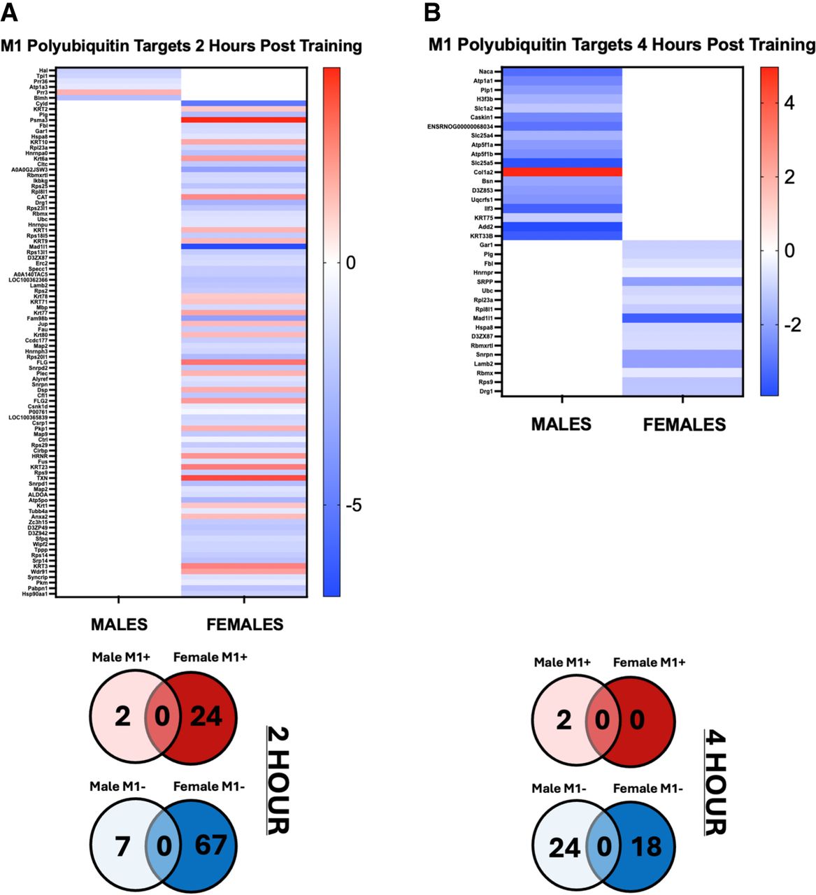
Sex-specific M1 polyubiquitin targets in the amygdala at 2 and 4 h after contextual fear conditioning. Utilizing the same BLA samples collected from Figure 1, protein extracts were purified with M1 polyubiquitin TUBE and analyzed by liquid chromatography mass spectrometry and compared to naive age- and sex-matched controls. M1 polyubiquitin targets in males and females 2 h (A) and 4 h (B) after contextual fear conditioning (n = 5–6 per group). Data are displayed as heat maps with targeted proteins on the left and a colorimetric log fold change indicator on the right with sex being displayed on the bottom. Below the heat maps, data are summarized as Venn diagrams separated by either a significant positive (M1+) or negative (M1−) log2 value when compared to naive controls by sex. Data analysis conducted using FragPipe-Analyst with a DE log2 fold change >1 and P-value <0.05 being considered significant.



