


Social isolation during adolescence differentially affects spatial learning in adult male and female mice
- Sadiyah Hanif1,
- Mia Sclar2,
- Jinah Lee1,3,
- Caleb Nichols1,
- Ekaterina Likhtik2,4 and
- Nesha S. Burghardt1,3
- 1Psychology Department, Hunter College, City University of New York, New York, New York 10065, USA
- 2Biology Department, Hunter College, City University of New York, New York, New York 10065, USA
- 3Psychology Program, The Graduate Center, City University of New York, New York, New York 10016, USA
- 4Biology Program, The Graduate Center, City University of New York, New York, New York 10016, USA
- Corresponding authors: nb844{at}hunter.cuny.edu, elikhtik{at}genectr.hunter.cuny.edu
Abstract
Social isolation is a risk factor for cognitive impairment. Adolescents may be particularly vulnerable to these effects, because they are in a critical period of development marked by significant physical, hormonal, and social changes. However, it is unclear if the effects of social isolation on learning and memory are similar in both sexes or if they persist into adulthood after a period of recovery. We socially isolated male and female 129Sv/Ev mice throughout adolescence (postnatal days 29–56), provided a 2-week resocialization recovery period, and then tested spatial learning and cognitive flexibility in the active place avoidance task. After behavioral testing, mice were injected with 5′-bromo-2′-deoxyuridine (BrdU) so that lasting effects of social isolation on cell proliferation in the dentate gyrus could be examined. Tissue was also stained for doublecortin (DCX). We found that in males, isolation led to a modest impairment in the rate of initial spatial learning, whereas in females, initial learning was unaffected. However, when the location of the shock zone was switched during the conflict variant of the task, cognitive flexibility was impaired in females only. Similarly, social isolation reduced cell proliferation and the number of immature neurons in the ventral dentate gyrus only in females. Together, these findings indicate that social isolation during adolescence differentially impairs spatial processing in males and females, with effects that persist into adulthood.
Humans are a social species whose cognitive function is negatively affected by social isolation (Wilson et al. 2007; Cacioppo and Hawkley 2009). This was exemplified by the COVID-19 pandemic, during which social isolation resulting from the global lockdown significantly impaired cognitive performance in children and adults around the world (Ingram et al. 2021; Betthauser et al. 2023). Students’ test scores not only declined during the lockdown, but remained low a year after schools reopened (Breit et al. 2023; Di Pietro 2023; Fahle et al. 2023). It remains unclear how long-lasting these effects will be or the extent to which they are caused by the stress of social isolation alone.
Across species, adolescence is a critical period of development characterized by dramatic changes in social behavior (Thor and Holloway 1984; Blakemore and Mills 2014), ongoing brain development, and increases in circulating levels of sex hormones. Puberty is also marked by a shift in the regulation of hypothalamic-pituitary-adrenal (HPA) axis function by sex hormones (McCormick and Mathews 2007), all of which could increase the impact of social isolation stress during this period of development. Based on the dramatic differences between males and females in the hormonal changes that occur during puberty, it is possible that social isolation during adolescence affects each sex differently. The finding that there are sex differences in HPA axis activity in adulthood is in line with this possibility (Sencar-Cupovic and Milkovic 1976; Bangasser and Wiersielis 2018).
The hippocampus is a stress-sensitive brain structure (McEwen et al. 2016) that continues to develop during adolescence (Spalding et al. 2013; Hueston et al. 2017) and is critical for learning and memory (Bird and Burgess 2008). Different types of social stressors have been shown to affect hippocampal-dependent learning. For example, social instability stress during adolescence impairs spatial memory in an object spatial location test (McCormick et al. 2010, 2012) and maternal deprivation impairs spatial learning in the Morris water maze, but increases contextual fear conditioning (Oomen et al. 2010). Studies of social isolation stress during adolescence have primarily focused on spatial learning in the Morris water maze, but the results have led to contradictory findings. While some groups show that adolescent isolation impairs spatial learning and memory (Hellemans et al. 2004; Ibi et al. 2008), others show no effect (Schrijver et al. 2004; Voikar et al. 2005; Kulesskaya et al. 2011), or improvements in acquisition (Wongwitdecha and Marsden 1996). In addition, almost all social isolation studies were conducted in male rodents and sex differences have not been explored.
We tested the effects of socially isolating adolescent male and female 129Sv/Ev mice (PND 29-56) on active place avoidance, a spatial learning task that is sensitive to minor hippocampal dysfunction (Cimadevilla et al. 2001b; Wesierska et al. 2005). During place avoidance, animals on a rotating arena learn to avoid a region of the room designated as a shock zone. In addition to evaluating spatial learning and memory, we used a conflict variant of the task to test cognitive flexibility, which we have previously shown to be dependent on dentate gyrus (DG) function (Kheirbek et al. 2013). We also examined the effects of social isolation on adult hippocampal neurogenesis, which undergoes dynamic changes during adolescence (Sousa et al. 1998; Spalding et al. 2013; Hueston et al. 2017; Boldrini et al. 2018) that could be altered by stress. Behavioral testing and cell quantification took place following a 2-week recovery period so that the long-lasting effects of social isolation could be evaluated. We found sex differences in both the behavioral and cellular effects of social isolation, which included a modest impairment in spatial learning without changes in hippocampal neurogenesis in males, but impaired cognitive flexibility and fewer immature neurons in the ventral DG of females. This work highlights the importance of considering sex when investigating mechanisms mediating the long-term effects of social isolation and when developing interventions.
Results
Social isolation stress during adolescence does not have long-lasting effects on anxiety-like behavior
While the focus of our study was on spatial learning, social isolation has been widely reported to affect mood and anxiety in humans (Cacioppo et al. 2006; Leigh-Hunt et al. 2017) and animals (Fone and Porkess 2008; Lukkes et al. 2009). To investigate whether there are sex differences in the long-lasting effects of isolation on anxiety-like behavior, we used exploration of the novel rotating arena during pretraining as a modified open field test. A two-way ANOVA on total distance traveled revealed no sex × stress interaction (F(1,29) = 0.17, P = 0.68) or main effects of sex (F(1,29) = 7.29 × 10−005, P = 0.99) or stress (F(1,29) = 1.22, P = 0.28) (Fig. 1A), indicating that isolation stress did not affect exploration of the arena in either sex. Interestingly, a two-way ANOVA on the percent of total distance traveled within the center half of the arena revealed a significant effect of sex (F(1,29) = 6.49, P = 0.02), with females walking in the center more than males (Fig. 1B). However, there was no sex × stress interaction (F(1,29) = 1.72, P = 0.20) or main effect stress (F(1,29) = 1.98, P = 0.17), indicating that adolescent stress did not affect anxiety-like behavior in adulthood in either sex when measured in this way.
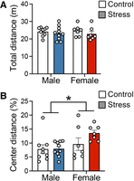
Social isolation during adolescence does not have long-term effects on exploration or anxiety-like behavior. (A) Total distance traveled on the rotating arena during pretraining. (B) Percent of total distance traveled in the center half of the rotating arena during pretraining. n = 9 control males; n = 10 stress males; n = 7 control females; n = 7 stress females. Data are represented as mean ± SEM. (*) P < 0.05.
Social isolation during adolescence leads to a modest impairment in the rate of spatial learning in males
We next investigated the long-term effects of social isolation on spatial learning in male mice using the active place avoidance paradigm (Fig. 2A). Before training began (pretraining), stressed males entered the inactive shock zone as often as nonstressed males (t(17) = 0.23, P = 0.82) (Fig. 2B,C). Then during initial training, mice received a brief foot shock upon entering a stationary shock zone defined by cues within the room. Mice were trained to avoid this shock zone with three 10 min long trials a day for two consecutive days (day 1: trials 1–3; day 2: trials 4–6), during which the shock was always on and the shock zone was always in the same location. A two-way repeated measures ANOVA on the number of entrances into the initial shock zone across all six trials revealed a main effect of trial (F(3.87,65.70) = 7.80, P < 0.0001), indicating that performance improved with training. However, there was no trial × stress group interaction (F(5,85) = 0.83, P = 0.53) (Fig. 2B,C). To evaluate whether stress affected retention of this spatial memory, we measured latency to first enter the shock zone at the beginning of the second day of training (trial 4), which was 24 h after the last training trial. We found that stressed males entered the shock zone faster than controls (t(17) = 2.09, P = 0.05) (Fig. 2D), indicating worse spatial recall. While this might reflect impaired memory consolidation during the 24 h retention period, it could also be attributed to slower acquisition the previous day. To explore the latter possibility, we investigated learning during the first day of training (trials 1–3) more closely. We found that both groups learned to avoid the shock zone quickly, as demonstrated by an average of 2.56 (control group) and 3.0 (stressed group) entrances during the third 10 min training trial (trial 3). To better capture the rate at which learning occurred, we calculated the number of times mice entered the shock zone during the first half of each trial. While the repeated-measures ANOVA on entrances during the first 5 min of trials 1–3 revealed no significant stress × trial interaction (F(2,34) = 1.24, P = 0.30), planned comparisons of each trial with Bonferroni's multiple comparisons test revealed that stressed animals entered the shock zone significantly more than controls on trial 2 (P = 0.006) (Fig. 2E). Similarly, a two-way repeated measures ANOVA on latency to first enter the shock zone on trials 2–3 revealed an effect of trial that approached significance (F(1,17) = 3.91, P = 0.06), with Bonferroni's multiple planned comparisons indicating that latency was lower in stressed males than controls on trial 2 (P = 0.05) but not trial 3 (P = 0.95) (Fig. 2F). Similarly, the time-in-location map illustrating where each group spent their time during each training trial showed subtle group differences during trials 1–2 (Fig. 3A). Collectively, these results indicate that by the end of the first trial, stressed males had not acquired spatial learning as well as controls, but were able to reach levels comparable to controls by the end of trial 3.
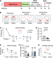
Social isolation during adolescence leads to a modest impairment in spatial learning in males. (A) Experimental time line. (B) Tracked behavior (gray) of a representative control and stressed male mouse before the shock was turned on (pretraining), during the last trial of initial training (trial 6), and during the first trial of conflict training. With the exception of pretraining, red circles indicate where the animal was shocked during each trial. (C) Social isolation did not affect the total number of times male mice entered the shock zone during pretraining or initial training. (D) The latency to first enter the shock zone after 24 h of retention was lower in stressed males than controls. (E) During initial training, stressed males entered the shock zone more than controls during the first 5 min of trial 2 and (F) entered the shock zone faster than controls on trial 2. (G) There were no group differences in the number of times males entered the new shock zone during conflict training. n = 9 control males; n = 10 stress males. Data are represented as mean ± SEM. (**) P < 0.01 versus trial 2 controls.
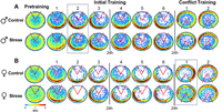
Social isolation affects patterns of avoidance behavior. Color-coded time-in-location maps for (A) males and (B) females of each group during pretraining, initial training (trials 1–6), and conflict training (trials 1–2). Colors represent group averages during each 10 min trial, with blue indicating the lowest dwell time and red indicating the highest dwell time. The time represented by each color was the same for all maps (color bar, 0–2 sec). Dashed lines indicate trials during which stress increased the number of entrances into the shock zone. n = 9 control males; n = 10 stress males; n = 7 control females; n = 7 stress females.
To evaluate cognitive flexibility, we used a conflict variant of the active place avoidance task, as previously described (Burghardt et al. 2012). This involved moving the shock zone 180° from its initial location, which challenged mice to flexibly adapt to a change in contingencies and suppress conditioned responses acquired during initial learning. A two-way repeated measures ANOVA on the number of entrances into the new location of the shock zone across two conflict trials revealed a significant effect of trial (F(1,17) = 21.31, P = 0002), but no trial × stress interaction (F(1,17) = 0.65, P = 0.43) or main effect of stress (F(1,17) = 0.06, P = 0.81) (Fig. 2G). Similarly, groups spent a similar amount of time within the new shock zone during the first conflict trial (t(17) = 0.40, P = 0.69) (Fig. 3A). Together, these results indicate that social isolation during adolescence did not affect cognitive flexibility in adult males.
Social isolation during adolescence impairs cognitive flexibility in females
To evaluate whether there are sex differences in the effects of social isolation on spatial learning, we tested female mice under the same conditions as males (Fig. 4A). During pretraining, we found that stressed females entered the inactive shock zone as often as female controls (t(12) = 0.70, P = 0.50) (Figs. 4B,C and 3B). When the shock was turned on, a repeated measures ANOVA on the number of entrances into the initial shock zone during all six trials revealed a significant effect of trial (F(2.875,34.49) = 4.44, P = 0.01), but no trial × stress interaction (F(5,60) = 0.55, P = 0.74) or main effect of stress (F(1,12) = 0.33, P = 0.58) (Fig. 4C). Both groups also exhibited a similar latency to first enter the shock zone on trial 4 (t(12) = 1.02, P = 0.33) (Fig. 4D), indicating that retention of this spatial memory was not affected by isolation stress during adolescence. Furthermore, the repeated measures ANOVA on the number of entrances during the first 5 min of trials 1–3 revealed no significant stress × trial interaction (F(2,24) = 0.29, P = 0.75) and planned comparisons of each trial confirmed that there were no group differences on any trial (P > 0.99). Despite being as successful as controls at avoiding the initial shock zone, the time-in-location maps showed that the stressed females were spending more time to the left of the shock zone than controls, indicating they learned to avoid the shock zone in a different way (Figs. 3B and 4B). Indeed, once the task was acquired (trials 2–6), stressed females spent significantly more time in this counterclockwise (CCW) position than controls (t(12) = 2.22, P = 0.047) (Fig. 4E). This strategy was risky, because stressed mice were effectively spending more time in closer proximity to the shock zone and were more likely to be rotated directly into that shock zone if they were inactive.
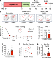
Social isolation during adolescence impairs cognitive flexibility in females. (A) Experimental time line. (B) Tracked behavior (gray) of a representative control and stressed female mouse before the shock was turned on (pretraining), during the last trial of initial training (trial 6), and during the first trial of conflict training. With the exception of pretraining, red circles indicate where the animal was shocked during each trial. (C) Social isolation did not affect the number of times female mice entered the shock zone during initial training. (D) There were no group differences in latency to first enter the shock zone after 24 h of retention. (E) During initial training, stressed females spent more time to the left of the shock zone (i.e., CCW) than controls. (F,G) During the first trial of conflict training, stressed females entered the new shock zone more and spent more time within that shock zone than controls. n = 7 per group. Data are represented as mean ± SEM. (*) P < 0.05.
When the position of the shock zone was moved to the opposite side of the room, stressed females entered the new shock zone more than controls. A two-way repeated measures ANOVA on the number of entrances during conflict training revealed a significant stress × trial interaction (F(1,12) = 4.78, P = 0.049) and the Bonferroni corrected posthoc test confirmed that stressed females entered the new shock zone more than controls during the first conflict trial (P = 0.03) (Fig. 4F). Stressed females also spent more time within the new shock zone than controls during conflict trial 1 (t(12) = 2.36, P = 0.036) (Figs. 3B and 4G), which is when flexibility was challenged the most. Together, these results demonstrate that unlike males, social isolation during adolescence in females impaired cognitive flexibility.
The effects of social isolation on spatial learning and cognitive flexibility are sex-specific
To verify that the effects of social isolation on place avoidance were sex-specific, we directly compared stressed males and females on each behavioral measure that we found to be affected by stress (see above). Data from stressed mice were calculated as a percentage of same-sex controls before the sexes were compared to each other. We found that during initial training, stressed males entered the shock zone faster on trial 2 (t(15) = 2.82, P = 0.01) (Fig. 5A) and more frequently during the first 5 min of trial 2 than stressed females (t(15) = 3.03, P = 0.009) (Fig. 5B). After a 24 h period of retention, stressed males also entered the shock zone faster than stressed females (t(15) = 2.24, P = 0.04) (Fig. 5C), which together confirm that stress led to a modest impairment in acquisition and memory retrieval in males only. In contrast, stressed females spent significantly more time in closer proximity to the shock zone than stressed males during initial training trials 2–6 (t(15) = 2.20, P = 0.04) (Fig. 5D), indicating that stress only affected avoidance strategy in females. When the shock zone was moved, stressed females entered the new location of the shock zone more frequently (conflict trial 1: t(15) = 2.19, P = 0.04) (Fig. 5E) and spent more time within that new shock zone than stressed males (conflict trial 1: t(15) = 2.06, P = 0.05) (Fig. 5F), demonstrating that stress specifically affected cognitive flexibility in females.
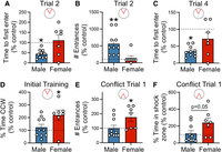
The effects of social isolation on spatial learning and cognitive flexibility are sex-specific. During initial training, stressed males (A) entered the shock zone faster on trial 2 and (B) entered the shock zone more frequently during the first 5 min of trial 2 than stressed females. (C) The latency to first enter the shock zone after 24 h of retention was lower in stressed males than stress females. (D) During initial training, stressed females spent more time to the left of the shock zone (i.e., CCW) than stressed males. (E,F) During the first trial of conflict training, stressed females entered the new shock zone more and spent more time within that new shock zone than stressed males. n = 10 stress males; n = 7 stress females. Data are expressed as a percentage of same-sex controls, and the dashed line indicates 100% of the corresponding control group. Data are represented as mean ± SEM. (*) P < 0.05, (**) P < 0.01.
Social isolation during adolescence reduces adult hippocampal neurogenesis in females
We next evaluated the long-lasting effects of social isolation on cell proliferation in the hippocampus. Upon completion of behavioral testing, mice of both sexes were injected twice with 5′-bromo-2′-deoxyuridine (BrdU) and perfused the next day (Figs. 2A and 4A). Based on known functional differences between dorsal and ventral hippocampus (Fanselow and Dong 2010), we analyzed BrdU-positive (+) cells (Fig. 6A) in the DG of each pole separately. In males, there was no effect of stress group (F(1,11) = 0.17, P = 0.69), pole (F(1,10) = 0.24, P = 0.64) or stress × pole interaction (F(1,10) = 1.67, P = 0.22), indicating that cell proliferation was not affected in this sex (Fig. 6B). In contrast, analysis of BrdU+ cells in females revealed a stress × pole interaction (F(1,12) = 5.85, P = 0.03), with Bonferroni's multiple comparisons test revealing a significant group difference in the ventral pole only (P = 0.036) (Fig. 6A,C). These results demonstrate a sex difference in the long-term effects of social isolation on cell proliferation and suggest that adult hippocampal neurogenesis may be reduced in the ventral hippocampus of females. To evaluate this possibility, we stained tissue from the same brains for doublecortin (DCX, Fig. 6D), a protein that is transiently expressed in immature neurons (Brown et al. 2003). Similar to the BrdU cell counts, our analysis of DCX+ cells in males revealed no effect of stress group (F(1,12) = 0.27, P = 0.62) or stress × pole interaction (F(1,11) = 3.37, P = 0.09), indicating that the number of immature neurons was not affected in this sex (Fig. 6E). However, in females, there was a stress × pole interaction (F(1,12) = 13.13, P = 0.004) and a significant effect of stress in the ventral pole only (Bonferroni's multiple comparisons, P = 0.02) (Fig. 6D,F), confirming that social isolation decreased the number of adult-born neurons in the ventral hippocampus of females.
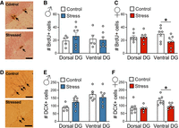
Social isolation decreases cell proliferation and adult hippocampal neurogenesis in females. (A) Representative images of BrdU immunostaining in the ventral DG of females in each group. Arrows indicate BrdU+ cells in the subgranular layer. (B) Social isolation did not affect the number of BrdU+ cells in the dorsal or ventral poles of males, but (C) decreased BrdU+ cells in the ventral pole of females. (D) Representative images of DCX immunostaining in the ventral DG of females in each group. (E) Social isolation did not affect the number of DCX+ cells in the dorsal or ventral poles of males, but (F) decreased DCX+ cells in the ventral pole of females. n = 7 control females, n = 7 stress females, n = 5–6 control males, n = 8 stress males. Data are represented as mean ± SEM. (*) P < 0.05. Scale bar, 200 μm.
Discussion
Using the active place avoidance paradigm, we tested the effects of social isolation throughout adolescence on hippocampal-dependent spatial learning in adulthood. We conducted our experiments in male and female 129Sv/Ev mice, a strain that exhibits high levels of anxiety-like behavior (Rodgers et al. 2002), is particularly vulnerable to social stress (Aubry et al. 2019), and is less aggressive than other commonly used strains (Abramov et al. 2008). This allowed us to rehouse males during a recovery period with minimal concerns about fighting, so that both sexes could be tested under identical conditions. We found that social isolation led to a modest impairment in spatial learning in males, but impaired cognitive flexibility and reduced adult neurogenesis in the ventral DG in females. Together, these results demonstrate that there are sex differences in the long-term effects of social isolation on learning.
Several previous studies have tested the effects of social isolation during adolescence on spatial learning in the Morris water maze. Those that report impairments tested males without a recovery period (Hellemans et al. 2004; Ibi et al. 2008), indicating that the modest deficit that we detect in males may represent a stress effect that is long-lasting rather than a delayed response to stress. However, numerous other studies in males of different species (rat or mice) report no effect of isolation on spatial learning in the Y-maze (Voikar et al. 2005) or water maze (Voikar et al. 2005; Han et al. 2011; Arakawa 2018). The discrepancy across findings could be attributed to a range of methodological differences, including variation in duration of isolation, mouse strain, or recovery period. Importantly, our study is the only one to assess spatial memory using the active place avoidance paradigm, which differs from the water maze in a number of ways. In the water maze, animals learn the location of an escape platform using visual cues in the room. In place avoidance, animals learn the location of a shock zone using visual cues in the room, while they are on a platform that is rotating. This rotation requires animals to segregate information about the stationary room and information about the rotating platform (Kubik and Fenton 2005), which is not required in the water maze where the environment is stable and lacks intramaze cues. Task demands are therefore different between the two paradigms. Learning is typically faster in the active place avoidance paradigm (Cimadevilla et al. 2001a,b), with place responses that are less precise than those required by the water maze but are more sensitive to subtle disruption in hippocampal function. For example, unilateral hippocampal inactivation with tetrodotoxin impaired acquisition, consolidation, and retrieval of place avoidance (Cimadevilla et al. 2001b), while the same manipulation impaired retrieval, but not acquisition, of water maze memory (Kubik and Fenton 2005). Interestingly, even bilateral removal of 40% of the dorsal hippocampus impaired only retrieval of water maze place learning (Moser and Moser 1998). Using active place avoidance may have therefore facilitated the detection of the subtle impairment we found in males. The two tasks also differ in the extent to which they stress test subjects. Swimming during water maze training leads to dramatic increases in plasma levels of corticosterone that are inversely correlated with spatial learning (Harrison et al. 2009). During place avoidance, animals receive mild foot shock but their corticosterone levels are similar to animals that were never shocked, indicating that stress is minimal in this paradigm (Lesburguères et al. 2016). This task difference is particularly relevant given that the current study tests mice following recovery from exposure to isolation stress, which could affect subsequent activation of the stress response during behavioral testing in ways that affect performance.
While fewer studies have tested the effects of adolescent isolation on spatial learning in females, previous work indicates that it does not affect performance in the Y-maze or water maze (Einon and Morgan 1978; Kulesskaya et al. 2011). Similarly, we found that isolation did not affect the ability of females to avoid the initial shock zone location in place avoidance, but it did affect the strategy that was used. The pattern of avoidance behavior shown in the time-in-location map demonstrates that isolated females were less likely than controls to walk against the direction of rotation (clockwise) to identify the right edge of the shock zone, resulting in limited movement that was concentrated within the left side of the room, and close to the shock zone. As a result, stressed females were more at risk of being rotated into the shock zone than controls, who were directly opposite the shock zone. These findings may reflect an effect of social isolation on response to uncertainty and willingness to explore, with isolated females being more reluctant to explore the right side of the room and risk being shocked, than controls. Instead, stressed females remained in a part of the room that they already learned was safe, despite being close to the shock zone.
Cognitive flexibility requires adapting to a change in contingencies by modifying a learned response. Several studies have evaluated cognitive flexibility in socially isolated animals using tasks that are dependent on the prefrontal cortex (Makinodan et al. 2012; Baarendse et al. 2013; Hinton et al. 2019). Other studies have tested cognitive flexibility by using reversal learning in the water maze, with results indicating no effect (Voikar et al. 2005) or a partial impairment in isolated males (Han et al. 2011), and an impairment in isolated females in the absence of recovery (Kulesskaya et al. 2011). Here, we tested cognitive flexibility using a variant of the active place avoidance task, which we have previously shown to be sensitive to changes in adult hippocampal neurogenesis. In that work, ablating adult neurogenesis from the DG with either focal x-irradiation or a genetic manipulation increased the number of times mice entered the new shock zone during conflict trials (Burghardt et al. 2012), which is similar to what we found in previously isolated females tested in the present study. Interestingly, removal of adult-born neurons did not impair the ability to learn the initial location of the shock zone, a form of learning that is more dependent on CA1 plasticity (Pavlowsky et al. 2017). The sex differences in behavior that we report here may therefore reflect sex-specific effects of social isolation on different hippocampal subregions. In males, the modest impairment in initial learning may be attributed to the long-lasting effects of social isolation on CA1 functioning, while the deficit in cognitive flexibility in females may reflect stress-mediated changes in DG function (Kheirbek et al. 2013). In addition to a reduction in cell proliferation and the number of immature neurons in the ventral DG, it is possible that there are other stress-induced changes within mature granule cells of the DG that contribute to the impairment in flexibility that we report in females.
Interestingly, the reduction in cell proliferation and the number of immature neurons that we find in isolated females is specific to the ventral DG. Similarly, social defeat, social submissiveness, maternal deprivation, and chronic unpredictable stress have all been reported to decrease adult neurogenesis in the ventral hippocampus only (Oomen et al. 2010; Anacker et al. 2018). Ventral hippocampus modulates HPA axis function via projections to the bed nucleus of the stria terminalis (Anacker et al. 2011, 2014, 2016) and suppression of adult hippocampal neurogenesis increases response of the HPA axis (Schloesser et al. 2009; Snyder et al. 2011). In addition, silencing adult-born neurons in ventral dentate promotes stress susceptibility (Anacker et al. 2018), which together suggests that the stress-induced changes in ventral hippocampus that we report in females could affect subsequent responses to stress. Our finding that adult hippocampal neurogenesis was intact in males indicates that females may be more vulnerable to these long-term effects of social isolation, potentially making them more susceptible to future stressors than males.
In conclusion, we tested the long-term effects of social isolation during adolescence in a stress-susceptible mouse strain. To our knowledge, this is the first study to test the effects of social isolation on spatial learning and memory in both males and females so that sex differences could be identified. Unlike previous studies, which were primarily conducted with the Morris water maze, we tested mice in the active place avoidance paradigm, which is particularly sensitive to minor hippocampal dysfunction. Overall, we find that isolation affects females more than males. Specifically, isolated males exhibit a subtle impairment in spatial learning, while isolated females exhibit impaired cognitive flexibility and reduced adult neurogenesis in the ventral DG. We suggest that these findings may reflect sex-specific effects of stress on different hippocampal subregions, potentially making females more vulnerable to stress later in life than males. This work demonstrates the importance of considering sex when evaluating the consequences of prolonged periods of isolation during adolescence on cognitive function.
Materials and Methods
Animals
129Sv/Ev mice were bred in-house using male and female breeders purchased at 8 weeks of age from Taconic Bioscience (Germantown, NY). Pups were weaned at postnatal (PND) 21, housed with littermates of the same sex, and maintained on a 12 h light–dark schedule (09:00–21:00) with free access to food and water. All experiments were conducted in accordance with NIH guidelines and approved by the Institutional Animal Care and Use Committee of Hunter College.
Social isolation
Mice were socially isolated throughout adolescence (PND 29-56) (Li et al. 2021) followed by a 2-week recovery period (Figs. 2A and 4A). On PND 29, mice were either singly housed (stress group) or remained group-housed with littermates (control group) in cages containing a nestlet. Resocialization began on PND 56, during which singly housed mice were housed together. Group-housed mice were also rehoused, but they were placed with novel cage mates, none of which were tested behaviorally. To minimize male aggression, which has been reported to increase with larger group size (Van Loo et al. 2001), males were always housed in groups of two and females were always housed in groups of three, unless they were individually housed during isolation. During the 2-week recovery period, all mice were placed in a new room in the animal facility in cages containing a nestlet and red igloo (Bio-Serve, Flemington, NJ).
Active place avoidance
Active place avoidance training began on PND 70 and was conducted as previously described (Burghardt et al. 2012). During a pretraining trial, mice walked freely on a circular platform (40 cm diameter) that rotated clockwise (1 rpm), where they were exposed to visual cues within the room in the absence of shock. Then initial training began, during which mice received a brief constant current foot shock (500 msec, 0.2 mA, 1.5 sec intershock interval) when they entered a 60° stationary shock zone that was defined by cues within the room. An overhead camera connected to a tracking program (Tracker, Bio-Signal Group) recorded the position of the mouse and delivered shocks accordingly. Mice were given three initial training trials (day 1: trials 1–3) followed by three additional initial training trials the next day (day 2: trials 4–6) with the shock zone in the same position on each trial. Twenty-four hours later, conflict training began, during which the location of the shock zone was moved 180° from its initial position (day 3: trials 1–2). This required mice to flexibly adapt to a change in contingencies. All trials (pretraining, initial training, conflict training) lasted 10 min, with an intertrial interval of 50 min. The following behavioral responses were computed by Track Analysis software (Bio-Signal Group): number of times mice entered the shock zone, percentage of time in each quadrant of the room, total distance traveled, and latency to first enter the shock zone.
Time-in-location maps
Heat maps were generated as previously described (Burghardt et al. 2012). For each trial, the arena was divided into 0.64 cm2 pixels and time spent within each pixel was averaged across all animals in the same group. The location with the lowest dwell time (0.1 sec) is represented in blue and the highest dwell time (2 sec) is represented in red.
BrdU labeling
5′-bromo-2′-deoxyuridine (BrdU) (Roche) was dissolved in 0.9% NaCl and injected intraperitoneally (i.p.) twice at a dose of 75 mg/kg, with 3 h between injections (150 mg/kg total). Twenty-four hours later, mice were deeply anesthetized with a mixture of ketamine (100 mg/kg, i.p.) and xylazine (7 mg/kg, i.p.) and transcardially perfused with cold PBS followed by cold 4% paraformaldehyde in PBS. Brains were removed, postfixed in 4% paraformaldehyde overnight, and then cryoprotected in 30% sucrose for at least 5 days in 4°C. Coronal sections (35 μm) were cut through the hippocampus on a cryostat and stored in 0.1% NaN3. The day before immunohistochemistry began, sections were rinsed in PBS, mounted on slides, and left to dry at room temperature. The next day, slides were immersed in 10 mM citrate buffer (pH 6.0) for 2 h at 95°C, rinsed in PBS, and then incubated in primary antibody (mouse anti-BrdU, 1:100, BD Biosciences, product number 555627) in 0.1% Triton X-100 overnight at room temperature. Slides were then rinsed in PBS, incubated in biotinylated secondary antibody (goat anti-mouse; 1:100, Jackson ImmunoResearch) in 0.1% Triton X-100 for 1 h, treated with avidin-biotin-peroxidase complex (ABC Elite Kit, Vector Laboratories), stained with 3,3′ diaminobenzidine (DAB Substrate Kit, Vector Laboratories), and counterstained with Nuclear Fast Red (Vector Laboratories).
DCX labeling
Free-floating sections were rinsed in PBS and then immersed in sodium citrate buffer (pH 8) for 30 min at 80°C. They were then rinsed in PBS, blocked with 10% normal donkey serum in PBS-0.2% triton for 2 h, and then incubated overnight at 4°C with anti-DCX antibody (rabbit polyclonal, 1:600, Cell Signaling, product number 4604S) in 10% normal donkey serum and PBS-0.2% triton. After being washed in PBS, tissue was incubated in biotinylated donkey anti-rabbit secondary antibody (1:250, Jackson ImmunoResearch) in PBS-0.2% triton for 2 h, treated with avidin-biotin-peroxidase complex (ABC Elite Kit, Vector Laboratories), and then stained with 3,3′ diaminobenzidine (DAB Substrate Kit, Vector Laboratories).
Cell quantification
BrdU+ cells and DCX+ cells in the granule cell layer of the DG were quantified manually on an Olympus DP73 microscope by an investigator blind to group. Cells were counted bilaterally from sections containing dorsal hippocampus (bregma −1.34 to −2.03 mm) and ventral hippocampus (bregma −2.94 to −3.51 mm) using both the 20× and 40× objectives.
Statistical analysis
Data were analyzed with Student's t-test for independent samples, mixed effects modeling, or an ANOVA with Bonferroni posthoc correction. All analyses were conducted using GraphPad Prism software (GraphPad).
Acknowledgments
We thank Dr. Victor Luna for guidance with doublecortin staining and Dr. Patricia Glennon, Barbara Wolin, and Sonia Acevedo for expert animal care. This work was supported by National Institutes of Health (NIH) grants R21 MH114182 (N.S.B. and E.L.), R21 MH135430 (N.S.B and E.L.), R01 MH118441 (E.L.), and PSC-CUNY Awards (N.S.B).
Footnotes
-
Article is online at http://www.learnmem.org/cgi/doi/10.1101/lm.054059.124.
- Received July 29, 2024.
- Accepted December 3, 2024.
This article is distributed exclusively by Cold Spring Harbor Laboratory Press for the first 12 months after the full-issue publication date (see http://learnmem.cshlp.org/site/misc/terms.xhtml). After 12 months, it is available under a Creative Commons License (Attribution-NonCommercial 4.0 International), as described at http://creativecommons.org/licenses/by-nc/4.0/.



