


Multiple changes in connectivity between buccal ganglia mechanoafferents and motor neurons with different functions after learning that food is inedible in Aplysia
- Itay Hurwitz1,
- Shlomit Tam1,
- Jian Jing2,
- Hillel J. Chiel3,4,5,
- Jeffrey Gill3 and
- Abraham J. Susswein1
- 1Gonda (Goldschmied) Brain Res Center and Goodman Faculty of Life Science, Bar Ilan University, Ramat Gan 52900, Israel
- 2State Key Laboratory of Pharmaceutical Biotechnology, Institute for Brain Sciences, School Life Sciences, Nanjing University, Jiangsu 210023, China
- 3Departments of Biology, Case Western Reserve University, Cleveland, Ohio 44106-7080, USA
- 4Neurosciences, Case Western Reserve University, Cleveland, Ohio 44106-7080, USA
- 5Biomedical Engineering, Case Western Reserve University, Cleveland, Ohio 44106-7080, USA
- Corresponding author: avy{at}biu.ac.il
Abstract
Changes caused by learning that a food is inedible in Aplysia were examined for fast and slow synaptic connections from the buccal ganglia S1 cluster of mechanoafferents to five followers, in response to repeated stimulus trains. Learning affected only fast connections. For these, unique patterns of change were present in each follower, indicating that learning differentially affects the different branches of the mechanoafferents to their followers. In some followers, there were increases in either excitatory or inhibitory connections, and in others, there were decreases. Changes in connectivity resulted from changes in the amplitude of excitation or inhibition, or as a result of the number of connections, or of both. Some followers also exhibited changes in either within or between stimulus train plasticity as a result of learning. In one follower, changes differed from the different areas of the S1 cluster. The patterns of changes in connectivity were consistent with the behavioral changes produced by learning, in that they would produce an increase in the bias to reject or to release food, and a decrease in the likelihood to respond to food.
Aplysia that attempt but consistently fail to swallow a tasty food learn that the food is inedible and decrease their response to the food (Susswein et al. 1986; Botzer et al. 1998). Memory after learning that a food is inedible may be retained for up to 3 weeks after learning (Schwarz et al. 1991). Aspects of long-term memory that a food is inedible in Aplysia are expressed in part by changes in the connections between the buccal ganglia S1 mechanoafferent neurons and their followers that were the subject of the previous report (Hurwitz et al. 2024). This was first shown by increases in the expression of the transcription factor CCAAT-enhancer-binding protein (C/EBP) in the buccal ganglia mechanoafferent clusters in the hours after such learning, when memory begins to be consolidated (Levitan et al. 2008, 2012). Changes in C/EBP expression are required for memory consolidation in other learning tasks in Aplysia (Alberini et al. 1994), as well as in the mammalian brain after learning (Taubenfeld et al. 2002). Prompted by this finding, a subsequent study (Tam et al. 2020) examined how long-term memory after learning that food is inedible may affect the connectivity of the S1 neurons to the five followers whose properties were examined in detail in the previous report (Hurwitz et al. 2024). Tam et al. (2020) found increases in net excitation from the S1 cluster to B4/B5 and to B61/B62 and a net increase in inhibition to B3, with no net changes in connectivity to B8a/b and to B31/B32. The changes in connectivity that were found are consistent with an increase in a bias to reject a nonfood object that was swallowed (Tam et al. 2020). However, the findings on synaptic changes after learning by Tam et al. (2020) were limited, because they examined in detail only a single fast postsynaptic potential (PSP) in the follower neurons elicited by a single action potential in the mechanoafferents. After having a richer description of the patterns of connectivity elicited by trains of action potentials from the S1 neurons to the five followers (Hurwitz et al. 2024), one can examine whether learning affected more complex properties of the connections. In particular, we are now able to examine the effect of learning on plasticity elicited by five stimuli within a stimulus train, and by plasticity elicited by the repetition of three stimulus trains, as well as possible effects on slow connections that are elicited only by a stimulus train but not by a single spike. Because we also have information on connectivity to the five followers from four separate subareas of the S1 cluster, we are able to examine whether changes due to learning are localized to specific subareas where identifiable mechanoafferent neurons with functions relevant to learning may be present.
The present report compares connections from S1 mechanoafferents to five followers in ganglia removed either from naive animals or from animals that learned that a food is inedible and displayed long-term memory 24 h after learning. In this learning task, the lips of Aplysia are stimulated with a tasty food wrapped in a plastic net. They are able to taste the food through the holes in the net, and they attempt to swallow the food, but the attempts fail. Over 20–30 min, attempts to swallow the food decrease, and animals stop trying. Twenty-four hours later animals display long-term memory expressed by fewer attempts to swallow and a shorter time to stop responding (Susswein et al. 1986; Schwarz et al. 1988).
The previous report (Hurwitz et al. 2024) suggested that the connectivity pattern of fast PSPs from the S1 neurons to their followers is consistent with the hypothesis that these connections may respond to the movement of food across the radula as the animals attempt to swallow the food. In addition, the pattern of connections of the fast PSPs to the followers is consistent with a bias to release of the food. We have found that memory after learning with inedible food amplifies the connection patterns that bias animals to release the food. This change is consistent with behavioral changes caused by learning: Animals that display long-term memory release food more readily, leading to less time in the mouth due to attempts to swallow food (Susswein et al. 1986; Botzer et al. 1998), before they eventually totally stop responding to the food. Unlike plasticity after learning in the abdominal and pleural ganglia, where the effects of learning are uniform in all followers, in the buccal ganglia qualitatively different synaptic changes are seen in different followers. For two followers, net fast excitation is increased, whereas for another, net fast excitation is decreased. For still another follower, net fast inhibition is increased, whereas for another follower, both fast excitation and inhibition increase, leading to no net change. Thus, learning produces different effects on the connectivity of a common sensory pool to different followers. In addition to an increase in bias to release food, the changes in connectivity of the S1 afferents to their followers may contribute to savings in the time to stop responding to food. The slow connections to the follower cells were unaffected by learning, suggesting that these connections may have unexplored functions not related to learning. Interestingly, for one follower, changes caused by learning were localized to specific areas of the S1 neurons, indicating the S1 neurons are not a homogeneous population and suggesting that different subareas of S1 may have different functions in learning.
Results
The experimental protocol was identical to that described in the previous paper (Hurwitz et al. 2024). However, in addition to examining synaptic connections in naive animals, connections were also examined in animals that had learned that a food is inedible 24 h previously and that displayed long-term memory. Data on the distribution of synaptic connections in naive animals and in animals that have learned is shown separately for each of the five follower neurons that were examined.
B3
B3 is a large neuron innervating the I1–I3 muscles, whose contraction produces most of the radula retraction (Ye et al. 2006). Retraction is the power phase in ingestion movements, and so B3 is recruited primarily in large ingestion responses, such as those in strong swallowing behaviors (Gill and Chiel 2020). The previous work (Tam et al. 2020) showed that learning causes an increase in net inhibition (the mean of all synaptic connections) from the S1 neurons to B3 in response to a single presynaptic spike. Because B3 must be inhibited during the onset of the retraction phase of rejection to ensure that the halves of the grasper do not reclose on the food that has been egested in the previous protraction (Ye et al. 2006), the net increase of inhibition of B3 was interpreted as a bias toward rejection.
Effect of previous learning on fast PSPs
The distribution of the fast PSPs recorded in B3 in naive animals and in animals that had previously learned that food is inedible is shown in Figure 1A,B. Data are shown separately for each of the six fast PSPs measured (first and fifth PSP within a stimulus train, from three stimulus trains). A cursory examination of the data from naive animals and from animals that had learned shows a general similarity between data from the two conditions, in that both excitatory postsynaptic potentials (EPSPs) and inhibitory postsynaptic potentials (IPSPs) are seen, with a preponderance of IPSPs. In addition, in both groups, the tendencies for PSP size to decrease within stimulus trains and to increase from the first to the second stimulus train are also apparent. As noted above, a previous report (Tam et al. 2020) showed that when averaging all synaptic connections in preparations from naive animals and from animals that had learned, inhibition is increased after learning in response to a single presynaptic spike. Figure 1 shows that in preparations from animals that had learned, the net inhibition increased in all six PSPs (first and fifth PSP in each of the three stimulus trains), with respect to the net inhibition in ganglia from naive animals. In addition, there were increases in the amplitude of the measured IPSPs in both the first and fifth IPSP from each stimulus train.
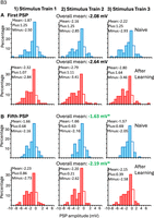
Distribution of fast PSP amplitudes in B3 from S1 neurons. Both EPSPs and IPSPs are present. There is a tendency for inhibition to increase after learning. Data are combined from all four subareas of the S1 cluster. Data are shown separately for the three stimulus trains of five spikes each (Stimulus Train 1, Stimulus Train 2, Stimulus Train 3) and for the first (A) PSP in a stimulus train and for the fifth (B) PSP in a stimulus train. Data are also shown separately for naive animals (blue) and for animals that had learned (red). The lack of a connection (PSP amplitude of zero) is indicated by an unshaded bar. In the upper left-hand corner of each histogram are shown the mean amplitudes of all measured PSPs including zeros, and also the mean amplitudes of the EPSPs (Plus) and of the IPSPs (Minus). Asterisks show a significant difference between naive animals and animals that had learned only for the fifth PSP (see the t-tests within the text). The overall means shown are the mean of all three measurements in the first or the fifth PSP elicited in a stimulus train. N = 88 connections were sampled in 14 preparations from naive animals, and N = 103 connections were sampled in 10 preparations from animals that had learned.
We used a four-way analysis of variance (mixed design, with learning and location as independent measures, and within stimulus train and between stimulus train plasticity as repeated measures) to examine the effects of (1) previous learning, (2) within-train plasticity, (3) between-train plasticity, and (4) location among the four subclusters of S1, as well as interactions between these variables (Table 1). The residuals in the ANOVA were close to normally distributed (Supplemental Fig. S1), justifying the use of the ANOVA for these data.
Results of a four-way ANOVA on data from synaptic connections to B3 as shown in Figure 1
The effect of previous learning as a main effect just approached significance (P = 0.053). The previously documented effects of repeated stimuli within a stimulus train and between stimulus trains (Hurwitz et al. 2024) were highly significant, with no interactions between these variables and previous learning. These data suggest that the effect of previous learning may be to increase inhibition to B3, although the effect is weak, without affecting PSP plasticity within a stimulus train or between stimulus trains, which remain as they are in naive animals. The possible effects of learning are not restricted to a particular subcluster of the S1 neurons, because there is no significant interaction between location and learning.
Source of the change in fast PSP amplitude
There was a significant increase in the mean resting potential of B3 after learning (Supplemental Fig. S2). This could lead to an increase in the amplitude of EPSPs, but such an increase was not seen. In contrast, an increase in net inhibition just missed being significant. An increase in net inhibition, if present, could arise from a larger number of inhibitory connections, at the expense of unconnected neurons, or of neurons with excitatory connections. Testing the distribution of excitation, inhibition, and lack of connection showed no significant difference between naive animals and animals that had learned (P = 0.465, χ2 test). Thus, the data suggested that if a net increase in inhibition is indeed present after learning, it affects the connectivity to B3 by increasing the amplitude of IPSPs. We tested this hypothesis directly by comparing the mean IPSP amplitudes of the three measures of the first IPSP and the three measures of the fifth IPSP. The mean difference of the first IPSP approached significance (P = 0.085, t = 1.74), and the mean difference of the fifth IPSP was significantly less after learning (P = 0.039, t = 2.09; df = 85; one-tailed t-tests) (Kolmogorov–Smirnov tests of normality for the first or the fifth IPSP [combined over the three stimulus trains], either in naive or in animals that had learned, showed that the distributions were not significantly different from normality [0.068 < P < 0.208]).
Effect of previous learning on slow PSPs
No significant differences were found in the amplitude of slow PSPs between ganglia from naive animals or from animals that had learned (for the average of the three stimulus trains: P = 0.32; for stimulus train 1: P = 0.87; for stimulus train 2: P = 0.83; for stimulus train 3: P = 0.98; two-tailed Mann–Whitney U-tests) (Supplemental Fig. S3A,B).
B4/B5
B4 and B5 are a pair of prominent neurons with widespread excitatory and inhibitory connections to other buccal ganglia neurons (Gardner 1977). B4/B5 inhibit motor neurons B3, B6, and B9, which innervate the I1/I3 muscles whose contraction effects much of the retraction. When animals reject food, they close and protract the radula on inedible material, pushing it out of the buccal cavity. They then open the radula to release the inedible material as they retract the radula back into the buccal cavity. During retraction, the radula passes through the lumen of the jaws, and if the jaws were to contract at that time, the jaws would force the radula closed, pulling the inedible material back into the buccal cavity. Thus, at the onset of retraction during rejection responses, B4/B5 fire strongly, inhibiting motor neurons B3, B6, and B9 that would otherwise close the jaws (Ye et al. 2006). B4/B5 also inhibits the B8a/b motor neurons (Gardner 1977) that close the radula (Morton and Chiel 1993a,b). Inhibition of B8a/b at the start of retraction contributes to radula opening at that time, contributing to a bias of motor activity away from ingestion, and toward rejection. B4/B5 fire more strongly during rejection than during ingestion (Warman and Chiel 1995; Jing and Weiss 2001, 2002; Ye et al. 2006; Sasaki et al. 2009). A previous report examining a single PSP from the S1 neurons to B4/B5 (Tam et al. 2020) showed that after learning with inedible food, the net excitation from the S1 neurons to B4/B5 is increased. This may be part of a mechanism to bias the response toward rejection after learning that food is inedible.
Effect of previous learning on fast PSPs
The distribution of the fast PSPs recorded in B4/B5 in preparations from naive animals and from animals that had previously learned that a food is inedible is shown in Figure 2A,B. As for B3, data are shown separately for each of the six fast PSPs measured (first and fifth PSP within a stimulus train, from three stimulus trains). The distributions of fast PSPs in B4/B5 were not normally distributed, showing large rightward tails. Data from preparations from naive animals and from animals that had learned are similar in many ways. In both, fast PSPs were exclusively excitatory. In addition, there were clear decreases in PSP amplitude from the first to the fifth PSP within a stimulus train, as well as tendencies for increases in PSP amplitude from the first to the second stimulus train (only for the first PSP within a stimulus train), but no further increase to the third stimulus train. Note that the rightward tails of the distributions are larger for preparations from animals that had learned.
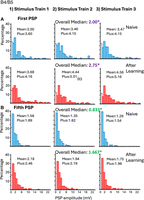
Distribution of fast PSP amplitudes in B4/B5 from S1 neurons. Only EPSPs are present. Excitation increases after learning. As above, data are combined from all four subareas of the S1 cluster, and data are shown separately for the three stimulus trains of five spikes each and for the first (A) and fifth (B) PSP in a stimulus train. Naive animals, blue; animals that had learned, red. The lack of a connection (PSP amplitude of zero) is indicated by an unshaded bar. Asterisks show significant effects between naive animals and animals that had learned (see statistics in the text). Note color coding of the median values and asterisks, showing the values that were compared and found to be significantly different. The overall medians shown are the median amplitudes derived from averaging all three measurements for the first or the fifth PSP elicited by a stimulus train. Medians are shown in this and in subsequent graphs, rather than means, to reflect the use of nonparametric statistics that measure differences in ranking, rather than parametric statistics measuring values. N = 127 connections were sampled in 25 preparations from naive animals, and N = 174 connections were sampled in 31 preparations from animals that had learned.
Because the distributions of fast PSPs in B4/B5 are not normally distributed, and because there are large differences between the first and fifth PSP within a stimulus train, data were analyzed separately for the first and fifth EPSP within a train, using nonparametric statistics. For each S1 neuron to B4/B5 connection, we calculated the mean amplitude of the three measurements of the first EPSP and the mean amplitude of the three measurements of the fifth EPSP. These values were then compared between connections recorded in naive animals and in animals that had learned. Unconnected cells were included in these comparisons and were assigned an amplitude of zero. For both the first and fifth EPSP, excitation was significantly larger in animals that had learned than in naive controls (first PSP: P = 0.0455; fifth PSP: P = 0.0257; two-tailed Mann–Whitney U-tests).
Source of the change in net fast PSP amplitude
An increase in excitation in animals that had learned could arise from an increase in the resting potential of B4/B5 after learning. However, such an effect was not seen (Supplemental Fig. S2). Increased excitation could arise from an increase in the percentage of connected S1 neurons to B4/B5 after learning (i.e., a decrease in unconnected neurons), or because the amplitude of EPSPs increases, or both. We examined these possibilities. There was a nonsignificant trend for fewer unconnected cells after learning (P = 0.073, χ2 test). In addition, there were significant increases after learning in EPSP amplitude for both the first and the fifth EPSP (first PSP: P < 0.0001; fifth PSP: P = 0.011; two-tailed Mann–Whitney U-tests). These findings indicate that increases in EPSP amplitude contribute to the increased excitation to B4/B5 and also suggest that increases in the number of excitatory connections might also contribute to the increased excitation.
Effects of learning on within stimulus train plasticity
In both preparations from naive animals, as well as those from animals that had learned, the amplitude of fast EPSPs decreases from the first to the fifth stimulus within a train of stimuli. Is within stimulus train plasticity affected by previous learning? The net reduction in EPSP amplitude (mean of the change in amplitude from the first to the fifth EPSP [fifth minus first] divided by the value of the first EPSP times 100) in naive animals was 48% of the initial amplitude; for animals that had learned, the mean decrease was 46% of the initial amplitude, indicating that previous learning is unlikely to affect within-train plasticity. Nonetheless, for each of the three stimulus trains, as well as for the average of the three stimulus trains, we compared the difference in amplitude between the first and fifth EPSP in naive animals to the difference between the first and fifth EPSP in animals that had learned. There were no significant differences between naive animals and animals that had learned in any of the three trains (first train: P = 0.936; second train: P = 0.290; third train: P = 0.165; false discovery rate [FDR]-corrected two-tailed Mann–Whitney U-tests) or in the average for all three trains (P = 0.271; Mann–Whitney U-test).
Effects of learning on between stimulus train plasticity
We also tested whether plasticity from one stimulus train to the next is affected by previous learning, and found no such differences (for the first PSP in a stimulus train: difference from first to second stimulus train: P = 0.19; difference from second to third stimulus train: P = 0.29; for the fifth PSP in a stimulus train: difference from first to second stimulus train: 0.93; difference from second to third stimulus train: P = 0.31; FDR-corrected two-tailed Mann–Whitney U-tests).
Subareas of the S1 cluster
We tested for differences in excitation produced by fast PSPs between ganglia from naive animals and from animals that had learned in each of the four subareas of S1 (Fig. 3A). The six measurements of connectivity (first and fifth EPSP in each of three stimulus trains, including zeros) were averaged, and we then tested for differences in the average between preparations from naive animals and from animals that had learned. There were significant increases in excitation after learning in S1-2 (P = 0.002) and in S1-3 (P = 0.010), with no significant differences in S1-1 (P = 0.795) or S1-4 (P = 0.912; two-tailed Mann–Whitney U-tests). The previous paper (Hurwitz et al. 2024) showed that excitation in preparations from naive animals from S1-1 to B4/B5 is larger than for the other three subareas of the S1 cluster. The lack of a significant effect of learning shows that this difference is maintained after learning. We then examined separately the first and fifth fast EPSP from each animal, to determine whether the increase was restricted to either of these. There were significant increases in excitation for the first (P = 0.004) and fifth (P = 0.038) fast EPSP in S1-2 and for the first (P = 0.045) and fifth (P = 0.045) fast EPSP in area S1-3, with no significant differences in either the first or the fifth fast EPSP in subareas S1-1 or S1-4 (P ≥ 0.776; FDR-corrected Mann–Whitney U-tests). Thus, the effects of learning are restricted to S1-2 and S1-3. Note that in area S1-1, excitation is higher than in other areas of the S1 cluster in naive animals. This increase was retained, but not changed, in animals that had learned.
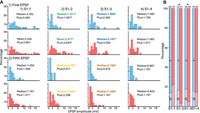
Connectivity to B4/B5 in the four subareas of the S1 cluster. Data are shown separately for ganglia taken from naive animals (blue) and from animals that had learned (red). (A) Distribution of the EPSP amplitudes in the four subareas of the S1 cluster. Data are shown separately for the first EPSP in the three trains, and of the fifth EPSP in the trains. Each graph shows the median amplitudes of all measured connections (median), including zeros (marked by an unfilled bar), and also median amplitudes of the EPSPs (plus), in which unconnected neurons are not included. Median amplitudes of EPSPs are increased after learning in S1-2 and S1-3. These increases are noted by bolded text and color-coded asterisks marking the bolded text. Significant increases in the amplitude of connected cells (plus) were restricted to the first EPSP in S1-2 (color-coded asterisk). (B) Distribution of excitatory connections (+) and unconnected cells (0). The number of connections in each bar is shown above the bar. There are significant decreases in the number of unconnected cells in subareas S1-2 and S1-3 in animals that had learned. Significant changes due to learning are noted by asterisks (see statistics in the text).
Increases of excitation in S1-2 and S1-3 in animals that had learned could arise because of an increase in the percentage of connected S1 neurons to B4/B5 after learning (i.e., a decrease in unconnected neurons), or because the amplitude of EPSPs increases, or both. We examined these possibilities. The distribution between connected and unconnected cells for each subarea is shown in Figure 3B. There were significant increases in the number of connected cells in areas S1-2 and S1-3 in animals that had learned, with no significant differences in the number of connected cells in areas S1-1 and S1-4 (for S1-1: P = 0.10; for S1-2: P = 0.045; for S1-3: P = 0.040; for S1-4: P = 0.862; χ2 tests).
We then examined whether there are differences in the amplitude of the EPSPs in the four subareas of the S1 cluster, by eliminating unconnected cells (plus amplitudes listed in Fig. 3A—distribution only of the filled bars). We examined separately the first and the fifth EPSP in the trains. The average amplitudes of the first EPSPs in the three trains and of the fifth EPSPs in the three trains were calculated for each of the four subareas of the S1 cluster and were compared. For the first EPSP in the train, there was a significant increase in EPSP amplitude for the EPSP in S1-2 (P = 0.033) with no significant differences in the other three subareas (for S1-1 and S1-4: P > 0.841; for S1-3: P = 0.10). There were no significant differences in EPSP amplitude between naive animals and animals that had learned for the fifth EPSP in the train (for S1-1: P = 0.470; for S1-2: P = 0.267; for S1-3: P = 0.161; for S1-4: P = 0.810; all comparisons are FDR-corrected two-tailed Mann–Whitney U-tests). Thus, for S1-2 there are both increases in the number of connected cells and in the amplitude of the first EPSP in a train. For S1-3 there is an increase in connectivity, with a trend to increased EPSP amplitude. Learning apparently does not affect connectivity in S1-1 or in S1-4. In S1-1: Connectivity is high in naive animals and remains high after learning.
Effect of previous learning on slow PSPs
There was no significant difference in the likelihood of an S1 neuron eliciting a slow PSP between ganglia from naive animals and ganglia from animals that had learned (P = 0.99; χ2 test). In both naive animals and in animals that had learned, slow PSPs were overwhelmingly excitatory, but nonetheless one S1 neuron from a naive animal and four S1 neurons from animals that had learned showed biphasic slow PSPs (i.e., the slow PSPs had both excitatory and inhibitory components).
Effects of learning were determined for each of the three trains, as well as the mean amplitudes for the three trains combined (average values of the three trains). In addition, separate tests were performed when unconnected cells were included (scored as zero), and when only connected cells were included. There were no significant differences between preparations from naive animals and from those that had learned (0.29 < P < 0.068; Mann–Whitney U-tests) (Supplemental Fig. S4A,B). Thus, previous learning apparently does not affect the slow PSPs.
We also tested for possible differences in slow PSPs in B4/B5 from the four subareas of the S1 cluster. For the averaged amplitude of the three stimulus trains, there were no significant differences between animals that had learned and naive animals in any of the four subareas of the S1 cluster (0.09 < P < 0.68; two-tailed Mann–Whitney U-tests) (not shown).
B8a/b
B8a/b are motor neurons innervating the radula closer muscle (Morton and Chiel 1993a,b). The neurons are active during different phases of feeding in different behaviors. In rejection, B8a/b are active during protraction, but in ingestion they are active during retraction. The previous work (Tam et al. 2020) showed that net connectivity in response to a single presynaptic spike is unchanged as a result of learning.
Effect of previous learning on fast PSPs
Figure 4A,B shows the distribution of the fast PSPs recorded in B8a/b from preparations from naive animals and from animals that had previously learned that food is inedible. In both preparations from naive animals and in animals that had learned, there were both excitatory and inhibitory connections, with the net connection (mean or median of all connections, including zero) being excitatory. In addition, the tendency for both excitation and inhibition to decrease in amplitude within stimulus trains was present in both naive animals and in animals that had learned. There was no tendency for changes among the three stimulus trains in either naive animals or in animals that had learned.
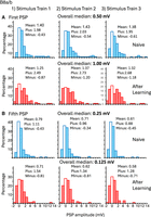
Distribution of fast PSP amplitudes in B8a/b from S1 neurons. Both excitation and inhibition are present. After learning, amplitudes of both excitation and inhibition increase, offsetting one another. Conventions within the figure are as those in Figures 1 and 2. Data are shown separately for the three stimulus trains of five spikes each and for the first (A) and fifth (B) PSP in a stimulus train. The overall medians shown are the median amplitudes derived from averaging all three measurements for the first or the fifth PSP elicited by a stimulus train. N = 142 connections were sampled in 28 preparations from naive animals, and N = 100 connections were sampled in 16 preparations from animals that had learned.
As for B4/B5, nonparametric statistics were used to test for differences between naive animals and animals that had learned, because of the long rightward tails, especially in ganglia from naive animals. As for B4/B5, we calculated the mean amplitude of the three measurements of the first EPSP, and the mean amplitude of the three measurements of the fifth EPSP. These values were then compared for ganglia from naive animals and from animals that had learned. Unconnected cells were included in these comparisons and were assigned an amplitude of zero. There was no net change in connectivity (EPSP, IPSPs, and zero combined) between ganglia from naive animals and animals that had learned for either the median of the first or the median of the fifth PSP in the stimulus train (first PSP: P = 0.912; fifth PSP: P = 0.347; two-tailed Mann–Whitney U-tests). Thus, there is no net change (EPSP, IPSPs, and zero combined) in connectivity from the mechanoafferents to B8a/b.
Changes in excitation and inhibition
Further analysis of the data showed that previous learning had affected synaptic connectivity. There was no significant change in the B8a/b resting potential after learning (Supplemental Fig. S2), so such a change could not contribute to changes in PSP amplitudes. The distribution between excitation, inhibition, and lack of connectivity was affected by learning (P = 0.021; χ2 test). Inhibitory connections after learning increased from ∼11% to ∼21% at the expense of excitatory connections, which decreased from ∼69% to ∼60%, with no net change in unconnected cells, which remained at ∼19%.
The amplitudes of both excitatory and inhibitory connections were also increased, essentially canceling one another, and therefore producing no net change. For excitatory synapses, there were significant increases for the first and fifth PSP in all three stimulus trains: (first stimulus train, first PSP: P = 0.007; first stimulus train, fifth PSP: P = 0.011; second stimulus train, first PSP: P = 0.007; second stimulus train, fifth PSP: P = 0.022; third stimulus train, first PSP: P = 0.007; third stimulus train, fifth PSP: P = 0.007; Mann–Whitney U-tests, with FDR correction). For inhibitory synapses, there were significant increases in the amplitude in both the first and second stimulus trains (first stimulus train, first PSP: P = 0.021; first stimulus train, fifth PSP: P = 0.050; second stimulus train, first PSP: P = 0.011; second stimulus train, fifth PSP: P = 0.071), with no increases in the amplitude of the inhibition in the third stimulus train (third stimulus train, first PSP: P = 0.156; third stimulus train, fifth PSP: P = 0.703; Mann–Whitney U-tests, with FDR correction).
In sum, for both excitation and inhibition, the amplitude of the connections increased, but the number of excitatory connections decreased, whereas the number of inhibitory connections increased.
Effects of learning on within stimulus train plasticity
We tested the possible effects of learning on within-stimulus train plasticity (i.e., the decrease in PSP amplitude from the first to the fifth PSP within a train). This was calculated as above for B4 (difference between the mean values of the first and fifth EPSP [fifth minus first] divided by the mean value of the first EPSP times 100). The decrement of the amplitude of EPSPs in preparations from animals that had learned was larger, with respect to the decrement in preparations from naive animals (P = 0.014), with no significant difference in the decrement of IPSP amplitudes (P = 0.11; two-tailed Mann–Whitney U-tests). In preparations from naive animals, the decrement in EPSP amplitude was 42.7%. In preparations from animals that had learned, the decrement was 52.0%.
Effects of learning on between stimulus train plasticity
We also tested whether the effects of the three repetitions of the stimulus trains are affected by previous learning. As above, we measured separately EPSPs and IPSPs, and differences from the first to the second stimulus train, and from the second to the third stimulus train, for both the first and the fifth PSPs with the stimulus trains. There were no significant differences as a result of previous learning (0.06 < P < 0.96; two-tailed Mann–Whitney U-tests).
Subareas of the S1 cluster
Because there were significant increases in the amplitude of both EPSPs and IPSPs, we attempted to determine whether these changes were localized to specific areas of the S1 cluster. For the IPSPs, there were too few samples to test, particularly for ganglia from naive animals. For EPSPs, as for B4/B5, we averaged the six values measured (first and fifth EPSP in the three stimulus trains), and the increase in EPSP amplitude in S1-1 approached significance (P = 0.056), but there were no significant differences in S1-2 (P = 0.424), in S1-3 (P = 0.285), or S1-4 (P = 0.105; two-tailed Mann–Whitney U-tests). Thus, we were unable to localize the changes in either EPSP or IPSP amplitude to a particular subarea of the S1 neurons.
Effect of previous learning on slow PSPs
There was no significant difference in the likelihood of an S1 neuron eliciting a slow PSP between ganglia from naive animals and ganglia from animals that had learned (P = 0.99; χ2 test). In addition, there were no significant differences in the amplitude of either slow EPSPs or slow IPSPs (averages of the three slow PSPs elicited in the three stimulus trains) between naive animals and those that had learned (EPSPs: P = 0.944; IPSPs: P = 0.992; two-tailed Mann–Whitney U-tests) (Supplemental Fig. S5A,B).
B31/B32
These neurons have a dual function (Hurwitz et al. 1994): In addition to innervating the primary muscle mediating radula protraction (Hurwitz et al. 1996), they decide on initiating a protraction–retraction consummatory feeding sequence (Hurwitz et al. 2008). Adequate depolarization initiates a plateau potential, which is the decision to initiate a bout of consummatory feeding behavior (Hurwitz et al. 2008). The previous work (Tam et al. 2020) showed that after learning, there are no changes in connectivity to B31/B32 in response to a single spike to S1 neurons.
Effects of previous learning on fast PSPs
The distribution of the fast PSPs recorded in B31/B32 in preparations from naive animals and in preparations from animals that had previously learned that food is inedible is shown in Figure 5A,B. Distributions of connections to B31/B32 were similar in ganglia from naive animals and in those from animals that had learned, in that there were large percentages of unconnected S1 cells in both, and connections were exclusively excitatory. As in the other follower cells, in ganglia from both naive animals and from animals that had learned, there were decreases in both net excitation and in the amplitude of EPSPs within stimulus trains. There was little difference in excitation or EPSP amplitude among the three stimulus trains. There was a general tendency for excitation to be decreased after learning that food is inedible, particularly for the rightward tail of the distribution to be decreased after learning.
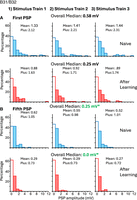
Distribution of fast PSP amplitudes in B31/B32 from S1 neurons. Only EPSPs are produced. Excitation is increased for the fifth PSP within a train after learning. Conventions within the figure are as those in Figures 1, 2, and 4. Data are shown separately for the three stimulus trains of five spikes each and for the first (A) and fifth (B) PSP in a stimulus train. Color-coded asterisks show significant effects between naive animals and animals that had learned (see statistics in the text). The overall medians shown are the median amplitudes derived from averaging all three measurements for the first or the fifth PSP elicited by a stimulus train. N = 85 connections were sampled in 12 preparations from naive animals, and N = 87 connections were sampled in 12 preparations from animals that had learned.
Initially, we compared the average amplitude of the first and the average amplitude of the fifth fast PSP in the three stimulus trains, in preparations from naive animals and from animals that had learned. Nonconnected neurons were given a value of zero. There was no significant difference in excitation for the first PSP in a stimulus train (P = 0.930), but the fifth PSP was significantly lower in animals that had learned (P = 0.019; two-tailed Mann–Whitney U-tests). There was a significant increase in the resting potential in B31/B32 after learning (Supplemental Fig. S2), which would lead to an increased excitation, rather than a decrease.
Source of the change in fast PSP amplitude
We tested whether the significant decrease in excitation for the fifth PSP after learning was caused by an increase of unconnected neurons after learning or by a decrease in the amplitude of connected neurons. There was a significant increase in unconnected neurons after learning (P = 0.024; χ2 test), with no significant differences in the amplitude of either the first (P = 0.258) or the fifth EPSP (P = 0.072; two-tailed Mann–Whitney U-tests), although the difference for the fifth EPSP approached significance.
Effects of learning on within stimulus train plasticity
A possible decrease in excitatory connectivity in the fifth, but not the first PSP in a stimulus train suggests that the within-train decrease in PSP amplitude may be larger in preparations from animals that have learned than in those from naive animals. A test of the percent decrease in EPSP amplitude from the first to the fifth EPSP within a train (averaging the values of the EPSPs over the three trains) showed a significantly larger decrease from the first to the fifth EPSP in preparations from animals that had learned (60.5% decrease), with respect to preparations from naive animals (55.6% decrease) (P = 0.01; two-tailed Mann–Whitney U-test), which is consistent with a reduced EPSP amplitude for the fifth EPSP in a stimulus train.
Effects of learning on between stimulus train plasticity
We also tested whether previous learning affected differences between the first and second and between the second and third stimulus trains. Differences from the first to the second stimulus train and from the second to the third stimulus train were calculated separately for first EPSPs and fifth EPSPs. There were no significant differences (0.26 < P < 0.842; two-tailed Mann–Whitney U-tests).
Subareas of the S1 cluster
We tested for differences in connectivity of fast PSPs between naive animals and animals that had learned (including zeros) in each of the four subareas of S1. As above, the measurements of connectivity (first and fifth PSPs in three stimulus trains) were averaged, and these values were compared over the four subareas of the S1 cluster. There were no significant differences in connectivity after training in any of the four subareas (0.147 < P < 0.952; two-tailed Mann–Whitney U-tests). However, because there is a significant difference only for the fifth PSP with a stimulus train between preparations from naive animals and from animals that had learned, we also examined possible changes in localization for the fifth PSP. A significant decrease in PSP amplitude was found in S1-4 (P = 0.038), and decreases approached significance in S1-3 (P = 0.057) and S1-1 (P = 0.087), with no indication of a difference in S1-2 (P = 0.397; one-tailed Mann–Whitney U-tests).
Effects of previous learning on slow PSPs
The effects of learning were determined for each of the three trains, as well as the mean values for the three trains combined. There were no significant differences between preparations from naive animals and from those that had learned (0.103 < P < 0.250; Mann–Whitney U-tests). Thus, previous learning apparently has no effect on the slow PSPs (Supplemental Fig. S6A,B).
We also tested whether slow PSPs were different from the four subareas of the S1 cluster. As above, for each preparation, the mean value of the slow PSP recorded in each of the three stimulus trains was calculated. We found no differences between naive animals and those that had learned (0.126 < P < 0.873; FDR-corrected two-tailed Mann–Whitney U-tests) in any of the four subareas of the S1 cluster (not shown).
B61/B62
B61/B62 are motor neurons innervating the I2 muscle, whose contraction causes protraction of the radula (Hurwitz et al. 1994, 1996). Protraction is the power phase in rejection (Morton and Chiel 1993a,b). A previous report examining a single PSP from the S1 neurons to B61/B62 (Tam et al. 2020) showed that net excitation from the S1 neurons to B61/B62 increases after learning with inedible food.
The distribution of the fast PSPs recorded in B61/B62 in naive animals and in animals that had previously learned that food is inedible is shown in Figure 6A,B, combined from all four of the subareas of the S1 cluster. Data are shown separately for the first and fifth PSP within the train, and for ganglia taken from naive animals (blue) and from animals that had learned (red). As in B3 and B8a/b, both excitatory and inhibitory connections were present, but unlike for B3, the connections are primarily excitatory. There is a consistent tendency for excitation to be increased after learning. Because of the large number of unconnected cells, and the relatively large number of cells of opposite signs, we used nonparametric statistics in examining the connectivity to B61/B62.
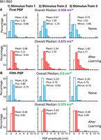
Distribution of fast PSP amplitudes in B61/B62 from S1 neurons. Both EPSPs and IPSPs are produced. Net excitation is increased, in part because there are few IPSPs after learning. Conventions within the figure are as those in Figures 1, 2, 4, and 5. Data are shown separately for the three stimulus trains of five spikes each and for the first (A) and fifth (B) PSP in a stimulus train. Color-coded asterisks show significant effects between naive animals and animals that had learned (see statistics in the text). The overall medians shown are the median amplitudes derived from averaging all three measurements for the first or the fifth PSP elicited by a stimulus train. N = 58 connections were sampled in eight preparations from naive animals, and N = 65 connections were sampled in eight preparations from animals that had learned.
Effects of previous learning on fast PSPs
As we did for the other followers, we compared the average amplitude of the first, and the average amplitude of the fifth fast PSP in the three stimulus trains, in preparations from naive animals and from animals that had learned. Nonconnected neurons were given a value of zero. For both the first PSP (P = 0.014) and the fifth PSP (P = 0.033; two-tailed Mann–Whitney U-tests), excitation was increased after learning.
Source of the change in fast PSP amplitude
The increased excitation after learning could arise from an increase in the resting potential of the B61/B62 neurons after learning. However, we found no significant change in the resting potential between neurons from naive ganglia and from those that had learned (Supplemental Fig. S2). The increase in excitation could arise from an increase in the number of neurons with excitatory connections, at the expense of neurons with either inhibitory connections or no connection, or to a relative increase in the amplitude of the excitatory connections and a relative decrease in the amplitude of inhibitory connections, or to a combination of processes. In ganglia removed from naive animals, 43.5% of the S1 neurons had excitatory connections to B61/B62. In contrast, in ganglia from animals that had learned, 56.9% of the neurons had excitatory connections with B61/B62. There was a corresponding decrease in the proportion of inhibitory connections in ganglia from animals that had learned from 12.9% to 3.1%, with a minimal change in the proportion of unconnected neurons, from 43.5% to 40.0%. The difference in distribution between the connections in preparations from naive animals and from animals that had learned was significant (P = 0.021; χ2 test). Thus, part of the increase in excitation stems from an increase in the number of excitatory connections, with an almost complete elimination of inhibitory connections.
We also examined the mean amplitude of the first and fifth excitatory connections within the stimulus trains, to determine whether learning affected EPSP amplitude, in addition to affecting their probability. We did not test possible changes in fast IPSP amplitude, because there were very few IPSPs in animals that had learned. For both the averaged first and the averaged fifth EPSP, the amplitudes were significantly increased after learning (for first EPSP: P = 0.014; for fifth EPSP: P = 0.033; two-tailed Mann–Whitney U-tests). Thus, part of the increased excitation also stems from an increase in the amplitude of both the first and fifth EPSPs.
Effects of learning on within stimulus train plasticity
For EPSPs, we tested whether there are differences in the relative decrease in amplitude from the first to the fifth EPSP within a stimulus train between naive animals and animals that had learned. As above, data from the three stimulus trains were combined. There was no significant difference in decreases in EPSP amplitude from the first to the fifth EPSP (P = 0.276; two-tailed Mann–Whitney U-tests). We did not test for changes in IPSP amplitude, because there were almost no IPSPs in animals that had learned.
Effects of learning on between stimulus train plasticity
For plasticity arising from the repetition of the three stimulus trains, we tested whether PSP amplitude increased, decreased, or stayed the same, from the first to the second stimulus train, and from the second to the third stimulus train. When examining changes in the first EPSP between the first and second stimulus trains, both preparations from naive animals and from animals that had learned showed significant increases in amplitude (naive: P = 0.003; learning: P < 0.0001; χ2 tests), with no significant difference between preparations from naive animals and from those that had learned (P = 0.368; two-tailed Mann–Whitney U-tests). Neither ganglia from naive animals nor from animals that had learned showed significant changes in the distribution of excitation and inhibition for the fifth EPSP within a stimulus train (naive: P = 0.905; learning: P = 0.717; χ2 tests), or for the first EPSP between the second and third stimulus trains (naive: P = 0.602; learning: P = 0.195; χ2 tests).
Subareas of the S1 cluster
We tested for differences in connectivity of fast PSPs between ganglia from naive animals and from animals that had learned (including zeros) in each of the four subareas of S1. As above, the six measurements of connectivity were averaged. There were significant differences in connectivity after training in S1-1 and S1-3 (S1-1: P = 0.015; S1-3: P = 0.018; two-tailed Mann–Whitney U-tests), with no significant differences in S1-2 (P = 0.200) or S1-4 (P = 0.548; two-tailed Mann–Whitney U-tests).
Effects of previous learning on slow PSPs
Both depolarizing and hyperpolarizing slow PSPs were elicited in B61/B62. There was no significant difference in the distribution of the slow PSP between preparations from naive animals and from animals that had learned. In addition, there was no significant difference in the likelihood of neurons displaying slow PSPs between ganglia from naive animals and from animals that had learned (P = 0.794; χ2 test), or in their overall average amplitude (P = 0.787; two-tailed Mann–Whitney U-tests), or for any of the three stimulus trains (for stimulus train 1: P = 0.726; for stimulus train 2: P = 0.749; for stimulus train 3: P = 0.976; two-tailed Mann–Whitney U-tests) (Supplemental Fig. S7A,B).
We also tested whether slow PSPs were different from the four subareas of the S1 cluster. As above, the mean of the three stimulus trains was calculated. We found no differences between naive animals and those that had learned (0.101 < P < 0.865; FDR-corrected two-tailed Mann–Whitney U-tests, not shown).
Summary of changes
There were no significant changes in connectivity of the slow connections to the five followers. In contrast, there were significant changes in fast connections to all of the five followers. However, changes differed for each of the followers. The significant changes in the connectivity of the fast connections from the S1 neurons to the five followers are summarized in Figure 7.
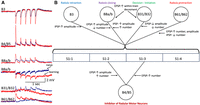
Summary of significant changes in fast connectivity from the S1 cells to the five followers. (A) Examples of recordings from preparations from naive animals (blue) and from animals that had learned (red). Note that fast EPSPs from B4/B5 and from B61/B62 increase in amplitude, and that fast EPSPs from B31/B32 decrease in amplitude. Fast IPSPs from B3 increase in amplitude. Both fast EPSPs and fast IPSPs in B8a/b increase in amplitude, and therefore recordings showing both effects are shown. (B) General summary. When there are no differences in connectivity from the four subareas of the S1 cluster, the connecting line from each of the followers is from the center of the bracket connecting the four subareas. Connecting lines to B4/B5 are shown below the bars depicting the four subareas of the S1 cluster, to illustrate the separate effects of learning from the different subareas. The function of each follower is shown above or below its name. Arrows pointing to a connecting line show the direction of change in a connection. Changes in EPSP or IPSP amplitude, or in the number of connections, are shown when these connections are significant. Note that changes in the amplitude of IPSPs in B3 and of the EPSPs in B31/B32 were found only for the last stimulus in the train of stimuli of the S1 neurons.
Discussion
We have expanded a previous study (Tam et al. 2020) that compared the properties of connections between the S1 mechanoafferents and five followers in naive animals and in animals that had learned that a food is inedible. The previous study (Tam et al. 2020) examined only the first PSPs in response to the first spike of the first stimulus train delivered to an S1 neuron. The immediately preceding report (Hurwitz et al. 2024) showed that connections between the S1 mechanoafferents and its followers in naive animals are rich and complex. The complexity is revealed largely because we have now examined multiple PSPs in followers in response to trains of stimuli, rather than to a single stimulus. After documenting this complexity in naive animals (Hurwitz et al. 2024), we were able to compare in greater detail how learning may change these connections. The additional data allowed us to determine possible changes in (1) the relative proportions of connected cells (excitatory, inhibitory, or no connection); (2) the amplitudes of excitatory and inhibitory connection; plasticity (3) within or (4) between stimulus trains; (5) changes in slow PSPs that are elicited only by a stimulus train; and (6) changes in connectivity from the different subareas of S1.
With respect to fast PSPs from S1 to their followers, this study is generally consistent with Tam et al. (2020), in finding increases in fast excitation to B4/B5 and to B61/B62, and a tendency to increased fast inhibition to B3, with no net change in connectivity to B8a/b. The earlier work indicated that these changes are consistent with an increased bias to reject food or nonfood objects after learning. Behavioral experiments confirmed that such a bias is present in animals that had learned (Tam et al. 2020).
Our new findings extend the previous work (Tam et al. 2020) by showing that the excitation to B31/B32 was decreased after learning. We did not find this previously, because the decrease was restricted to the fifth PSP in a stimulus train, which was not previously explored. One behavioral consequence of learning is a decreased likelihood to respond to food. A major function of B31/B32 is to initiate a consummatory feeding response (Hurwitz et al. 1994, 2008). After learning that food is inedible, animals stop responding to food more quickly. The decreased excitation to B31/B32 is consistent with a role of this synapse in contributing to this parameter of memory after learning. The new findings highlight the importance of using multiple trains of stimulus (a more physiologically relevant stimulus) to examine synaptic changes following learning.
Based on the strong decrement in the amplitude of fast PSPs from the first to the fifth PSP within a stimulus train, the previous paper (Hurwitz et al. 2024) suggested that the connections from S1 to its followers could signal the movement of food on the radula, perhaps outward slippage of food as animals attempt but fail to swallow food. In addition, the pattern of connections from the S1 mechanoafferents to their followers suggested that these connections bias toward rejection responses or the release of food. Increases in excitation to B4/B5 and to B61/B62, and of a tendency to increased inhibition to retractor motor neuron B3, are consistent with an amplification of this bias after learning. These changes are consistent with the behavioral effects of learning that food is inedible. In animals that have learned, memory is expressed in part by a reduction in the time that food remains within the mouth eliciting attempts to swallow (Susswein et al. 1986; Botzer et al. 1998; Schwarz et al. 1988), before animals completely stop responding to the food. Amplification of the patterns of connectivity to B3, B4/B5, and B61/B62 by learning may contribute to earlier release of food, and less time spent in the mouth.
Although there was no net change in connectivity to B8a/b, there were significant increases in both excitation and inhibition, which canceled one another. B8a/b differs from the other followers that we examined in that it is active during different phases of feeding in different behaviors. It is primarily active during protraction in rejection but is primarily active during retraction in other feeding behaviors (Morton and Chiel 1993b; Jing and Weiss 2001, 2002; Sasaki et al. 2009). If the effect of learning is to increase a bias to rejection or to the release of food, increases in excitation may reflect a tendency to drive B8a/b more strongly during protraction, and increases in inhibition may reflect a tendency to prevent B8a/b activity during retraction. If this speculation is correct, it implies that in natural behavior different populations of S1 neurons may be active in different phases of feeding behavior.
The amplitude of the synaptic connections to the followers was generally not normally distributed. Even in ganglia from naive animals, there were long rightward tails in predominantly excitatory connections, such as to B4/B5, B8a/b, B31/B32, and B61/B62. Learning enhanced these tails, suggesting that learning might act primarily on a subpopulation of particularly large connections. Future studies will be required to identify the relevant S1 neurons. Changes in connectivity after learning are likely to cause relatively subtle biases in behavior, rather than turning on or off a behavior.
Examining the effects of stimulus trains allowed us to determine whether previous learning affects within-train or between-train plasticity. In three of the five followers, within-train and between-train plasticity were present in both naive animals and in animals that had learned, with no significant differences between them. One exception was B31/B32, in which the within-train decrease in EPSP amplitude was larger after learning, thereby leading to a weakening in the synaptic connection from the S1 cells to B31/B32 late in a stimulus train. The second exception was B8a/b, in which within-train EPSP decrement was larger after learning, emphasizing the effect of learning on the earlier EPSPs within a train.
Examining the effects of three stimulus trains also provided us an opportunity to determine whether slow PSPs are affected by previous learning. We did not find the effects of learning on slow PSPs. However, it is possible that longer stimulus trains, or additional stimulus trains, might have elicited larger slow PSPs, and these might have been affected by the learning. Differences in effects of previous learning on synaptic plasticity between fast and slow PSPs may reflect differential effects of learning on the different receptor types for transmitters signaling fast and slow PSPs or may reflect differential effects on the presynaptic mechanisms underlying the release of transmitters that produce fast and slow PSPs, with only either the receptors or the release of transmitters for the fast PSPs affected by the learning. Mechanisms for restricting plasticity to a single type of synapse (fast or slow), when a connection may use more than one type, is of interest, which has rarely been described previously.
Given that S1 neurons consist of a cluster of neurons, dividing S1 into four subclusters allowed us to examine whether changes due to learning are localized to a specific subarea. When localization to a subcluster was not found, it is important to note that the number of connections sampled from each subcluster is less than the number of connections sampled overall, and therefore more samples of the connections might have shown localization or might have revealed within a subcluster a small number of neurons that are affected by learning. One follower, B4/B5, showed localization of learning-dependent plasticity in specific subareas of S1. B4/B5 was also unusual in that it was the only follower in which there were differences in connectivity from different subareas in ganglia from naive animals (Hurwitz et al. 2024). However, in preparations from naive animals, connectivity was largest in a different subcluster than those affected by learning. In addition, B31/B32 showed some localization of learning to area S1-4, and B61/B62 showed localization to S1-1 and S1-3. Differences in connectivity from different subareas of the S1 cluster are likely to reflect some difference in function. Future studies on the sensory responses of the S1 cells will be needed to understand why different areas produce different amplitude responses in both ganglia from naive animals and from animals that have learned, and why such differences are expressed only in some followers.
Comparison to learning-dependent changes in other mechanoafferents
The mechanoafferent populations in the abdominal and pleural ganglia of Aplysia have been intensively studied, because their synaptic outputs are regulated by experiences leading to short-term and long-term memories (Byrne and Kandel 1996; Byrne and Hawkins 2015). Molecular and synaptic mechanisms underlying short-term and long-term memory that were first identified in these synapses have subsequently been found to have a role in learning and memory in mammalian nervous systems (Kandel 2002; Pittenger and Kandel 2003). In the abdominal and pleural ganglia, learning causes changes in synaptic strength (Castellucci et al. 1970; Walters et al. 1983; Bailey and Chen 1988) or in the number of afferent neurons that are connected to a follower (Castellucci et al. 1978). Different types of learning have uniform effects on synaptic connections. The synaptic effects are consistent with changes in behavior: Sensitization that causes an increase in a withdrawal response causes increases in the amplitude of synapses and in the number of connections (Bailey and Chen 1988), whereas habituation causes a decrease in a withdrawal response and has opposite effects on synapses (Castellucci et al. 1978; Bailey and Chen 1988). In contrast, in our study, learning that leads to a decrease in response to food had different effects on different synapses: Net fast excitation to B8a/b and B4/B5 was increased, but net fast excitation to B31/B32 was decreased. In addition, there was a tendency for net fast inhibition to B3 and to B8a/b to increase. Thus, changes in connectivity after learning in the buccal ganglia are seemingly more complex than in the abdominal and pleural ganglia. The increased complexity is also reflected in the balance between increases in PSP amplitude and the number of connections. In B4/B5, both the amplitude of fast EPSPs and the number of connected neurons increased. In contrast, an opposite effect was seen in B31/B32: Both the amplitude and the number of EPSPs decreased. In B8a/b there were increases in the amplitude of both fast EPSPs and fast IPSPs. In B3 only the amplitude of fast IPSPs increased. However, even in the abdominal ganglion, short-term sensitization of the gill and siphon withdrawal reflex is associated with a number of parallel changes (Frost et al. 1988), including presynaptic facilitation of connections from mechanoafferents to their followers, presynaptic inhibition of a connection to a recurrent inhibitor, posttetanic potentiation of excitation to a key excitatory interneuron, and increased firing in a follower. Thus, the mechanisms of even simple learning are not restricted to a unitary effect at a single synapse.
Long-term learning that food is inedible also produced qualitatively different changes in connectivity that were not seen after long-term learning affecting withdrawal effects. One prominent effect was a change in the distribution between excitation and inhibition. Thus, in B61/B62, after learning, inhibitory connections virtually disappear, and the percentage of excitatory connections increases, suggesting that inhibition turns into excitation. In B8a/b there was an opposite effect: After learning, there was a significant increase in the number of inhibitory connections at the expense of excitatory connections. These changes could occur postsynaptically, by placing different receptors into postsynaptic sites, or presynaptically, by using different transmitters for excitation and inhibition and differentially regulating the expression of the different transmitters in synaptic terminals after learning. It is important to note that the same population of S1 neurons is involved in the different types of synaptic change seen in the different followers, implying that mechanisms of synaptic change may be different in different axonal branches of the same presynaptic neuron. Mechanisms for producing different changes in different branches of a neuron might be postsynaptic or might be presynaptic. Branch-specific plasticity has been documented in Aplysia mechanoafferents (Clark and Kandel 1993; Martin et al. 1997). Opposite effects in different branches of the same Aplysia neuron after learning have not been previously shown. However, such effects have been observed in the release of acetylcholine (ACh) from Kenyon cells in the Drosophila mushroom body to different mushroom body output neurons, in which learning causes increases and decreases in release to different output neurons (Stahl et al. 2022).
Changes in the buccal ganglia mechanoafferent to motor neuron synapses after learning are only part of the story
In addition to expanding our information on synaptic plasticity in mechanoafferent outputs caused by learning, this study provides important information on another aspect of understanding the neural basis of learning and memory. The learning task that we have examined is behaviorally more complex than a simple withdrawal reflex. Learning that food is inedible causes a number of different changes in behavior. Memory is expressed by (1) a shorter time to stop responding to inedible food (Susswein et al. 1986; Botzer et al. 1998; Schwarz et al. 1991); (2) specificity of memory to the taste and texture of the inedible food (Susswein et al. 1986; Schwarz et al. 1988); (3) changes in motor pattern before animals stop responding to inedible food (Susswein et al. 1986); and (4) a bias in animals that had learned to reject a nonfood stimulus (Tam et al. 2020). In the Aplysia CNS, both the buccal and the cerebral ganglia are involved in controlling feeding. The buccal ganglion contains a pattern-generating network, which includes sensory, inter-, and motor neurons, whereas the cerebral ganglion contains higher-order neurons that are activated by sensory afferents responding to food and in turn project to the buccal ganglion to initiate or modulate its activity. We have presented evidence that the various changes in behavior arise at different neural sites in different ganglia (McManus et al. 2019; Levy et al. 2023) by different mechanisms. Thus, the changes in the synaptic output of the buccal S1 neurons documented in this report are only a fraction of the neural changes that lead to an integrated change in behavior after learning. Savings in the time to stop responding to food, and food specificity, are likely to arise as a result of a decrease in response of command-like neurons in the cerebral ganglion to ACh released by afferents responding to food (McManus et al. 2019; Levy et al. 2023). The increased bias to reject food can be partially explained by some of the changes between the S1 neurons and its followers that were documented previously, as well as in this report. We have now presented data suggesting that some of the behavioral changes in response to food that occur before Aplysia stop responding to food may be explained by the changes in synaptic output documented in this report.
Our finding that different connections from the mechanoafferents to their followers are affected in different ways is consistent with those previously found in both vertebrates and invertebrates, in that learning and memory arise via multiple neural mechanisms acting in parallel. Parallel processing occurs at many levels. Within a specific synapse modified by learning, both presynaptic and postsynaptic changes can contribute to memory in vertebrates and invertebrates (Bekkers and Stevens 1990; Lechner and Byrne 1998; Roberts and Glanzman 2003; Nicoll 2017; Pribbenow et al. 2022), and multiple parallel processes can contribute to memory formation even on one side of the synapse (Byrne and Kandel 1996). In addition, multiple parallel changes at a number of neural sites can contribute to a specific behavioral change (Frost et al. 1988). Because learning may also produce a number of integrated behavioral changes, learning and memory may be localized to different neural sites, each responsible for different aspects of behavioral change (Cohen and Squire 1980; Milner et al. 1998). In particular, the Aplysia learning paradigm we are studying is an advantageous model system for studying changes in multiple neural sites affected by learning.
Materials and Methods
Animals
The population examined in this study is the same as in a previous study (Tam et al. 2020), but has been reanalyzed from the original raw data. In addition to examining more connections from more preparations, the new analysis allowed us to examine the effects of learning on stimulus trains, rather than on single spikes. Examining the effects of stimulus trains allowed the examination of plasticity within and between stimulus trains. In addition, the use of stimulus trains, rather than single spikes, allowed us to examine the possible effects of learning on slow PSPs elicited only by stimulus trains. The reanalysis also allowed examining the possible differences of connectivity from different subareas of the S1 mechanoafferent clusters.
Aplysia californica weighing 50–250 g were purchased from Marinus Scientific, and were stored in 600 L tanks filled with natural Mediterranean seawater maintained at 17°C. The animals were fed two to three times weekly with Ulva lactuca gathered from the Mediterranean coasts of Israel, or purchased from Seakura (https://www.seakura.co.il/en/), and stored frozen until used. A total of 501 connections from S1 cells to followers in the buccal ganglia from 80 naive animals, and 529 connections in the buccal ganglia from 54 animals that had learned were used in this study.
Training and testing memory
As in numerous previous studies examining learning that food is inedible in Aplysia (Susswein et al. 1986; Katzoff et al. 2002, 2006; Levitan et al. 2012), 24 h before being trained, animals were transferred to 10 L experimental aquaria that were maintained at room temperature (23°C). They were kept two to an aquarium, with the two animals separated by a partition allowing the flow of water. As in previous studies (Susswein et al. 1986), the animals were trained with inedible food, the seaweed Ulva wrapped in a plastic net. The food induced biting, leading to food entering the buccal cavity, where it induced attempts to swallow. Netted food cannot be swallowed, and it produces repetitive failed swallows. When the unswallowed food subsequently leaves the buccal cavity, the experimenter continues holding it touching the lips, inducing further bites, entries into the buccal cavity, and failed swallows. As training proceeds, many bites fail to cause entry of food into the mouth. When food does enter the mouth, it stays within the mouth for progressively shorter periods, eliciting fewer attempted swallows. Training proceeded until the animals stopped responding to food, which was defined as a lack of entry of food into the mouth for 3 min. Data were included only from animals in which food in the mouth elicited failed attempts to swallow for at least 130 sec. Animals displaying less time attempting to swallow are relatively unresponsive to food and are often not well trained and therefore display poor memory. Previous experience (Levitan et al. 2012) showed that the use of a criterion of even 100 sec within the mouth almost always generates long-term memory. We used an even more stringent criterion to increase the likelihood of producing memory. Approximately 80% of the animals satisfied this criterion. A full training session until animals stop responding to food requires 10–25 min of training. Such a training session causes long-term memory measured after 24 h. Animals that stopped responding in <5 min were discarded.
To be certain that ganglia from animals that were fully trained and that exhibited long-term memory were examined, 1 h after training the animals were trained a second time with a procedure identical to that during the first training. The double training was used to increase the likelihood of obtaining animals displaying memory. The next day, before examining the buccal ganglia, memory was tested by training the animals again. Only animals displaying long-term memory, shown by a decrease in the time to stop responding to the food with respect to the first training, were retained for further study. About 10% of the animals did not satisfy this criterion and were not used in the subsequent electrophysiological analysis.
Electrophysiology
Techniques for preparing the ganglia and for recording were identical to those described in the previous report (Hurwitz et al. 2024).
Terminology
Net or overall excitation or inhibition refers to the combined amplitude of all connections: excitatory, inhibitory, and no connection. Net increase or decrease in PSP amplitude refers to changes in the combined connections (excitatory, inhibitory, or no connection) as a result of a treatment. Within-stimulus changes refer to changes in the PSP amplitude from the first to the fifth PSP within a stimulus train. Between-stimulus changes refer to changes in PSP amplitudes from the first to the second and from the second to the third stimulus train.
Statistics
For four of the five postsynaptic neurons, distributions of the amplitude of fast PSPs were not normally distributed, and therefore nonparametric statistics were used. Mann–Whitney U-tests (https://www.socscistatistics.com/tests/mannwhitney/default2.aspx) were used to compare differences in the amplitude between naive animals and animals that had learned. Chi-square tests were used to compare the distributions of excitation, inhibition, and lack of a connection, between ganglia from naive and trained animals. In these tests, the distribution in naive animals was used as the expected distribution, and the goodness of fit in animals that had learned was tested against this distribution. Chi-square tests also were used to compare the likelihood of increases, decreases, or lack of change of the size of a PSP as a result of within-train or between-train repetition. The distribution observed was tested against the expectation that increases and decreases and absence of change (when it applies) are equally likely. Where relevant, P values were corrected for FDR, using an online calculator (https://www.sdmproject.com/utilities/?show=FDR) using the method proposed by Benjamini and Hochberg (1995). For B3, the distribution of fast PSPs was normally distributed, and so a four-way analysis of variance was used to analyze the data.
Acknowledgments
We thank Dr. Jennifer Benichou Israel-Cohen for statistical help. This work was funded by U.S.–Israel Binational Science Foundation grant no. 2017624 (to A.J.S.); Israel Science Foundation grant no. 2396/18 (to A.J.S.); U.S. National Institutes of Health (NIH) grant no. 1R01NS118606-01 (to A.J.S. and H.J.C.); the National Natural Science Foundation of China grant nos. 32171011, 31861143036, 31671097, 31371104 (to J.J.); and the U.S. National Science Foundation grant IOS-1754869 (to H.J.C.).
Author contributions: I.H. planned the experiments and supervised them. S.T. gathered the data. A.J.S. analyzed the data, prepared the figures, and wrote the paper. J.G. helped analyze the data. I.H., J.J., and H.J.C. revised the paper. A.J.S., J.J., and H.J.C. obtained funding.
Footnotes
-
[Supplemental material is available for this article.]
-
Article is online at http://www.learnmem.org/cgi/doi/10.1101/lm.053882.123.
- Received September 4, 2023.
- Accepted April 19, 2024.
This article is distributed exclusively by Cold Spring Harbor Laboratory Press for the first 12 months after the full-issue publication date (see http://learnmem.cshlp.org/site/misc/terms.xhtml). After 12 months, it is available under a Creative Commons License (Attribution-NonCommercial 4.0 International), as described at http://creativecommons.org/licenses/by-nc/4.0/.



