


Dysregulating mTORC1-4E-BP2 signaling in GABAergic interneurons impairs hippocampus-dependent learning and memory
- Ziying Huang1,2,6,
- Shane Wiebe1,2,6,
- Anmol Nagpal1,2,3,
- Junghyun Choi1,2,
- Caleb Walters2,3,
- Niaz Mahmood1,2,
- Arkady Khoutorsky4,
- Jean-Claude Lacaille5 and
- Nahum Sonenberg1,2
- 1Department of Biochemistry, McGill University, Montreal, Quebec, Canada H3G 1Y6
- 2Goodman Cancer Institute, Montreal, Quebec, Canada H3A 1A3
- 3Integrated Program in Neuroscience, McGill University, Montreal, Quebec, Canada H3A 2B4
- 4Department of Anaesthesia and Faculty of Dental Medicine and Oral Health Sciences, McGill University, Montreal, Quebec, Canada H3G 1Y6
- 5Department of Neuroscience and CIRCA, University of Montreal, Montreal, Quebec, Canada H3C 3J7
- Corresponding author: nahum.sonenberg{at}mcgill.ca
-
↵6 These authors contributed equally to this work.
Abstract
Memory formation is contingent on molecular and structural changes in neurons in response to learning stimuli—a process known as neuronal plasticity. The initiation step of mRNA translation is a gatekeeper of long-term memory by controlling the production of plasticity-related proteins in the brain. The mechanistic target of rapamycin complex 1 (mTORC1) controls mRNA translation, mainly through phosphorylation of the eukaryotic initiation factor 4E (eIF4E)-binding proteins (4E-BPs) and ribosomal protein S6 kinases (S6Ks). mTORC1 signaling decreases throughout brain development, starting from the early postnatal period. Here, we discovered that in mice, the age-dependent decrease in mTORC1 signaling occurs selectively in excitatory but not inhibitory neurons. Using a gene conditional knockout (cKO) strategy, we demonstrate that either up- or downregulating the mTORC1-4E-BP2 axis in GAD65 inhibitory interneurons, but not excitatory neurons, results in long-term object recognition and object location memory deficits. Our data indicate that the mTORC1 pathway in inhibitory but not excitatory neurons plays a key role in memory formation.
Learning and memory are fundamental mechanisms that support diverse and complex cognitive functions, such as reasoning, decision-making, and social interaction. With an aging population, dementia has become an alarming health concern. According to the World Health Organization, dementia affects more than 55 million people worldwide (World Health Organization 2023). Deciphering the genes, proteins, and signaling pathways underlying memory processes should lead to better therapeutics for memory-related disorders, such as Alzheimer's disease and Frontotemporal dementia.
Synaptic plasticity underlies the neuronal activity-dependent changes in synapse strength in response to learning, a process inextricably linked to memory formation. Neuronal activity engenders neuron connectivity and synaptic plasticity, which necessitates de novo protein synthesis (Costa-Mattioli et al. 2009; Shrestha and Klann 2022).
The mechanistic target of rapamycin complex 1 (mTORC1) is a Ser/Thr kinase, whose subunit protein Raptor (regulatory-associated protein of mTOR) acts as an adaptor to recruit phosphorylation target proteins. mTORC1 stimulates protein synthesis, and in the brain, promotes synaptic plasticity, and facilitates long-term memory formation (Bekinschtein et al. 2007; Myskiw et al. 2008; Stoica et al. 2011; Pereyra et al. 2018). Pharmacological mTORC1 inhibition causes deficits in hippocampus-dependent spatial and recognition memory and in hippocampal long-term synaptic plasticity (Tang et al. 2002; Myskiw et al. 2008; Qi et al. 2010). Surprisingly, excitatory neuron-specific deletion of Raptor (encoded by the Rptor gene)—an essential component of mTORC1—had no impact on object learning and memory (Zhu et al. 2018). However, deletion of Raptor in somatostatin interneurons caused impairment in spatial memory (Artinian et al. 2019).
mTORC1 regulates translation initiation primarily through phosphorylation of S6 kinase (S6K) and eukaryotic initiation factor (eIF) 4E-binding proteins (4E-BPs) (Hara et al. 2002; Kim et al. 2002; Hay and Sonenberg 2004). Phosphorylated 4E-BPs disassociate from the mRNA 5′ cap-binding protein eIF4E, which allows the formation of eIF4F (a complex consisting of eIF4E-eIF4G-eIF4A) to promote translation initiation (Gingras et al. 2001; Prévôt et al. 2003; Hay and Sonenberg 2004). There are three mammalian paralogs of 4E-BP: 4E-BP1, 4E-BP2, and 4E-BP3. 4E-BP2 (encoded by the gene Eif4ebp2) is the predominant isoform in the brain, while expression of 4E-BP1 is low, and 4E-BP3 is undetectable (Banko et al. 2005; Aguilar-Valles et al. 2021). Notably, deletion of 4E-BP2 in mice results in abnormal synaptic plasticity and deficits in hippocampus-dependent long-term memory (Banko et al. 2005, 2007; Biever et al. 2015).
General mTORC1 activity dramatically declines in the forebrain with age (Bidinosti et al. 2010; Kouloulia et al. 2019). This raises the important question of how mTORC1 inhibition causes deficits in hippocampus-dependent spatial and recognition memory. We therefore investigated how mTORC1 signaling in excitatory and inhibitory neurons changes during postnatal development. We corroborated the earlier finding that hippocampal mTORC1 activity sharply declines in excitatory pyramidal neurons during postnatal development but strikingly is maintained in GABAergic neurons. To study the role of mTORC1 in memory as a function of excitatory versus inhibitory neurons, we generated conditional knockout (cKO) mouse models that recapitulate abnormal mTORC1 activity in the brain. Specifically, deletion of Raptor impairs mTORC1 signaling (Godale et al. 2022; Khlaifia et al. 2022), and deletion of 4E-BPs in the brain mimics a constitutively active mTORC1-4E-BP-eIF4E axis. We observed that either overactivating (Eif4ebp2 cKO) or suppressing (Rptor cKO) this axis in inhibitory GABAergic neurons impaired spatial and recognition memory. However, disrupting mTORC1-4E-BP2 activity in pyramidal excitatory neurons, by deleting either Raptor or 4E-BP2, did not affect hippocampus-dependent memory.
Taken together, these findings demonstrate that precise regulation of mTORC1 activity in inhibitory neurons, but not excitatory neurons, is crucial for adult long-term hippocampus-dependent memory formation.
Results
mTORC1 signaling declines during postnatal development in excitatory but not inhibitory neurons
mTORC1 activity in the mouse forebrain decreases with age (Bidinosti et al. 2010; Kouloulia et al. 2019). However, control of mRNA translation by mTORC1 in the adult hippocampus is crucial for long-term memory formation (Bekinschtein et al. 2007; Pereyra et al. 2018). To examine the developmental changes in mTORC1 activity in the hippocampus, we compared mTORC1 protein levels in mice of different ages. Immunoblotting of hippocampal lysates from early postnatal period (P10–15) and adulthood (P80–120, the age used in the memory tests) revealed a decrease in phosphorylation of the major mTORC1 targets, p-S6 (Ser240/244, Ser235/236), and p-4E-BPs (Thr37/46) (reduced by 78.62%, 84.33%, and 72.39%, respectively) (Fig. 1A–D).
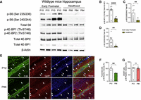
mTORC1 activity in excitatory neurons versus inhibitory neurons during postnatal development to adulthood. (A) Representative immunoblots of hippocampus lysates taken from early postnatal (P10–15) and adult (P84–88) mice. (B–D) Quantification of (a). Number of mice, n = 8 per group, (**) P < 0.01, (****) P < 0.0001, calculated with an unpaired t-test. (E) Confocal microscopy images (scale bar, 50 μm, magnification 20×) of mouse hippocampus immunostained for GAD67 (red) and p-S6 (Ser240/244) (green) at P12 and P86. (F) Quantification (integrated density) of p-S6 (240/244) (green) from the CA1 pyramidal neuron layer of P12 and P86 mice, (**) P < 0.01, unpaired t-test. Data are presented as mean ± SEM. Number of mice, n = 4 per group. (G) Quantification (integrated density) of p-S6 (240/244) (green) from GAD67-positive neurons (red) of P12 and P86 mice; (ns) not significant. Data are presented as mean ± SEM. Number of mice, n = 4 per group.
We next examined mTORC1 signaling in different neuron subtypes in the hippocampus, assessing phosphorylation of rpS6 at Ser240/244 as a readout of mTORC1 activity (Roux et al. 2007). Immunofluorescence analysis of hippocampal sections from early postnatal (P12) to adult (P86) mice showed a 40% decrease in p-S6 (Ser240/244) levels in the pyramidal cell layer (mainly excitatory neurons) of the CA1 region with age (Fig. 1E,F), while p-S6 levels in GABAergic neurons (colabeled by GAD67) remained stable (Fig. 1E,G). Thus, during postnatal development, from early postnatal to young adulthood, mTORC1's activity is maintained in GABAergic neurons but declines in pyramidal neurons.
We next conducted behavioral experiments on adult animals to investigate the cell-type-specific role of mTORC1-4E-BP2 in learning and memory processes. We generated transgenic mouse models of Raptor depletion in excitatory CaMK2A+ neurons (driven by Camk2a-Cre, referred to as Rptorflx/flx:Camk2a-Cre) or inhibitory GAD65+ neurons (driven by Gad65-Cre, Rptorflx/flx:Gad2-Cre), and 4E-BP2 depletion in excitatory CaMK2A+ neurons (Eif4ebp2flx/flx:Camk2a-Cre) or inhibitory GAD65+ neurons (Eif4ebp2flx/flx:Gad2-Cre).
We first used Rptorflx/flx:Camk2a-Cre and Rptorflx/flx:Gad2-Cre mice to investigate memory. To confirm cell-type-specific knockout of Raptor, we analyzed p-S6 (Ser240/244) in CaMK2A+ and GAD67+ neurons (detected in the cytoplasm of GABAergic neurons, mostly coexpressed with GAD65) (Kaufman et al. 1991). We observed a 55.13% decrease in p-S6 intensity in CaMK2A+ neurons (Fig. 2A,B) and a 62.88% decrease in GABAergic neurons (Fig. 2C,D) in Rptorflx/flx:Camk2a-Cre and Rptorflx/flx:Gad2-Cre cKO mice, respectively, indicating effective mTORC1 activity loss. Next, we used Eif4ebp2flx/flx:Camk2a-Cre and Eif4ebp2flx/flx:Gad2-Cre mouse models to mimic the activation of mTORC1 signaling (4E-BP2 phosphorylation by mTORC1 causes the disassociation of 4E-BPs from eIF4E) in CaMK2A+ excitatory neurons and GAD65+ inhibitory neurons, respectively. We verified conditional deletion of 4E-BP2 in CaMK2A+ excitatory neurons (Fig. 2E,F) with a 72.26% decrease of 4E-BP2 fluorescence, and in GAD67+ inhibitory neurons of the brain (Fig. 2G,H, respectively) with a 79.58% decrease of 4E-BP2 fluorescence intensity ratio.
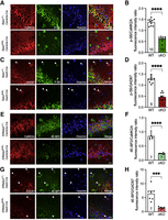
Validation of Raptor and 4E-BP2 deletion in CaMK2A and GAD67 positive neurons. (A) Immunofluorescence analysis of p-S6 (S240/244) (green) in CaMK2A+ cells (red) in Rptorflx/flx:Camk2a-Cre versus Rptor+/+:Camk2a-Cre (n = 5). (B) Quantification (mean integrated density) of p-S6 (240/244)/CaMK2A ratio from Rptorflx/flx:Camk2a-Cre versus Rptor+/+:Camk2a-Cre mice (cell number = 10). (C) Immunofluorescence analysis of p-S6 (240/244) (green) in GAD67-positive cells (red) in Rptorflx/flx:Gad2-Cre versus Rptor+/+:Gad2-Cre (mice = 5). (D) Quantification (mean integrated density) of p-S6 (240/244)/Gad67 ratio from Rptorflx/flx:Gad2-Cre versus Rptor+/+:Gad2-Cre mice (cell number = 9,10, respectively). (E) Immunofluorescence analysis of 4E-BP2 (green) in CaMK2A+ cells (red) in Eif4ebp2flx/flx:Camk2a-Cre versus Eif4ebp2+/+:Camk2a-Cre. (F) Quantification (mean integrated density) of 4E-BP2 protein/CaMK2A ratio from Eif4ebp2flx/flx:Camk2a-Cre versus Eif4ebp2+/+:Camk2a-Cre (cell number = 7). (G) Immunofluorescence analysis of 4E-BP2 (green) in GAD67-positive cells (red) in Eif4ebp2flx/flx:Gad2-Cre versus Eif4ebp2+/+:Gad2-Cre. (Η) Quantification (mean integrated density) of 4E-BP2 protein/GAD67 ratio from Eif4ebp2flx/flx:Gad2-Cre versus Eif4ebp2+/+:Gad2-Cre (cell number = 7). Confocal microscopy images: scale bar, 20 µm; magnification 60×. Cell nuclei are stained with Hoechst (blue). Statistical significance: (***) P < 0.001, (****) P < 0.0001 calculated with an unpaired t-test. Data are presented as mean ± SEM.
The mTORC1-4E-BP2 axis in GABAergic interneurons is required for hippocampus-dependent learning and memory
Since mTORC1 activity is maintained in hippocampal GABAergic interneurons, but not excitatory neurons during postnatal development, it is likely important for adult hippocampus-dependent memory. To determine whether interneuron-specific loss of Raptor or 4E-BP2 leads to defects in learning and memory, we performed novel object location (NOL), object place learning (OPL), and novel object recognition (NOR) tests on adult mice with cell-type-specific conditional deletions (Fig. 3A). These tasks are highly dependent on forebrain function, especially hippocampus (Cohen and Stackman 2015; Chao et al. 2022).
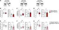
Inhibitory neurons require optimal mTORC1 activity to promote memory formation. (A) Schematics of the hippocampus-dependent memory tests used for the study. (Left panel) Novel object location (NOL). (Middle panel) OPL. (Right panel) NOR. (B–D) Memory measured by DI of NOL, OPL, and NOR, respectively, during testing in Rptorflx/flx:Gad2-Cre (NOL n = 10; OPL n = 12; NOR n = 13) versus Rptor+/+:Gad2-Cre (NOL n = 12; OPL n = 11; NOR n = 13). (E–G) Memory measured by DI of NOL, OPL, and NOR, respectively, during testing in Eif4ebp2flx/flx:Gad2-Cre (NOL n = 15; OPL n = 15; NOR n = 15) versus Eif4ebp2+/+:Gad2-Cre (NOL n = 16; OPL n = 16; NOR n = 16). Statistical significance: (*) P < 0.05, (**) P < 0.01; (ns) not significant, calculated by an unpaired t-test. (###) P < 0.001; (ns) not significant, calculated by a one-sample t-test. Data are presented with box and whisker plots where “+” indicates the mean. Dashed line indicates no discrimination of objects (i.e., memory impairment).
We observed long-term memory impairment in mice with Raptor and 4E-BP2 deletion in GABAergic inhibitory neurons. Mice with ablation of Raptor or 4E-BP2 in GABAergic neurons exhibited memory impairment as discrimination index (DI) change was not different from baseline in the NOL (Fig. 3B, Rptor flx/flx:Gad2-Cre; 3E, Eif4ebp2flx/flx:Gad2-Cre), OPL (Fig. 3C, Rptor flx/flx:Gad2-Cre; 3F, Eif4ebp2flx/flx:Gad2-Cre) and NOR (Fig. 3D, Rptor flx/flx:Gad2-Cre; 3G, Eif4ebp2flx/flx:Gad2-Cre) tasks, while the total exploration time was not altered (Fig. 3B–D, Rptor flx/flx:Gad2-Cre; 3E–G Eif4ebp2flx/flx:Gad2-Cre, right panels, respectively). These results indicate that the mTORC1-4E-BP2 pathway in inhibitory neurons plays an important role in long-term memory. This is in agreement with a previous study showing that loss of Raptor in somatostatin-expressing inhibitory neurons results in spatial memory and plasticity impairments (Artinian et al. 2019).
To investigate whether the phenotypes described above are specific to 4E-BP2, we assessed hippocampus-dependent memory in mice lacking 4E-BP1 in GAD65+ cells (Eif4ebp1flx/flx:Gad2-Cre). We confirmed the conditional deletion of 4E-BP1 in GAD67+ neurons using immunofluorescence (Supplemental Fig. S1A). In NOL and NOR tests, we did not observe any memory impairments or exploration time differences (Supplemental Fig. S1B,C). Thus, the mTORC1-4E-BP2 axis in inhibitory neurons is important for memory.
The mTORC1-4E-BP2 axis in excitatory neurons is dispensable for hippocampus-dependent learning and memory
We next performed memory tests on Raptor and 4E-BP2 cKO mice targeting excitatory CaMK2A+ neurons (Fig. 4A). Consistent with previous findings that Rptor deletion in CaMK2A+ neurons did not affect hippocampus-dependent memory (Zhu et al. 2018), we did not observe changes in memory in Raptor excitatory neuron cKO mice (Fig. 4B–D, Rptorflx/flx:Camk2a-Cre). Likewise, memory was maintained in mice with 4E-BP2 knockout in excitatory neurons (Fig. 4E–G, Eif4ebp2flx/flx:Camk2a-Cre). Total exploratory behavior was not altered, except for Rptorflx/flx:Camk2a-Cre mice, which had reduced total exploration by 40% during the NOL task (Fig. 4B, right panel). However, despite this reduction, long-term memory remained intact (Fig. 4B, left panel).
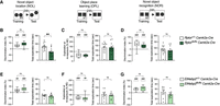
mTORC1-4E-BP2 signaling in excitatory neurons is dispensable for memory formation. (A) Schematics of the hippocampus-dependent memory tests used for the study. (Left panel) NOL. (Middle panel) OPL. (Right panel) NOR. (B–D) Memory measured by DI of NOL, OPL, and NOR, respectively, during testing in Rptorflx/flx:Camk2a-Cre (NOL n = 14; OPL n = 13; NOR n = 13) versus Rptor +/+:Camk2a-Cre (NOL n = 13; OPL n = 14; NOR n = 14). (E–G) Memory measured by DI of NOL, OPL, and NOR, respectively, during testing in Eif4ebp2flx/flx:Camk2a-Cre (NOL n = 11; OPL n = 10; NOR n = 11) versus Eif4ebp2+/+:Camk2a-Cre (NOL n = 10; OPL n = 10; NOR n = 10), (**) P < 0.01; (ns) not significant, calculated by an unpaired t-test. Statistical significance: (##) P < 0.01, (###) P < 0.001; (ns) not significant, calculated by a one-sample t-test. Data are presented with box and whisker plots where “+” indicates the mean. Dashed line indicates no discrimination of objects (i.e., memory impairment).
Camk2a-driven Cre is expressed postnatally in excitatory neurons (Dragatsis et al. 2000). Thus, it is possible that an earlier developmental deletion of 4E-BP2 is required to engender a memory phenotype. To investigate this possibility, we deleted 4E-BP2 in neurons under the expression of the excitatory neuron-specific promoter, EMX1, which expresses during embryonic development (Supplemental Fig. S2A,B; Gorski et al. 2002). We did not observe memory deficits in 4E-BP2-EMX1 cKO mice (Supplemental Fig. S2C,D), indicating that mTORC1 signaling via 4E-BP2 in excitatory neurons is dispensable for hippocampus-dependent long-term memory.
Deletion of 4E-BP2 in distinct subtypes of inhibitory neuron does not affect NOL and NOR memory
In the neocortex, parvalbumin (PValb), somatostatin (SST), and 5HT3a receptor-expressing inhibitory neurons—including vasoactive intestinal peptide (VIP) and non-VIP neurons—comprise nearly the entire population of the GABAergic neurons (Rudy et al. 2011). Thus, we investigated the impact of 4E-BP2 deletion on memory in these major classes of inhibitory neuron subtypes. We generated cKO mice lacking 4E-BP2 in either PValb, SST, or VIP+ neurons using the Cre-lox system. The immunofluorescent analysis confirmed cell-type-specific knockout of 4E-BP2 in PValb, SST, and VIP+ neurons (Fig. 5A,C,E). The mean fluorescence intensity ratio of 4E-BP2 compared to respective neuronal markers decreased by 71.3%, 86.3%, and 71.8% in PValb, SST, and VIP+ neurons, respectively, in each 4E-BP2 cKO mouse model (Fig. 5B,D,F).
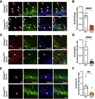
Confirmation of 4E-BP2 cKO in inhibitory neuron subclasses. (A) Immunofluorescence analysis of 4E-BP2 (green) in PValb-positive cells (red) in Eif4ebp2flx/flx:Pvalb-Cre versus Eif4ebp2+/+:Pvalb-Cre mice (n = 5). (B) Quantification (mean integrated density) of 4E-BP2 protein/PValb ratio from Eif4ebp2flx/flx:Pvalb-Cre versus Eif4ebp2+/+:Pvalb-Cre (cell number = 7). (C) Immunofluorescence analysis of 4E-BP2 (green) in SST-positive cells (red) in Eif4ebp2flx/flx:Sst-Cre versus Eif4ebp2+/+:Sst-Cre. (D) Quantification (mean integrated density) of 4E-BP2 protein/SST ratio from Eif4ebp2flx/flx:Sst-Cre versus Eif4ebp2+/+:Sst-Cre (cell number = 7). (E) Immunofluorescence analysis of 4E-BP2 (green) in VIP-positive cells (red) in Eif4ebp2flx/flx:Vip-Cre versus Eif4ebp2+/+:Vip-Cre. (F) Quantification (mean integrated density) of 4E-BP2 protein/VIP ratio from Eif4ebp2flx/flx:Vip2-Cre versus Eif4ebp2+/+:Vip-Cre (cell number = 7). Statistical significance: (**) P < 0.01, (****) P < 0.0001, calculated using an unpaired t-test. Confocal microscopy images: scale bar, 20 µm, magnification 60×. Cell nuclei were stained with Hoechst (blue).
Mice were subjected to NOL and NOR tests (Fig. 6A). The deletion of 4E-BP2 in PValb, SST, or VIP-expressing neurons alone did not impair memory. Three groups of cKO mice (Eif4ebp2flx/flx:Pvalb-Cre, Eif4ebp2flx/flx:Sst-Cre, and Eif4ebp2flx/flx:Vip-Cre) showed a significant DI above baseline in the NOL test (Fig. 6B–D, left panel), and in the NOR test (Fig. 6B–D, right panel). However, the deletion of 4E-BP2 in PValb inhibitory neurons led to a reduction (41.77% in NOL and 25.37% in NOR) in exploratory behavior. Specifically, cKO mice displayed a 41.77% reduction in mean total exploration time in the NOL test and a 25.37% reduction in mean total exploration time in the NOR test. Next, we conducted light–dark box and self-grooming tests on Eif4ebp2flx/flx:Pvalb-Cre and Eif4ebp2+/+:Pvalb-Cre mice; no significant differences in exploratory behavior or anxiety levels were detected (Supplemental Fig. S3A,B). Despite the reduced exploratory behavior, mice lacking 4E-BP2 in PValb+ neurons exhibited intact NOL and recognition memory (Fig. 6B). The mean DI for cKO mice in the NOL and NOR tests were 27.49 and 31.38, respectively. Both values were significantly above 0, demonstrating a preference for the novel object.
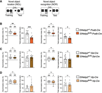
Analysis of NOL and NOR memory in inhibitory neuron subclass-specific 4E-BP2 knockout mice. (A) Schematics of the hippocampus-dependent memory tests used for the study. (Left panel) NOL. (Right panel) NOR. (B) DI and total exploration time for Eif4ebp2flx/flx:Pvalb-Cre (n = 12) versus Eif4ebp2+/+:Pvalb-Cre (n = 12) in the NOL task (left panel) and NOR task (right panel). (C) DI and total exploration time for Eif4ebp2flx/flx:Sst-Cre (n = 14) versus Eif4ebp2+/+:Sst-Cre (n = 14) in the NOL task (left panel) and NOR task (right panel). (D) DI and total exploration time for Eif4ebp2flx/flx:Vip-Cre (n = 11) versus Eif4ebp2+/+:Vip-Cre (n = 9) in the NOL task (left panel) and NOR task (right panel). Statistical significance: (*) P < 0.05, (**) P < 0.01, (***) P < 0.001; (ns) not significant, calculated with an unpaired t-test. Data are presented as mean ± SEM. (#) P < 0.05, (##) P < 0.01, (###) P < 0.001; (ns) not significant, calculated by a one-sample t-test. Data are presented with box and whisker plots where “+” indicates the mean. Dashed line indicates no discrimination of objects (i.e., memory impairment).
Our results suggest that NOL and recognition memory are not affected in mice with conditional deletion in SST or VIP-expressing interneurons. Although memory deposits were reduced in Eif4ebp2flx/flx:Vip-Cre mice, their memory deposits remain intact (indicated by DI). We thus conclude that there is redundancy among inhibitory neuron subtypes to mediate memory via the 4E-BP2-eIF4E axis, or alternatively, another subpopulation of inhibitory neurons (Pelkey et al. 2017) not tested in this study is responsible for mediating memory, via 4E-BP2.
Taken together, our results demonstrate that either increasing or decreasing mTORC1 pathway activity in GAD65+ neurons, but not excitatory neurons, diminish long-term NOL and NOR memory formation. We conclude that precise regulation of 4E-BP2-dependent translation, primarily in GABAergic inhibitory neurons, is required in long-term memory formation in the hippocampus.
Discussion
mTORC1 plays a cardinal role in brain development and memory formation (Hoeffer and Klann 2010). We used transgenic mouse models to gain insight into the role of the mTORC1-4E-BP2 axis in excitatory versus inhibitory neurons in adult hippocampus-dependent learning and memory. We demonstrated that mTORC1 activity, in sharp contrast to declining in hippocampal excitatory pyramidal neurons, maintains its activity in inhibitory neurons during postnatal development. Furthermore, the requirement of mTORC1 activity for maintaining memory in the adult brain is specifically and sufficiently orchestrated by inhibitory neurons. We also show that either hypoactivity or hyperactivity of the mTORC1 pathway is detrimental to hippocampus-dependent memory mnemonic functions. Specifically, loss of Raptor and the downstream translation initiation repressor protein, 4E-BP2, in GABAergic interneurons impaired NOL, OPL, and NOR memory.
mTORC1 activity generally decreases during development in the mouse cortex (Bidinosti et al. 2010; Kouloulia et al. 2019). We observed a similar decrease in mTORC1 signaling in the mouse hippocampus area, resulting in lower phosphorylation of rpS6 and 4E-BPs in adulthood. In contrast, mTORC1 activity, as measured by S6(S240/244) phosphorylation, remains high in GABAergic interneurons during development to adulthood, suggesting that active mTORC1 plays an important role in inhibitory neurons in adult mice. A plausible explanation for the decreased mTORC1 activity in hippocampal excitatory neurons of adult brains, relative to young, may involve age-related gene expression regulation affecting mTORC1 components or changes in upstream signaling pathways. These mechanisms are not well understood. Another plausible explanation is the age-related decline in neurogenesis and proliferation. Stimulation of mTORC1 using ketamine (an N-methyl-D-aspartate receptor [NMDAR] antagonist) restored age-related decline in proliferation, thereby enhancing neurogenesis in the hippocampus of aged mice (Romine et al. 2015). This suggests that age-related changes in cellular processes impact mTORC1 activity, which affects neuronal function. In our study, we observed that mTORC1 activity does not decline in total inhibitory neurons during development. This finding aligns with Amegandjin et al. (2021), who reported an upregulation of mTORC1 in parvalbumin interneurons during adolescence (Amegandjin et al. 2021). Given that the activation of inhibitory neurons in the adult brain regulates neurogenesis, circuit activity, and synaptic integration, these findings support a critical role of interneurons in adult hippocampus-related behaviors (Ge, et al. 2006; Song, et al. 2013; Wang et al. 2020).
The mTORC1 pathway plays an important role in hippocampus-dependent learning and memory, as rapamycin, an mTOR-specific inhibitor, impaired NOL and NOR memory (Tang et al. 2002; Jobim et al. 2012, Pereyra et al. 2018). Mice with Raptor deletion in excitatory neurons did not exhibit learning and memory impairment in the OPL and NOR tasks (Zhu et al. 2018). Consistent with this finding, we did not observe altered spatial and recognition memory in mice in which mTORC1 is inhibited in excitatory neurons (Rptorflx/flx:Camk2a-Cre mice) (Fig. 4B–D). In contrast, we observed NOL, OPL, and NOR memory impairment in mice with GABAergic inhibitory neuron disruption of mTORC1 (Rptorflx/flx:Gad2-Cre cKO mice). The finding is consistent with results showing that deletion of Raptor in somatostatin-expressing neurons impairs spatial memory in the Barnes maze (Artinian et al. 2019; Honoré and Lacaille 2022) and prevents the acquisition of reward-related activity in the goal-directed spatial learning task (Michon et al. 2023). mTORC1 deletion in somatostatin neurons, but not parvalbumin neurons, also disrupts contextual memory (Artinian et al. 2019; Khlaifia et al. 2022). We conclude that mTORC1 in inhibitory neurons, but not excitatory neurons, is required for hippocampus-dependent object recognition and spatial memory.
The consequences of mTORC1 overactivation in memory are less consistent than its impairment. For instance, deleting TSC1, a repressor of mTORC1, in somatostatin neurons resulted in enhanced contextual memory in the fear conditioning test and spatial memory in the Barnes maze task (Artinian et al. 2019). Moreover, deletion of FK506-binding protein 12 (FKBP12, a regulator of mTORC1 signaling) in CaMK2A+ excitatory neurons increased mTORC1 activity and enhanced contextual and Y-maze memory (Hoeffer et al. 2008; Schreiber et al. 2015). However, heterozygous deletion of PTEN, another mTORC1 repressor, did not affect contextual or NOR memory in male mice (Chen et al. 2019; Clipperton-Allen et al. 2022).
In this study, we found that mimicking mTORC1-4E-BP2 overactivation in GABAergic neurons by selectively deleting 4E-BP2 in inhibitory neurons, but not in excitatory neurons, impairs memory formation. Our findings are consistent with the observations of impaired memory in the full-body 4E-BP2 KO mice (Banko et al. 2005). However, we did not observe memory loss in 4E-BP2 GABAergic interneuron subpopulations (PValb, SST, and VIP+ neurons) cKO mice. It is plausible that deletion of 4E-BP2 in only one inhibitory neuron cell type is not sufficient to impact memory. This insufficiency could also be attributed to the complementary roles played by different populations of interneurons (Antonoudiou et al. 2020). Given that both PValb and SST neurons are implicated in various behaviors, further studies should investigate the exploratory behavior, locomotor activity, and fear memory of Eif4ebp2flx/flx:Pvalb-Cre mice and Eif4ebp2flx/flx:Sst-Cre mice more comprehensively (Udakis et al. 2020). The involvement of additional interneuron subtypes, such as CCK (cholecystokinin)- and NPY (Neuropeptide Y)-expressing neurons, may also be important in 4E-BP2-regulated long-term memory (Kelsom et al. 2013; Saffari et al. 2016; Nguyen et al. 2020).
Besides 4E-BP2, mTORC1 regulates multiple additional target proteins, such as S6Ks, which control protein synthesis (Shahbazian et al. 2006). S6K1 and S6K2 mutant mice display mild spatial memory deficits (Antion et al. 2008). Additionally, the loss of S6K1 is associated with increased anxiety and reduced exploratory behaviors in open-field, light–dark box, and elevated plus maze tests (Koehl et al. 2021; David et al. 2024). We observed impaired long-term memory in Rptorflx/flx:Gad2-Cre mice (Fig. 3B–D) and altered exploratory behavior in Rptorflx/flx:Camk2a-Cre mice, specifically in the NOL task (Fig. 4B). These findings indicate that distinct mTORC1 signaling in different neuronal populations may specifically affect memory and exploratory behaviors.
In summary, mTORC1-4E-BP2 pathway under- or overactivation in inhibitory neurons cause hippocampus-dependent memory deficits in adult mice. Our data provide evidence that translational control via mTORC1 signaling in learning and memory is mediated by inhibitory neurons. The optimal mTORC1 activity level and inhibitory neuron cell-type-specific synaptic mRNA translation required for learning and memory remains to be determined. Future studies should identify selective mRNAs regulated by mTORC1-4E-BP2 signaling in hippocampal GABAergic neurons.
Significance statement
The mechanistic target of rapamycin complex 1 (mTORC1) controls the activity of the translational repressor protein 4E-BP2 to affect synaptic plasticity and memory formation. The role of mTORC1-4E-BP2-dependent translational control in different neuronal subpopulations in memory formation is unknown. We show that mTORC1-4E-BP2 signaling in GABAergic interneurons, but not excitatory neurons, is required for memory formation in adult mice. mTORC1 signaling in the brain declines during postnatal development in excitatory, but not inhibitory neurons. Therefore, mTORC1-4E-BP2 signaling in adult inhibitory neurons constitutes a crucial node for long-term memory formation and may have implications in aging-related cognitive decline.
Materials and Methods
Animal husbandry
All animals were housed in the Goodman Cancer Institute mouse facility at McGill University. Room temperature (RT) was maintained at 20–22°C, and mice were given access to food and water ad libitum. Mice were kept on a standard 12 h light–dark cycle (7:00–19:00 light period), weaned at postnatal Day 21 (P21), and housed according to sex (2–5 mice per cage) with environmental enrichment. At weaning, mice were ear-notched for identification, and the ear tissue was used for genotyping by PCR.
Animal models
All experiments were approved by the McGill University Animal Care Committee following guidelines from the Canadian Council on Animal Care. The Eif4ebp2flx/flx mice were previously described (Wiebe et al. 2019). Cre-expressing mice were purchased from the Jackson Laboratory (JAX) on C57BL/6J background or backcrossed to C57BL/6J mice for at least 10 generations. Eif4ebp2flx/flx mice were crossed to each Cre-expressing line to generate the following lines: Eif4ebp2flx/flx:Camk2a-Cre, Eif4ebp2flx/flx:Gad65-Cre, Eif4ebp2flx/flx:Pvalb-Cre, Eif4ebp2flx/flx:Sst-Cre, Eif4ebp2flx/flx:VIP-Cre, and Eif4ebp2flx/flx:Emx1-Cre. Mice were previously validated for loss of protein expression in the appropriate cells (Wiebe et al. 2019; Aguilar-Valles et al. 2021; Sharma et al. 2021). 4E-BP2 wild-type (WT) and cKO alleles were genotyped by PCR with primers BP2-F (5′-GTCGGTCTTCTGTAGATTGTGAGT) and BP2-R (5′-GGCGATCCCTAGAAAATAAAGCCT-3′). Eif4ebp1flx/flx mice were crossed to the Cre-expressing line to generate Eif4ebp1flx/flx:Gad65-Cre and were previously validated (Aguilar-Valles et al. 2021). 4E-BP1 WT and cKO alleles were detected by PCR with primers BP1-F (5′-CACATTTCAGGGAGAGGGTGATG-3′) and BP1-R (5′-GCTGGGTTCTAAGAGTGGTACTTT-3′). Rptorflx/flx:Camk2a-Cre and Rptorflx/flx:Gad65-Cre were previously validated (De Gregorio et al. 2021). Rptor WT and cKO alleles were detected by PCR with primers Raptor-F (5′-CTCAGTAGTGGTATGTGCTCA-3′) and Raptor-R (5′-GGGTACAGTATGTCAGCTCAG-3′). Cre recombinase expression was confirmed by PCR with primers CreF (5′-GATTGCTTATAACACCCTGTTACG-3′) and CreR (5′-GTAAATCAATCGATGAGTTGCTTCA-3′). Primers were purchased from Integrated DNA Technologies or BioCorp. All experiments were performed on WT mice expressing Cre recombinase in the same cell type to normalize for any confounding effects due to the expression of Cre. Adult (3–4 months) male mice were used for behavioral experiments.
Western blotting
Western blotting was performed as previously described (Wiebe et al. 2020). The hippocampus isolated from male mice was homogenized in ice-cold radioimmunoprecipitation assay (RIPA) buffer (R0278, Sigma) containing proteinase and phosphatase inhibitors (Roche). Samples were incubated for 30 min on ice and centrifuged at 16,000g for 20 min at 4°C. Supernatants were collected and quantified and then 25 µg proteins were diluted with 2× loading buffer. Proteins were separated by SDS-PAGE (12%) and transferred onto nitrocellulose membranes. Membranes were blocked in 5% BSA in Tris-buffered saline with 0.1% Tween 20 (TBST) for 1 h at RT and incubated at 4°C with primary antibodies overnight. The following antibodies were purchased from Cell Signaling Technology Laboratories and used at the indicated dilutions: 4E-BP2 (#2845, 1:500), 4E-BP1(#9452, 1:500), p-4E-BP1/2 Thr37/46 (#2855, 1:1000), p-S6 Ribosomal Protein Ser240/244 (#5364, 1:1000), S6 5G10 (#2217, 1:1000). GAPDH was purchased from Abcam (#9842, 1:20,000). Membranes were washed with TBST before 1 h incubation at RT in horseradish peroxidase (HRP)-conjugated secondary antibody (Jackson Immuno #111-035-003, 1:20,000). After washing with TBST, membranes were treated with chemiluminescence and exposed against an X-ray film.
Immunofluorescence
Immunofluorescence was performed as previously described (Wiebe et al. 2019). Briefly, mice were anesthetized, followed by perfusing with ice-cold 1× phosphate-buffer saline (PBS) and 4% paraformaldehyde (PFA). Brains were fixed in PFA at 4°C overnight and transferred to 30% sucrose in PBS for cryoprotection. Mouse hippocampal coronal sections (20 µm) were prepared from PFA-fixed brains. Sections were then placed in 10 mM boiling sodium citrate buffer for antigen retrieval. Sections were washed with PBS and blocked for 1.5 h at RT, followed by primary antibody incubation at 4°C overnight (4E-BP1, Cell Signaling Technology #9644, 1:100; 4E-BP2, Cell Signaling Technology #2845, 1:50; CAMK2A, Cell Signaling Technology #50049, 1:800; GAD67, Millipore MAB5406, 1:5000; p-S6 Ribosomal Protein Ser240/244, Cell Signaling Technology, #5364, 1:200; PVALB, Millipore #1572 1:100; SST, GeneTex #71935, 1:400). Sections were washed with PBS, then incubated in Alexa Fluor 488 goat anti-rabbit IgG (Thermo Fisher Scientific, #11034, 1:400), Alexa Fluor 546 donkey anti-mouse IgG (Thermo Fisher Scientific, #10036, 1:400), Alexa Fluor 647 donkey anti-rabbit IgG (Thermo Fisher Scientific, #31573, 1:400) and Hoechst 33342, trihydrochloride, trihydrate (Life Technologies, #3570, 1:200) in blocking buffer for 1.5 h at RT. Brain slices were mounted with DAKO and imaged with a ZEISS LSM880 laser scanning confocal microscope.
Behavioral testing
Adult male mice (3–4 months) were handled for 2 min each day for 3 days before habituation. Behavioral experiments were performed in a soundproof room between 7 a.m. and 3 p.m. Mice were allowed to rest for 2 days between each behavioral experiment. For each memory test, mice were habituated to the room for 30 min in dim lighting before each session (in the morning and in the afternoon). Four floor lamps were used for even distribution of lighting over the boxes. All objects and boxes were cleaned with odorless disinfectant after each use. Mice were counterbalanced to the boxes and objects to ensure there was no bias to a given location in the box or a particular object. The order of animal testing was randomized to avoid confounding effects, such as time of day and box/object preference, on memory performance. The experimenter was blinded to mouse genotype during data analysis and scoring (manual). Sniffing of and climbing on the objects were scored as exploration time, while sitting and standing on the objects were not included. The DI equals to novel object exploration time minus familiar object exploration time divided by the total exploration time, times 100.
Novel object location
Mice were first habituated twice to the empty (i.e., without objects) white-colored square box (50 × 50 × 30 cm). Habituation occurred once in the morning (between 7 a.m. and 11 a.m.) and again in the afternoon (12 noon–3 p.m.) for 10 min. The following day, mice were trained twice in the same boxes to the location of the two identical objects. Similar to habituation, training occurred once in the morning (between 7 a.m.–11 a.m.) and again in the afternoon (12 noon–3 p.m.) for 10 min. Training was repeated a second day for a total of four training sessions. The next day, mice were tested for memory retention by moving one of the objects to a corner of the box, leaving a 10 cm space around it, and allowing the mice to freely explore the objects for 10 min. The testing session was recorded by an overhead camera.
Novel object recognition
Using the same setup as NOL, mice were first habituated once in the morning (between 7 a.m. and 11 a.m.) for 10 min. The following day (training Day 1), mice were trained once in the morning (between 7 a.m. and 11 a.m.) to recognize two identical objects by 10 min of exploration. The training was repeated the next day (training Day 2) using the same configuration for a total of 2 training sessions. On the last day, mice were tested for NOR by replacing one object with an unfamiliar novel object that has a different appearance. Mice were allowed to explore the objects for 10 min. Training and testing sessions were recorded by an overhead camera.
Object place learning
OPL was measured using data from training sessions of Day 1 and Day 2 of NOR. If mice retain memory of objects, total exploration will be reduced on Day 2 (i.e., training 2) versus Day 1 (i.e., training 1). The index of novelty exploration (NE) = Day 2 exploration time/Day 1 exploration time×100.
Statistical analysis
The graphing and statistical analysis of data was performed using GraphPad Prism 9. Data in bar graphs are presented as mean ± the standard error of the mean (SEM). DIs are graphed using a box and whisker plot where the box indicates the interquartile range (the bottom of the box is the lower quartile Q1 and the top of the box is the upper quartile Q3), the line in the middle indicates the median, and the “+” indicates the mean. The whiskers of the plot indicate the minimum and maximum data points. The # symbol represents the P-value calculated from a one-sample t-test, which compares the mean of the data set to a hypothetical value (indicated by a grid line). The asterisk represents the P-value calculated from an unpaired Student's t-test. A Welch's corrected t-test was used when the difference in standard deviation between groups was significantly different (f test). P-values <0.05 were considered statistically significant [ns: P ≥ 0.05, (*) P < 0.05, (**) P < 0.01, (***) P < 0.001, (****) P < 0.0001].
Data deposition
The research data are available from the corresponding author on reasonable request.
Supplemental material
Supplemental material is available for this article.
Acknowledgments
We thank Karim Nader (McGill University) for providing access to behavioral testing facilities and Wayne Sossin (McGill University) for critical comments. We extend our gratitude and our respects to the late Dr. Jerry Pelletier (McGill University) for his help with statistical analysis. Funding was provided by Canadian Institutes of Health Research (CIHR) foundation grant (FDN-148423) and Natural Sciences and Engineering Research Council of Canada (NSERC) Discovery Grants (RGPIN-2021-02764). J.C.L. was supported by a CIHR project grant (PJT-153311), a FRQS Research Center grant (Centre Interdisciplinaire de Recherche sur le Cerveau et l'Apprentissage [CIRCA]), and was the recipient of the Canada Research Chair in Cellular and Molecular Neurophysiology (CRC-950-231066).
Author contributions: Z.H. and S.W. conceptualized the project, performed experiments, analyzed, and interpreted data, and wrote the manuscript. A.N., J.C., C.W., and N.M. performed experiments and analyzed data. A.K. and J.C.L. provided conceptual support. N.S. provided supervision and oversaw the study. S.W. and N.S. secured funding for the study. All authors assisted in editing the manuscript and approved the final version.
Footnotes
-
[Supplemental material is available for this article.]
-
Article is online at http://www.learnmem.org/cgi/doi/10.1101/lm.054018.124.
- Received April 22, 2024.
- Accepted September 15, 2024.
This article is distributed exclusively by Cold Spring Harbor Laboratory Press for the first 12 months after the full-issue publication date (see http://learnmem.cshlp.org/site/misc/terms.xhtml). After 12 months, it is available under a Creative Commons License (Attribution-NonCommercial 4.0 International), as described at http://creativecommons.org/licenses/by-nc/4.0/.



