


Chronic stress enhances threat responding and impacts fear extinction
- Corresponding author: jamie.maguire{at}tufts.edu
Abstract
Chronic stress is a major risk factor for psychiatric disorders. Previous work from our laboratory has demonstrated that chronic stress exposure disrupted network activity in the basolateral amygdala (BLA) and altered the activity of projection-specific neurons in the BLA to bias information processing to favor negative valence processing. Although the BLA is well established as a critical hub for fear memory encoding and retrieval, it remains unclear how chronic stress alters ensemble recruitment within the BLA and its downstream circuit dynamics to shape fear and extinction learning. To answer this question, we used retrograde tracing combined with immunohistochemistry in adult male mice to examine how chronic unpredictable stress (CUS) during adulthood alters the activity of the BLA and its projections to the nucleus accumbens (NAc) or bed nucleus of the (BNST) following fear extinction. Here we demonstrate that CUS produced a shift toward persistent threat responding and alters extinction learning, accompanied by reduced recruitment within the lateral nucleus of the BLA (LA) and in BLA-NAc projecting neurons. These findings provide circuit-level evidence for how chronic stress disrupts information flow from BLA, biasing valence processing toward extinction-resistant fear states and potentially contributing to the persistence of maladaptive fear in stress-related psychiatric disorders.
Chronic stress is an enduring and major neuropathological contributor to many psychiatric illnesses such as depression, anxiety disorders, and posttraumatic stress disorder (PTSD) (McEwen 2004; Maren and Holmes 2016). It impacts all categories of psychiatric disorders defined by the six Research Domain Criteria (RDoC) dimensions proposed by the National Institute of Mental Health (NIMH) including negative and positive valence, cognition, sociality arousal, and sensorimotor function. Chronic stress has a major impact on valence processing (Antonoudiou et al. 2023), an internal cognitive computational process for assigning binary value (positive or negative) to sensory inputs and experiences, guiding approach or avoidance behaviors accordingly (Tye 2018). Chronic stress disproportionately impacts valence processing by favoring negative valence and dampening positive valence (Antonoudiou et al. 2023). For example, chronic stress is associated with negative bias (Braund et al. 2019), or a tendency toward negative valence processing. Clinical studies have also revealed that individuals experiencing elevated levels of chronic stress exhibit reduced reward responsiveness and symptoms of anhedonia (Berenbaum and Connelly 1993; Pizzagalli et al. 2007). These data demonstrate that chronic stress is capable of influencing both negative and positive valence processing.
The basolateral amygdala (BLA) is a critical brain region involved in valence processing (Pignatelli and Beyeler 2019). Substantial evidence supports that heterogeneous neuronal populations within the BLA are activated by aversive and appetitive stimuli and can mediate positive and negative valence behaviors (Stuber et al. 2011; Beyeler et al. 2018; Russell et al. 2020; Han et al. 2024). The BLA sends dense projections to multiple downstream targets, including the nucleus accumbens (NAc) and the bed nucleus of the stria terminalis (BNST). These projection-defined pathways support distinct behavioral functions. BLA neurons are highly sensitive to stress. For example, acute restraint stress selectively increases FOS expression in basolateral amygdala to nucleus accumbens (BLA-NAc) pathway without affecting BLA-CeA (central amygdala) neurons (Aukema et al. 2024). Chronic stress reduced discriminative reward learning and reward-to-effort valuation through opposing changes in reward and aversion neurons in BLA-NAc pathway (Madur et al. 2023). Recent work from our laboratory also demonstrated that chronic stress facilitated activation of basolateral amygdala to bed nucleus of the stria terminalis pathway (BLA-BNST) while suppressing the activity of BLA-NAc pathway to promote avoidance and stress-induced helplessness behaviors (Antonoudiou et al. 2024). These studies demonstrate that stress can differentially shape activity across distinct BLA subpopulations depending on their cell type, downstream targets, and behavioral functions, ultimately influencing valence processing. However, less is known about how chronic stress influences the allocation of extinction-related ensembles within BLA circuits and their downstream projections.
Prior exposure to stressful events can facilitate fear learning and lead to exaggerated fear responses. Studies using the stress-enhanced fear learning (SEFL) paradigm have shown that both acute and chronic stress can potentiate later fear conditioning and promote fear generalization to novel contexts (Rau et al. 2005; Long and Fanselow 2012; Conoscenti et al. 2024). These effects of stress are associated with changes in amygdala ensemble recruitment. For example, acute stress–induced fear generalization was associated with a larger engram size in the lateral amygdala (LA) (Lesuis et al. 2025), and chronic stress enhanced fear generalization corresponded with increased neuronal activity in the BLA (Hoffman et al. 2014, 2015). These findings suggest that stress alters how fear memories are allocated within amygdala circuits and potentially bias behavior toward generalized fear states. Critically, the circuit imbalances that drive generalization are likely to interfere with extinction, which depends on forming a new “safety” memory to suppress fear memory (Lacagnina et al. 2019). Extinction has been conceptualized as a reward-like learning process requiring the engagement of positive valence pathways such as the BLA-NAc projection (Correia et al. 2016), while avoidance-related projections such as BLA-BNST promote anxiety and sustained fear. Thus, if stress promotes generalized fear through biased ensemble recruitment, it may also impact extinction by shifting projection-specific balance away from safety/reward circuits. Yet, it remains unclear how chronic stress alters the allocation of these projection-defined ensembles during extinction. We address this gap by using a chronic unpredictable stress (CUS) model to examine how chronic stress alters BLA subnucleus and projection-specific recruitment after extinction learning.
Here, we employed retrograde tracing combined with immunohistochemistry to examine how chronic unpredictable stress (CUS) alters the activity of BLA projection-specific neurons projecting to either the NAc (BLA-NAc) or BNST (BLA-BNST) using C57 adult male mice. CUS produced a shift toward persistent threat responding and altered extinction evidenced by increased intertrial freezing, cue-specific freezing, and reduced recruitment of BLA-NAc neuronal ensembles. These findings provide circuit-level evidence for how chronic stress disrupts information processing in the BLA in a circuit-specific manner, thereby biasing valence processing toward extinction-resistant fear states and potentially contributing to the persistence of maladaptive fear in psychiatric disorders associated with chronic stress.
Results
Chronic unpredictable stress does not affect fear acquisition
To investigate the effect of chronic unpredictable stress (CUS) on auditory fear acquisition, percent freezing was quantified during cued fear conditioning in control (CTL) and CUS mice (N = 16 mice per group). The fear conditioning paradigm consisted of four tones (20 sec, 4 kHz) coterminating with a shock (2 sec, 0.7mA), followed by a 40 sec intertrial intervals (ITIs) (Fig. 1A). Equivalent fear learning was observed in both CUS and CTL groups as measured by the magnitude of freezing across the four tone presentations (CS+) (Fig. 1B). Average freezing during habituation (Fig. 1C), CS+ (Fig. 1D), and ITIs (Fig. 1E) were also not significantly different between groups, further indicating no difference in fear learning due to CUS exposure. Collectively, auditory fear conditioning resulted in robust fear memory acquisition in both the CUS and CTL groups.
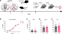
Chronic unpredictable stress does not affect fear acquisition. (A) Experimental time line of the fear conditioning paradigm. Mice (N = 32) were randomly assigned to either 4 weeks of chronic unpredictable stress (CUS) or nonstress (CTL) groups. After 4 weeks of CUS protocol, all mice underwent fear conditioning followed by 2 days of extinction protocols. N = 16 mice underwent surgeries prior to the behavioral paradigms to retrogradely label projection-specific neurons (either BNST or NAc) in the BLA. Mice were sacrificed 90 min after the final extinction session for subsequent analyses. (B) Average percent freezing (±SEM) during tone presentations on the fear acquisition day. Freezing increased with repeated tone-shock presentations but was not impacted by previous CUS exposure (two-way repeated-measures ANOVA: tones: F(2.745,82.34) = 123.7, P < 0.0001; stress: F(1,30) = 0.8169, P = 0.3733; tone × stress: F(2.745,82.34) = 0.8553, P = 0.4593). (C) Average freezing during habituation was not impacted by CUS (Mann–Whitney test, U = 125, P = 0.9260). (D) Average freezing during exposure to the tone cue did not differ between groups (unpaired t-test, t(30) = 0.9872, P = 0.3314). (E) Average freezing during intertrial intervals (ITIs) did not differ between groups (unpaired t-test, t(30) = 0.5957, P = 0.5559). Bars are plotted as mean ± SEM. (*) P < 0.05.
Chronic stress enhances threat responding and impacts fear extinction
To evaluate the impact of CUS on fear recall and extinction learning, CTL and CUS mice underwent extinction training to the tones in a novel context (context B). Extinction consisted of 5 min of habituation followed by 10 CS+ presentations with a 40 sec ITIs repeated over 2 days (Fig. 2A). During day 1 extinction, freezing during habituation to the novel context was equivalent between CTL and CUS mice (Fig. 2B). Across the 10 tones trials and ITIs trials, mice froze more to the tones than to the ITIs and freezing declined over time, demonstrating cued recall to the CS+. CUS mice froze more overall than CTL mice over extinction training (Fig. 2C). To specifically assess whether these group differences reflected altered cue recall, freezing during the first three CS+ presentations was examined and showed no difference between groups (data not shown), indicating similar retrieval of fear memory. To further evaluate within-session extinction learning, average freezing during the first five tone presentations was compared to last five tones. Freezing during last five tones was significantly lower than the first five tones, indicating that extinction learning occurred within the session for both groups (Fig. 2D). Despite within-session reduction in freezing to tone exhibited in both groups, there was a group difference over the entire training session (Fig. 2C). To determine whether this group difference was specific to cue periods or non-cue periods, freezing was averaged separated across tone presentations and ITIs. When averaging freezing across the 10 trials during either tone presentation or ITIs, this group difference was present regardless of cue type (CS+ vs. ITIs)/cue presentation (Fig. 2E). Post hoc comparisons indicated that the effect of CUS was driven by higher freezing to the ITIs, whereas freezing to the tones did not significantly differ between groups. Together, these results suggest that group differences on extinction day 1 reflect elevated freezing in CUS mice during the non-cue periods rather than exaggerated fear recall to cue. To explore whether this reflected an inability to disengage from freezing, we measured the duration of freezing bouts. Bout duration was not different between groups during fear conditioning for either CS+ or ITIs (Supplemental Fig. S1A), but significantly longer in CUS mice (Supplemental Fig. S1B,D) and elevated during CS+ compared to ITIs during extinction day 1. Overall, CUS mice exhibited longer freezing bouts after the habituation period (Fig. 2F), consistent with reduced flexibility in terminating fear responses.
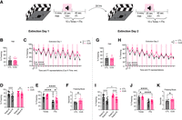
Chronic stress impacts fear extinction. (A) Experimental time line of the 2 day extinction paradigm. On each day, mice were habituated to context B for 5 min and then exposed to ten 20 sec tones with 40 sec intertrial intervals. (B) CUS and CTL groups had similar levels of freezing during habituation on extinction day 1 (unpaired t-test, t(30) = 0.3058, P = 0.7619). (C) The percent freezing across 10 CS+ tones and ITIs on extinction day 1 declined over repeated presentations (three-way ANOVA; time: F(9,270) = 9.689, P < 0.0001). Freezing was greater during tones than ITIs (cue: F(0.6971,20.91) = 233.1, P < 0.0001), declined faster to ITIs (time × cue: F(6.979,209.4) = 3.034, P = 0.0047), and was enhanced by CUS (stress: F(1,30) = 5.185, P = 0.0301). (D) Both groups exhibited with-session extinction learning on extinction day 1, as average percentage of freezing during the last five tone presentations was significantly reduced compared to the first five tones (two-way repeated-measures ANOVA; time: F(1,30) = 35.22, P < 0.0001; stress: F(1,30) = 3.689, P = 0.0643; time × stress interaction: F(1,30) = 0.7933, P = 0.3802). Post hoc Sidak's tests revealed significantly lower freezing during the last five tones in both CUS (P = 0.0025) and CTL (P < 0.0001) groups. (E) Average percent freezing during tones and ITIs on extinction day 1 was greater for tones than ITIs (two-way repeated-measures ANOVA; cue: F(1,30) = 233.1, P < 0.0001) and elevated in CUS mice (stress: F(1,30) = 5.185, P = 0.0301). Although there was no significant cue and stress interaction (F(1,30) = 1.549, P = 0.2229), post hoc Sidak's tests were significant in ITIs CTL versus CUS (P = 0.0252), CTL tones versus ITIs (P < 0.0001), CUS tones versus ITIs (P < 0.0001), but not in tones CTL versus CUS (P = 0.1838). (E) CUS did not affect fear recall measured by average freezing during the first three tones (unpaired t-test, t(30) = 1.160, P = 0.2552). (F) CUS had longer freezing bouts than CTL during extinction day 1 posthabituation period (unpaired t-test, t(30) = 2.638, P = 0.0131). (G) The average percent time freezing was similar between CUS and CTL groups during habituation on extinction day 2 (unpaired t-test, t(30) = 0.3839, P = 0.7038). (H) The percent time freezing across 10 tones and ITIs on extinction day 2 declined over repeated presentations (three-way ANOVA; time: F(9,270) = 19.61, P < 0.0001) was greater during tones (cue: F(0.6866,20.60) = 215.4, P < 0.0001), and increased in CUS mice (stress: F(1,30) = 4.421, P = 0.0440). (I) Both groups exhibited within-session extinction learning on extinction day 2, as the average percentage of freezing during the last five tone presentations was significantly reduced compared to the first five tones (two-way repeated-measures ANOVA; time: F(1,30) = 71.40, P < 0.0001; stress: F(1,30) = 6.177, P = 0.0187; time × stress interaction: F(1,30) = 0.2258, P = 0.6381). Post hoc Sidak's tests revealed significantly lower freezing during the last five tones in both CUS (P < 0.0001) and CTL (P < 0.0001) groups. In addition, the CUS group had significantly higher freezing than the CTL group in the last five tone presentations (P = 0.0429). (J) Average percent freezing during tones and ITIs on extinction day 2 showed that mice froze more during tones than ITIs (two-way repeated-measures ANOVA; cue: F(1,30) = 215.4, P < 0.0001), and CUS mice froze more overall (stress: F(1,30) = 4.421, P = 0.0440). Although there was no significant cue × stress interaction (F(1,30) = 1.614, P = 0.2137), post hoc Sidak's tests were significant in tones CTL versus CUS (P = 0.0385), CTL tones versus ITIs (P < 0.0001), and CUS tones versus ITIs (P < 0.0001), but not in ITIs CTL versus CUS (P = 0.2264). (K) The average duration of freezing bouts were longer in CUS mice compared to CTL during the extinction day 2 posthabituation period (unpaired t-test, t(30) = 2.574, P = 0.0152). Data are presented as mean ± SEM; N = 16/group; (*) P < 0.05.
On extinction day 2, freezing during habituation again did not differ between groups (Fig. 2G). Across the extinction session, freezing declined over repeated presentations, was higher in the CS+ than during the ITIs, and remained elevated in the CUS group compared to CTL (Fig. 2H). During the first three CS+ presentations, both CTL and CUS showed a high level of freezing, suggesting that extinction memory was not yet fully expressed and retained at the beginning of the session. Notably, CUS mice froze more than CTL mice (data not shown) during these early trials, indicating either a weaker consolidation of extinction learning from the previous extinction training day or a stronger CS+ evoked fear memory. More importantly, freezing decreased from the first five tones to the last five tones in both groups, and CUS maintained higher freezing levels than CTL mice during the last five tones (Fig. 2I), suggesting that although extinction occurred in both groups, CUS mice exhibited persistently elevated freezing. To determine whether group difference was specific to cue-evoked freezing or non-cue period, freezing was averaged separately across CS+ and ITIs. When freezing was averaged across the trials, CUS mice continued to freeze more than CTL, with greater freezing to the CS+ than the ITIs in both groups (Fig. 2J). Although the effect of CUS did not vary between CS+ and ITIs, post hoc comparisons revealed that the main effect of CUS was driven by greater freezing to the tones rather than the ITIs, a reversal of the pattern observed on extinction day 1. In addition, CUS presented longer freezing bouts after habituation on extinction day 2 (Fig. 2K). Further analysis revealed that this effect was driven by prolonged freezing bouts during tone presentations. CUS mice showed longer bout duration than CTL mice, and freezing bouts were longer during tones than ITIs (Supplemental Fig. S1C,E). Overall, these results suggest that CUS impacts extinction learning by reducing the ability to suppress cue-evoked freezing.
Chronic unpredictable stress refines ensemble activation
To investigate how CUS altered extinction-related neuronal activity, we collected brains 90 min after extinction day 2 and quantified the expression of immediate early gene c-Fos in the BLA, a key region for fear learning and extinction (Fig. 3A; Herry et al. 2008; Maren and Holmes 2016). To capture potential variation along the BLA's anterior–posterior (AP) axis, which has been shown to exhibit functional specificity in the context of valence (Kim et al. 2016; Zhang et al. 2020), we obtained three measurements for each mouse (anterior, central, and posterior BLA), such that each mouse contributed three data points spanning the full AP axis (Fig. 3B). Since we did not observe differences in c-Fos density across the anterior–posterior axis (Supplemental Fig. S2A), we combined all measurements for analysis. CUS mice showed a marginal reduction in overall c-Fos density (# cells/µm2) compared to CTL mice (Fig. 3C), suggesting a small but not statistically significant reduction of BLA recruitment after extinction training. Given the evidence that the lateral (LA) and basal (BA) nuclei within the BLA have distinct roles, with the LA more associated with cued fear memories and the BA more involved in contextual fear (Calandreau et al. 2005; Reijmers et al. 2007; Davis and Reijmers 2018), we next analyze these subregions separately. This revealed that CUS mice had significantly lower c-Fos density in the LA compared to the CTL mice (Fig. 3D), while BA activity did not differ between groups (Fig. 3E), indicating altered neuronal activity in the LA associated with the observed changes in cue-related extinction (Fig. 2).
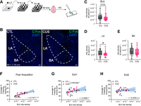
Chronic stress refines memory ensemble recruitment. (A) Experimental time line for postextinction ensemble identification. Brains were collected after extinction day 2, and c-Fos immunohistochemistry on BLA tissue was performed. (B) Representative images of c-Fos expression in the BLA. (C) CUS group had a trend to a lower BLA c-Fos expression compared to CTL group (Mann–Whitney test, U = 845, P = 0.0702, # data points = 46–47/group, # outliers = 1/group removed). (D) CUS led to lower c-Fos expression in the lateral amygdala (LA) compared to CTL (Mann–Whitney test, U = 797, P = 0.0199, # data points = 46–48/group, # outliers = 0–1/group removed). (E) CTL and CUS had comparable c-Fos expression in the basal amygdala (BA) (Mann–Whitney test, U = 946, P = 0.3877, # data points = 45–47/group, # outliers = 1–2/group removed). (F) Average % freezing during entire fear conditioning training session was significantly correlated with BLA c-Fos density (Spearman's correlation, r = 0.7371, P < 0.0001). Simple linear regression slopes and intercepts were not different between CUS and CTL groups (slopes: F(1,28) = 0.187, P = 0.6687; Y-intercepts: F(1,29) = 4.04, P = 0.0539). Regression lines for CTL (black, Y = 557,737 X + 6.39), CUS (pink, Y = 638,544 X + 8.35), and combined (blue, Y = 555,324 X + 8.65) were shown. (G) Average % freezing during entire extinction day 1 was significantly correlated with BLA-cFos density (Spearman's correlation, r = 0.4506, P = 0.0097). Simple linear regression slopes were not different between CUS and CTL mice, but intercepts were significant (slopes: F(1,28) = 0.8916, P = 0.3531; Y-intercepts: F(1,29) = 5.327, P = 0.0283). Regression lines for CTL (black, Y = 330,820 X + 31.80), CUS (pink, Y = 626,092 X + 31.26), and combined (blue, Y = 378,481 X + 34.39) were shown. (H) Average % freezing during entire extinction day 2 was not significantly correlated with BLA-cFos density (Spearman's correlation, r = 0.3035, P = 0.0912). Simple linear regression slopes and intercepts were not different between CUS and CTL groups (slopes: F(1,28) = 0.112, P = 0.7407; Y-intercepts: F(1,29) = 2.12, P = 0.1563). Regression lines for CTL (black, Y = 161,175 X + 40.80), CUS (pink, Y = 262,520 X + 42.90), and combined (blue, Y = 160,792 X + 43.40) were shown. Data are presented as a box-and-whisker plot with whiskers showing minimum and maximum, (*) P < 0.05. Scale bar, 100 µm.
Extinction learning is thought to be an active learning process that suppresses a conditioned fear response by forming a new memory, rather than erasing the original fear memory. We therefore asked how BLA activity after extinction day 2 related to freezing across different phases of learning. Using Spearman rank-order correlations, we found that overall BLA c-Fos density was positively correlated with freezing during fear acquisition (Fig. 3F) and extinction day 1 (Fig. 3G) but not with freezing during extinction day 2 (Fig. 3H), which may be due to the lack of extinction memory retention across days. Overall, this pattern suggests that neurons recruited during extinction day 2 still carry a memory trace of prior fear expression rather than fully consolidated extinction memory, such that greater recruitment is associated with stronger freezing during acquisition and early extinction, phases when the CS-shock associations are strongest.
We next asked whether these relationships between BLA activity and freezing differed between CTL and CUS mice. To do this, we fit separate linear regressions for each group and compared the slopes and intercepts of the regression lines. For both acquisition and extinction days 1 and 2, slopes did not differ between groups, indicating that the strength and directionality of the relationship was similar. However, for extinction day 1, the y-intercept differed (Fig. 3G), indicating that at equivalent levels of BLA activity, CUS mice froze more than CTL mice. This parallel upward shift suggests that CUS increases the behavioral output for a given level of BLA recruitment. By extinction day 2, there was no significant relationship between BLA activity and freezing (Fig. 3H). These data demonstrate an effect of CUS on activation patterns in the BLA, which correlates with fear and extinction learning.
Chronic unpredictable stress shifts ensemble activation
Stress and chronic corticosterone administration have been shown to bias BLA output toward specific downstream targets by enhancing the activation of BLA neurons projecting to BNST (BLA-BNST) while suppressing those projecting to the nucleus accumbens (BLA-NAc) (Antonoudiou et al. 2024; Bigot et al. 2024). To determine whether CUS alters the engagement of these pathways following fear extinction, we injected a retrograde mCherry virus into either the NAc or BNST (Supplemental Fig. S3) prior to CUS exposure and collected BLA tissue 90 min after extinction day 2 (Fig. 4A). The percentage of mCherry+ neurons coexpressing c-Fos were then quantified in the BLA, with three data points per mouse distributed along the AP-axis (Fig. 4B). As no differences in the percentage of mCherry neurons coexpression c-Fos were observed across the AP-axis for either pathway (Supplemental Fig. S2B,C), all slices were combined for analysis. Extinction-related activity of BLA-BNST projection neurons were comparable between CUS and CTL mice (Fig. 4C), indicating that CUS did not alter activation of this anxiety-linked pathway. In contrast, the recruitment of BLA-NAc projection neurons was reduced in CUS compared to CTL (Fig. 4D), suggesting a reduced engagement of this pathway following CUS. Moreover, while BLA-BNST activity was not correlated with freezing during extinction day 2 (Fig. 4E), BLA-NAc activity was negatively correlated with freezing (Fig. 4F), indicating that greater engagement of this projection is associated with more effective suppression of fear expression. These findings demonstrate that CUS selectively suppresses the recruitment of BLA-NAc neurons during extinction, potentially impairing the activation of safety-associated circuits and contributing to the observed deficits in extinction learning.
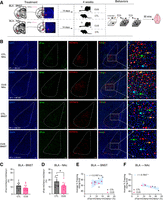
Chronic stress shifts ensemble activation. (A) Experimental time line of retrograde labeling and ensemble identification. Stereotaxic injection and target validation of pAAV-hSyn-mCherry retrograde virus into either BNST (CUS N = 5, CTL N = 4) or NAc (CUS N = 3, CTL N = 4); adapted from Paxinos and Franklin (2019). (B) Representative images of DAPI, c-Fos expression, and m-Cherry in BLA-NAc neurons are shown. Within the merged image, blue arrow indicates a DAPI+ cell, green arrow indicates cFos+ cell, red arrow indicates mCherry+ cell, and orange arrow indicates a mCherry+ cell colocalized with cFos+. (C) There was no statistically significant difference in the activation of BLA-BNST neurons between the CUS and CTL groups (unpaired t-test, t(24) = 1.940, P = 0.0643, # data points = 12–14/group, # outlier = 0–1/group removed). (D) The percent activation of BLA-NAc neurons is decreased in mice exposed to CUS compared to CTL (unpaired t-test, t(19) = 2.095, P = 0.0498, # data points = 9–12/group, # outlier = 0/group removed). (E) There is no significant correlation between activation of the BLA-BNST pathway with freezing during extinction day 2 (Spearman's correlation, r = 0.3167, P = 0.4101). Regression line: Y = 0.676 X + 48.70. (F) There is a negative correlation between BLA-NAc pathway activation with freezing during extinction day 2 (Spearman's correlation, r = −0.7857, P = 0.0480). Regression line: Y = −0.562 X + 56.70. Data are presented as mean ± SEM; N = 3–5/group, three data points/mouse along BLA AP-axis; (*) P < 0.05. Scale bar, 100 µm.
To evaluate the composition of the presumably extinction-training-activated c-Fos+ neuronal ensemble, we quantified the percentage of c-Fos+ neurons coexpressing mCherry for each BLA-projecting pathway. Two-way ANOVA revealed a significant main effect of stress (F(1,42) = 11.33, P = 0.0016) but no main effect of pathway (F(1,42) = 2.806, P = 0.1014) nor interaction between stress and pathway (F(1,42) = 3.291, P = 0.0768). Post hoc Sidak's multiple comparison test indicated that in CUS mice, BLA-BNST neurons contributed to a smaller fraction to the c-Fos+ ensemble (P = 0.0008) compared to CTL, whereas within the CUS group, BLA-NAc neurons accounted for greater fraction of activated neurons compared to BLA-BNST (P = 0.0418) (data not shown).
Discussion
Here we demonstrated that chronic unpredictable stress (CUS) disrupts extinction learning, which is associated with biased BLA activation in a subnucleus- and projection-specific manner. These effects were evident through elevated freezing and longer freezing bouts during extinction, and reduced extinction-related neuronal activity specific to the LA and BLA-NAc projection neurons. Together, our results suggest that CUS biases BLA network dynamics toward extinction-resistant fear states by disrupting cue-specific encoding in the LA and reducing engagement of safety-promoting BLA-NAc projections.
CUS-exposed mice showed elevated freezing during extinction day 1, particularly during intertrial intervals (ITIs), and engaged in longer bouts of freezing, patterns suggestive of difficulty disengaging from defensive states between cues. On extinction day 2, CUS mice exhibited heightened cue-evoked freezing during both early trials and later trials and continued to express longer freezing bouts. These patterns extend prior reports that chronic stress disrupts fear extinction by sustaining conditioned fear expression (Hoffman et al. 2014; Conoscenti et al. 2024) and blunting the capacity to encode and consolidate extinction as a distinct associative memory. These alterations may bias behavior away from flexible cue–outcome updating thereby biasing behavior toward more rigid habit-oriented stimulus-response strategies (Schwabe et al. 2008; Dias-Ferreira et al. 2009). Indeed, the persistence of freezing beyond cue presentations further suggests a maladaptive shift toward generalized threat responding, possibly linked to altered hypothalamic–pituitary–adrenal (HPA) axis regulation and amygdala plasticity under chronic stress (Maren and Holmes 2016), which may be a general characteristic of animals that have undergone chronic stress exposure.
Neuronal ensembles that are recruited during fear conditioning and are reactivated during recall are termed engrams (Josselyn et al. 2015; Tonegawa et al. 2015; Denny et al. 2017). The size of the engram is thought to reflect the strength of the memory, with larger hippocampal engrams associated with more precise and reliable learning and smaller engrams associated with less detailed learning, overgeneralization, and errors (Leake et al. 2021). Extending this framework to extinction, we found that a larger extinction-recruited ensemble in the BLA was associated with increased freezing during fear conditioning and extinction 1 training, but not during extinction 2. This pattern suggests that greater BLA ensemble recruitment during extinction may reflect persistent reactivation of the original fear memory rather than successful formation of a new “safety” association. Further, the correlation between overall BLA activity and freezing during early extinction revealed a parallel upward shift in the y-intercept for CUS mice, without a change in slope. Thus, for a given level of BLA activity, CUS mice froze more than controls, indicating that chronic stress amplifies the behavioral outcome of BLA neuronal output. This shift may reflect the observed bias in downstream network engagement, which may facilitate fear responding and suppress safety responding. Consistent with this, CUS mice also showed elevated ITI freezing, prolonged freezing bouts, and heightened cue-evoked freezing on day 2, which may be behavioral markers of an amygdala state that is less precisely tuned and more fear-based. These findings suggest that chronic stress may bias extinction-related ensemble recruitment in the BLA toward persistent fear encoding by increasing the behavioral outcome of BLA activity.
The reduced activation within the LA observed in CUS mice suggests a disruption in cue-specific processing during extinction. LA neurons have been implicated in encoding, retrieving, and updating conditioned stimulus–unconditioned stimulus (CS–US) associations (Repa et al. 2001; An et al. 2012) and contribute to discrimination between threat and safety cues (Grosso et al. 2018). During extinction, partial reactivation of these cue-specific ensembles facilitates updating of CS value, allowing animals to suppress fear to a now-safe cue (Keefer and Petrovich 2022). In CUS mice, reduced LA recruitment may reflect impaired retrieval or updating of the original cue–outcome association, limiting the ability to distinguish between threat and safety signals and biasing behavior toward generalized fear. These findings suggest that chronic stress compromises extinction not only by amplifying the behavioral outcome of BLA-driven activity but also by reducing LA-mediated cue discrimination, which may together foster an extinction-resistant state.
The projection-defined analyses provide a circuit-level mechanistic basis for the extinction deficits in CUS mice. Extinction is increasingly recognized as a form of safety learning, requiring the recruitment of positive valence pathways that can suppress conditioned fear (Zhang et al. 2025). Within the BLA, engram allocation has been proposed to reflect a competitive process in which neurons with higher baseline excitability are more likely to be incorporated into the memory trace (Han et al. 2007; Josselyn and Tonegawa 2020; Delamare et al. 2024). Chronic stress can influence ensemble recruitment by selectively altering the activity patterns of projection-specific neurons in the BLA (Antonoudiou et al. 2024). This study showed that CUS selectively suppressed the recruitment of BLA-NAc neurons, a pathway associated with positive valence signaling and in facilitating extinction by suppressing conditioned fear. Studies have shown that optogenetic activation of this projection during fear extinction reduces the spontaneous recovery of conditioned fear (Correia et al. 2016). Here, reduced activity of this projection was correlated with enhanced freezing on extinction day 2, suggesting that weakened engagement of this safety-promoting projection contributes to extinction resistance. These findings complement prior work from our group showing that CUS can change the intrinsic electrophysiological profiles of projection-specific neurons in the BLA, depolarizing BLA-BNST neurons, and hyperpolarizing BLA-NAc neurons (Antonoudiou et al. 2024), thereby supporting a shift toward favoring circuits that sustain defensive states at the expense of those promoting safety.
Given the reduction in activation of BLA projections to NAc under chronic stress, it is plausible that stress biases the engagement of BLA ensembles toward alternative downstream targets within the broader amygdala circuits. One prominent candidate is the central nucleus of the amygdala (CeA), a key component of the extended amygdala circuit that receives input from BLA and projecting to many downstream structures, including the BNST, to regulate fear-related behaviors (Ciocchi et al. 2010; Asok et al. 2018). Persistent CeA activation can also reinstate extinguished fear memory (Jo et al. 2020). Notably, fear-related functions of CeA are also mediated by anatomically and functionally distinct subnuclei. Fear conditioning strengthens synaptic inputs from BLA to medial CeA (CeM), and optogenetic inhibition of CeM projecting BLA neurons decreases freezing and increases reward seeking (Namburi et al. 2015), suggesting that this pathway is involved in negative valence processing. On the other hand, optogenetic silencing of lateral CeA projections in dorsolateral BNST during contextual fear conditioning suppresses freezing during later time points of fear retention test (Asok et al. 2018), suggesting that this pathway may involve in fear consolidation or induce fear extinction learning. In the present study, we did not observe a significant difference in overall CeA c-Fos density between CUS and CTL groups in tissues collected on extinction day 2 (Supplemental Fig. S4). This absence of a population-level difference may reflect the engagement of distinct CeA subnuclei and projection-defined neuronal populations during extinction. Together, these findings suggest that stress-related alterations in fear processing may arise from pathway- and subnuclei-specific modulation within the entire amygdala circuit.
One limitation of this study is that female mice were not included in this study. Women are more susceptible to some mental disorders such as mood and anxiety disorders (Seedat et al. 2009). Previous work from our laboratory using the same CUS protocol elicited comparable behavioral deficits in open field, tail suspension, and forced swim tests in both male and female mice (Walton et al. 2023), and the activation of BLA-BNST pathway and inactivation of BLA-NAc pathway in nonstressed mice induced aversive behaviors in both sexes (Antonoudiou et al. 2024). Other studies have shown that female rats were more vulnerable to chronic mild stress than male rats, as evidenced by reduced sucrose intake and open-field activity (Dalla et al. 2005). Indeed, sex differences have been reported in stress-induced dendritic morphology changes as discussed in review literature (Farrell et al. 2013), as well as in recruitments of associated brain regions during memory retrieval and context generalization (Keiser et al. 2017), suggesting that the effects of stress on fear conditioning and extinction and preferential downstream circuits recruitment may vary in males and females.
Collectively, the current study points toward a CUS-induced reorganization of BLA network dynamics, with heightened behavioral outcome from overall BLA activity, reduced cue representation of LA neurons and reduced activation of safety-promoting BLA-NAc. These neural changes limited the ability of CUS mice to update extinction-related cue associations and promoted persistent fear responses. Together, this work identifies potential local and circuit-level mechanisms whereby chronic stress promotes persistent maladaptive fear, with the BLA-NAc pathway emerging as a candidate target for interventions aimed at restoring extinction-resistance in stress-related disorders.
Materials and Methods
Animals
Adult male C57BL/6J (Stock 000664) mice (N = 32; 10–12 weeks old), were purchased from Jackson Laboratory and group-housed in a humidity and temperature-controlled vivarium on a 12 h light–dark cycle (lights on 7 a.m.) with ad libitum access to food and water. Animals were handled in accordance with protocols approved by the Tufts University Institutional Animal Care and Use Committee (IACUC).
Stereotaxic surgery
All mice undergoing surgery were anesthetized with ketamine (90 mg/kg, i.p.)/xylazine (5–10 mg/kg, i.p.) cocktail and treated with sustained release buprenorphine (0.5–1.0 mg/kg, s.c.). For retrograde labeling, mice were stereotaxically injected with 220 nL of rgAAV-hSyn-mCherry (Addgene 114472-AAVrg) into the NAc (target both core and shell) (AP: +1.30 mm; ML: ±0.75 mm; DV: −3.95 mm) or BNST (AP: +0.02 mm; ML: ±0.6 mm; DV: −4.00 mm) using a 33-gauge Hamilton syringe at an infusion rate of 100 nL/min. Mice were allowed to recover for 2 weeks before experimentation to allow for optimal viral expression.
Chronic unpredictable stress (CUS) protocol
Adult mice were subjected to a 4 week CUS protocol as previously described (Antonoudiou et al. 2022; Walton et al. 2023). Briefly, each week, mice experienced 4 consecutive days of random, alternating stressors during the dark period, including cage tilt, restricted cage, food and water restriction, soiled cage, and unstable cage. On the fifth day, mice underwent an acute stressor (either 30 min restraint stress, 2 h cold stress exposure, or 7 min tail suspension). Mice in the non-CUS condition were subjected to minimal handling and served as controls in parallel.
Fear conditioning and extinction
For auditory fear conditioning, mice were individually placed into fear conditioning chambers (Coulbourn Instruments, H10-11R-TC, 12″W × 10″D × 12″H) with a scent presented (either almond or lemon). Five minutes after habituation in context A, mice received a series of four 20 sec tones (85 dB, 4 kHz, pure tone) that coterminated with footshocks (2 sec, 0.7 mA) separated by a 40 sec interval over a 9 min testing period. Extinction occurred in 2 days and 24 h after the previous experimental session. During extinction days, mice were placed in a novel environment (context B), a rectangular plastic container with black and white stripes along the sides with a novel scent (counter-balanced between conditioning and extinction days). Five minutes after habituation, mice were subjected to ten 20 sec tones separated by 40 sec intertrial interval for a period of 10 min without shocks. Freezing behavior was analyzed using the Actimetrics FreezeFrame software (Coulbourn Instruments, bout length 0.75 sec). The average percentage of freezing, defined as the average of total freezing across the entire training session or during specific time intervals, and bout durations were calculated. Mice were sacrificed, and the brains were extracted 90 min after the last session of extinction.
Immunohistochemistry
Mice were anesthetized with isoflurane, decapitated, and their brains were carefully removed. The brains were immersion-fixed in 4% paraformaldehyde at 4°C for 24 h before being transferred and stored in 1× PBS until sectioning. Coronal slices, 50 μm in thickness, were obtained using a vibratome (VT1000 S, Leica, Germany). The free-floating slices were rinsed with 1× PBS and blocked for 1 h in a blocking solution (1× PBS, 5% normal goat serum, and 0.2% Triton X-100) at room temperature. The slices were then incubated with rabbit anti-c-Fos antibody (1:1000; Synaptic Systems 226008) for 48 h at 4°C, rinsed, and incubated with a goat antirabbit secondary antibody conjugated to Alexa Fluor 488 nm (1:200; Invitrogen A-11008) for 2 h at room temperature. Finally, sections were rinsed, mounted, and coverslipped with an antifade hardset mounting medium with DAPI (VECTASHIELD H1500-10). Fluorescence imaging was performed using a wide-field fluorescence microscope (Keyence BZ-X700). Multiple studies have reported spatial gradients of positive- and negative-valence encoding neurons across the BLA (Kim et al. 2016; Piantadosi et al. 2024). For example, Kim's paper shows that neurons expressing Rspo2 gene, which are enriched in the anterior BLA, promote freezing behavior when optogenetically activated. In contrast, neurons expressing Ppp1r1b gene, preferentially located in the posterior BLA, support reward-related behaviors. Here, for each mouse, at least one image was captured from the anterior, central, and posterior regions of BLA (except for one control brain lacking anterior BLA) to capture any potential heterogeneity along the A/P axis. The number of c-Fos-positive cells, mCherry-positive cells, and the colocalized cells were quantified using CellProfiler software (version 4.2.6, Broad Institute, Cambridge, MA) and averaged for each region along the AP axis.
Statistical analyses
Data sets that met the criteria for the Shapiro–Wilk parametric test and F test to compare variances were analyzed using a two-tailed unpaired t-test; otherwise, they were analyzed with a two-tailed Mann–Whitney test or Welch's test accordingly. Three-way ANOVAs with the Geisser–Greenhouse correction (matching by factors: time and cue) were used to assess differences in freezing during extinction days 1 and 2 due to stress (CTL vs. CUS), cue type (CS+ and ITIs), and time (10 presentations of each) as repeated-measures and two-way repeated-measures ANOVAs with the Geisser–Greenhouse correction, if applicable, were used to test the effects of stress and cue type on freezing averaged across extinction. ANOVAs were followed by post hoc multiple comparisons tests when appropriate. The c-Fos and pathway-activation data sets were evaluated by ROUT method with aggressive level Q = 1% to identify and remove significant outliers. Spearman's r was used to assess the relationship between overall BLA c-Fos density and freezing behavior during fear acquisition, extinction day 1, and extinction day 2. To compare these relationships between control and CUS mice, separate simple linear regressions were fit for each group. Slopes were compared using ANCOVA-based test for equality of regression slopes, and intercepts were compared when slopes were not significantly different. Spearman's r was also used to evaluate the relationship between pathway-activation and freezing during extinction day 2. The α criterion was set to 0.05. Statistical analyses were performed using Prism software (GraphPad Software v10.5.0). Data are expressed as mean ± standard error of the mean (SEM), and c-Fos data are expressed using box and whisker plots.
Competing interest statement
J.L.M. had a sponsored research agreement and previously served as a member of the Scientific Advisory Board for SAGE Therapeutics, Inc. and currently serves on the Scientific Advisory Board for Ovid Therapeutics for work unrelated to this project. The remaining authors declare no competing interests.
Acknowledgments
This work was supported by the National Institutes of Health (grant nos. R01AA026256, R01MH128235, and P50MH122379).
Author contributions: Y.H. and J.L.M. contributed to the conceptualization of the project; Y.H. contributed to investigation; Y.H., C.D., P.A., and J.L.M. contributed to analyses and interpretation of the results; Y.H. and J.L.M. contributed to writing—original draft; Y.H., C.D., P.A. and J.L.M. contributed to writing—review and editing; J.L.M. obtained the funding for the project.
Footnotes
-
[Supplemental material is available for this article.]
-
Article is online at http://www.learnmem.org/cgi/doi/10.1101/lm.054214.126.
-
Freely available online through the Learning & Memory Open Access option.
- Received February 4, 2026.
- Accepted March 8, 2026.
This article, published in Learning & Memory, is available under a Creative Commons License (Attribution-NonCommercial 4.0 International), as described at http://creativecommons.org/licenses/by-nc/4.0/.



