


Using a socially enriched environment test to evaluate the effects of different therapeutic approaches on social behavior in a mouse model of social anxiety disorder
- Zineb Boudjafad1,3,
- Chaima Ihsan1,3,
- Ilias Chaibi1,
- Fatima-Zahra Lamghari Moubarrad1,
- Mohamed Bennis1,
- René Garcia2 and
- Saadia Ba-M'hamed1
- 1Laboratoire de Pharmacologie, Neurobiologie, Anthropologie, et Environnement, Université Cadi Ayyad, Marrakech 40000, Marocco
- 2Laboratoire Interdisciplinaire Récits Cultures et Sociétés, Université Côte d'Azur, 06204 Nice, France
- Corresponding author: rene.garcia{at}univ-cotedazur.fr
-
↵3 These authors contributed equally to this work.
Abstract
Social anxiety disorder (SAD) stands as a prevalent psychiatric condition characterized by the apprehension of scrutiny and embarrassment in social settings, leading to anxiety symptoms, avoidance behaviors, and impaired social and occupational functioning. Despite the efficacy of various evidence-based treatments, a substantial portion of patients remain unresponsive. In this study, we used the socially enriched environment test (SEE, developed in our laboratory) to assess behaviors associated with social anxiety disorder after intervention, using various therapeutic strategies. We tested, in male mice, the effects of acute oxytocin injection, behavioral extinction, and high-frequency stimulation of the infralimbic (IL) cortex on social anxiety induced by a social fear conditioning paradigm. The SEE test revealed three behavioral changes, including reduced social interaction, reduced collective object exploration, and increased freezing behavior. Oxytocin and high-frequency stimulation of the IL cortex affected all these behavioral changes, while extinction training affected two (social interaction and freezing behavior). In conclusion, the SEE test is a reliable tool for exploring social anxiety behaviors in mice. Moreover, it can be used to evaluate different therapeutic approaches, providing valuable information on innovative therapeutic strategies for the effective treatment of SAD.
The understanding of social anxiety disorder (SAD) has undergone a transformative journey, progressing from a mere recognition that it extends beyond mere shyness to a more nuanced comprehension of its prevalence, chronic nature, and neurobiological underpinnings. It is positioned as the most prevalent anxiety disorder, potentially manifesting as early as the conclusion of childhood (Stein and Stein 2008), with a 12 month and lifetime prevalence of 6.8% and 12.1%, respectively (Kessler et al. 2005). While various trials have evidenced the treatability of SAD through cognitive-behavioral therapy (including exposure therapy, involving repeated confrontation with feared stimuli in the absence of the feared outcome), pharmacotherapy, or their combination (Ipser et al. 2008), a significant number of patients only attain partial remission of symptoms, with some showing resistance to these interventions (Stein and Stein 2008). Consequently, numerous studies have attempted to identify more efficacious treatments for SAD.
The relevance of oxytocin research surged notably during the 2020 COVID-19 crisis, owing to the social isolation-induced inactivation of the oxytocin system (Grinevich and Neumann 2020). This endogenous neuropeptide exerts prosocial effects, particularly in attachment behaviors, social exploration, and recognition (Winslow and Insel 2004; Meyer-Lindenberg and Tost 2012). Moreover, activation of the ventromedial prefrontal cortex (the infralimbic [IL] cortex, in rodents), which has been identified as a key player in anxiety regulation (Minami et al. 2017), plays a crucial role in facilitating long-term suppression of conditioned fear (Hamann et al. 2022). Therefore, a pharmacological approach with oxytocin and deep brain stimulation activating the ventromedial prefrontal cortex may hold the promise of unveiling novel therapeutic avenues for the treatment of SAD.
Laboratory rats and mice provide ideal animal models partially mimicking the stress-induced pathophysiological and behavioral changes seen in humans (Jaggi et al. 2011; Bryda 2013). In this context, our work aspires to contribute to the understanding of diverse treatment approaches for social anxiety. To achieve this, we induced SAD in male Swiss mice using a social fear conditioning (SFC) paradigm developed by Toth et al. (2013). Subsequently, we focused on evaluating critical aspects: the impact of acute oxytocin treatment and the effect of high-frequency stimulation (HFS) of the IL cortex. We also tested extinction through repeated exposure on social anxiety. Employing the socially enriched environment (SEE) test, we aimed to assess behaviors associated with SAD. This study sheds light on innovative pathways for enhancing the therapeutic options that hold the potential to provide relief and empower individuals grappling with SAD.
Results
Evaluation of SFC-induced social fear
Following the induction of social anxiety in male mice using the SFC paradigm, we examined behavioral changes using the SEE test 1 day after conditioning (Fig. 1).
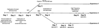
Timeline of the three experimental procedures. Experiment 1: behavioral effects of acute oxytocin treatment. Experiment 2: behavioral effects of HFS. Experiment 3: behavioral effects of social fear extinction. (SFC) Social fear conditioning, (SEE) socially enriched environment, (EXT) extinction.
The measurements and statistical analyses of various test parameters are presented below, including social interaction with stimulus mice, freezing time, collective and individual object exploration, and index of preference for the time spent exploring an object outside a proximity zone (OPZ) and inside a proximity zone (IPZ) to conspecifics.
First, we evaluated the duration of object exploration in the arena (Fig. 2A). A two-way ANOVA performed on SFC, and exploration type data showed a significant effect of SFC (F(1,16) = 12.53, P < 0.001) and SFC × exploration interaction (F(1,16) = 41.64, P < 0.001), but not for exploration alone (F(1,16) = 4.44, P = 0.051). The Holm-Šídák post hoc analysis showed a decreased IPZ object exploration displayed by SFC+ mice compared with SFC− mice (t = 7.06, P < 0.001). The two groups did not differ from each other regarding OPZ object exploration time (t = 2.06, P = 0.10). Hence, the conditioned mice spent more time in OPZ exploration than in IPZ exploration (t = 6.05, P < 0.001).
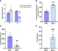
Behavioral responses to SFC in the SEE test during experiment 1. (A) Duration of object exploration. (B) Preference for OPZ object exploration. (C) Social interaction time. (D) Freezing duration. Results are presented as means ± SEM. Post hoc Holm-Šídák test. n(SFC−) = 5, n(SFC+) = 5. (*) P < 0.05; (***) P < 0.001 versus OPZ exploration; (###) P < 0.01 versus SFC−. (OPZ) Outside a proximity zone, (IPZ) inside a proximity zone.
To better illustrate the preference for OPZ object exploration over IPZ object exploration, the values were expressed as a percentage preference (Fig. 2B). The t-test analysis revealed a highly significant increase in preference for OPZ exploration in the SFC+ group compared to the SFC− group (t = 8.33, P < 0.001).
Furthermore, t-test analysis revealed that SFC significantly reduced interaction time (t = 5.67, P < 0.001; Fig. 2C) and increased freezing time (t = 5.63, P < 0.001; Fig. 2D) in the SFC+ group compared to the SFC− group.
Effects of oxytocin
We next evaluated the effect of acute oxytocin treatment on behaviors related to social anxiety induced by SFC.
Three-way ANOVA analysis indicated a significant effect of the SFC factor on the duration of object exploration (F(1,40) = 9.36, P = 0.003) (Fig. 3A). However, the factor type of exploration and treatment did not have a significant effect on this parameter (F(1,40) = 3.75, P = 0.059; F(1,40) = 1.63, P = 0.208, respectively). Moreover, the interactions type of exploration × SFC and type of exploration × treatment revealed significant effects (F(1,40) = 14.12, P < 0.001; F(1,40) = 6.73, P = 0.013, respectively). The Holm-Šídák post hoc test showed a significant increase of IPZ exploration, which was observed in the SFC+/OXT group compared to the SFC+/Veh group (t = 3.36, P = 0.007). It is also worth noting that the comparison between the unconditioned and conditioned groups revealed a significant decrease in IPZ exploration only for the SFC+/Veh group compared to the SFC−/Veh group (t = 4.83, P < 0.001).
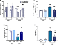
Effect of acute oxytocin treatment on SFC-induced social fear in male mice. (A) Duration of object exploration. (B) Individual exploration preference index. (C) Time of social interaction. (D) Freezing time. Results are presented as means ± SEM. Post hoc Holm-Šídák test. n(SFC−/Veh) = 5, n(SFC−/OXT) = 8, n(SFC+/Veh) = 5, n(SFC+/OXT) = 6. (*) P < 0.05; (**) P < 0.01 versus OPZ exploration. (μ) P < 0.05, (μμμ) P < 0.001 versus SFC−/Veh. ($) P < 0.05, ($$) P < 0.01, ($$$) P < 0.001 versus SFC+/Veh. (###) P < 0.001 versus SFC−/OXT. (OPZ) Outside a proximity zone, (IPZ) inside a proximity zone, (Veh) vehicle, (Oxt) oxytocin.
Furthermore, a two-factor ANOVA analysis (SFC and treatment) demonstrated significant effects of SFC (F(1,20) = 37.5, P < 0.001), treatment (F(1,20) = 20.0, P < 0.001), and SFC × treatment interaction (F(1,20) = 13.7, P = 0.001) on the preference for OPZ object exploration (Fig. 3B). The Holm-Šídák post hoc test showed a higher preference for OPZ object exploration in SFC+/Veh mice than in the SFC−/Veh group (t = 6.46, P < 0.001). Interestingly, the SFC+/OXT group showed a significant decrease in the preference for OPZ object exploration compared to SFC+/Veh (t = 5.61, P < 0.001).
Subsequently, we evaluated the effect of oxytocin on the social interaction parameter (Fig. 3C). Two-way ANOVA analysis (SFC and treatment) showed significant effects of SFC (F(1,16) = 18, P < 0.001) and SFC × treatment interaction (F(1,16) = 8.7, P < 0.01), but not treatment (F(1,16) = 1.6, P = 0.2202). The results of multiple comparisons indicated that the duration of social interaction in the SFC+/Veh group decreased considerably compared to the SFC−/Veh group (t = 5.08, P < 0.001). In contrast, the SFC+/OXT group showed a slight decrease, statistically not significant, of social interaction duration similar to that of the SFC−/OXT group (t = 0.9, P = 0.375). Moreover, the SFC+/OXT group exhibited a significant increase in this parameter compared to the SFC+/Veh group (t = 2.989, P < 0.05).
Finally, evaluation of the effect of oxytocin on freezing time (Fig. 3D) was performed using a two-way ANOVA analysis (SFC and treatment). This analysis revealed significant effects of SFC (F(1,20) = 110, P < 0.001) and treatment (F(1,20) = 22.1, P < 0.001) as well as their interaction (F(1,20) = 23.1, P < 0.001). The Holm-Šídák post hoc test showed a longer freezing time in the SFC+/Veh group compared to SFC−/Veh (t = 10.1, P < 0.001), and in the SFC+/OXT group compared to the SFC−/OXT group (t = 4.39, P < 0.001). Interestingly, the freezing time was significantly decreased in the SFC+/OXT group compared to the SFC+/Veh group (t = 6.52, P < 0.001).
Effects of IL HFS
To examine the impact of HFS of the IL cortex on SAD, we conducted the SEE test before and after applying HFS to male mice.
The three-factor ANOVA analysis (stimulation, SFC, and type of exploration) showed that the duration of object exploration significantly varied based on SFC and type of exploration (F(1,32) = 13.76, P < 0.001; F(1,32) = 24.25, P < 0.001, respectively) (Fig. 4A). However, stimulation did not have a significant effect on this parameter (F(1,32) = 1.90, P = 0.1775). Moreover, the interactions of the type of exploration × SFC and the type of exploration × stimulation revealed significant effects (F(1,32) = 49.34, P < 0.001; F(1,32) = 10.29, P = 0.003, respectively). However, there was no statistical significance regarding the SFC × stimulation interaction (F(1,32) = 1.43, P = 0.2398). Using the Holm-Šídák post hoc analysis, we observed that after stimulation, there was a significant decrease of OPZ exploration time in conditioned mice (t = 2.58, P = 0.0324). We also noted a significant increase in IPZ exploration time in both the SFC− and SFC+ groups during SEE+HFS compared to SEE (t = 4.14, P = 0.0015; t = 3.12, P = 0.0281, respectively). However, SFC− mice spent more time exploring collectively after stimulation than SFC+ mice (t = 6.64, P < 0.001).
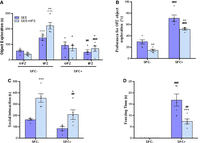
Effect of high-frequency brain stimulation of the IL cortex on SFC-induced social fear in male mice. (A) Duration of object exploration. (B) Individual exploration preference index. (C) Time of social interaction. (D) Freezing time. Results are presented as means ± SEM. Post hoc Holm-Šídák test. n(SFC−) = 5, n(SFC+) = 5. (***) P < 0.001 versus OPZ exploration, (##) P < 0.01, (###) P < 0.001 versus SFC−. (+) P < 0.05; (++) P < 0.01; (+++) P < 0.001 versus SEE1. (OPZ) Outside a proximity zone, (IPZ) inside a proximity zone, (HFS) high-frequency stimulation.
Moreover, two-way ANOVA analysis revealed significant effects of SFC (F(1,16) = 123.2, P < 0.001) and stimulation (F(1,16) = 22.98, P < 0.001) on the preference for OPZ object exploration (Fig. 4B). However, no significant interaction effect between the two factors was observed (F(1,16) = 0.39, P = 0.5402). Multiple comparisons indicated that the preference for OPZ object exploration in SFC+ mice was higher than in SFC− mice during SEE (t = 8.29, P < 0.001). However, this preference significantly decreased in both the SFC− and SFC+ groups after stimulation compared to before stimulation (t = 2.94, P = 0.0095; t = 3.832, P = 0.0029, respectively). Nevertheless, this preference was much higher for SFC+ mice compared to SFC− mice during SEE+HFS (t = 7.40, P < 0.001).
Furthermore, a two-way ANOVA analysis revealed main effects of the SFC (F(1,16) = 14.06, P < 0.01) and the stimulation (F(1,16) = 27.49, P < 0.001) on the duration of social interaction (Fig. 4C). However, SFC × stimulation interaction was not significant (F(1,16) = 1.28, P = 0.2744). Regarding the unconditioned group, the Holm-Šídák post hoc analysis confirmed that social interaction in this group was significantly higher after stimulation (t = 4.50, P < 0.001), as was the case in the SFC+ group (t = 2.90, P = 0.0103). However, during SEE+HFS, SFC− mice interacted more than SFC+ mice (t = 3.45, P = 0.0066).
On the other hand, the two-factor ANOVA analysis (SFC and stimulation) of freezing time data revealed significant effects of SFC and stimulation factors in addition to the SFC × stimulation interaction (F(1,16) = 68.27, P < 0.001; F(1,16) = 10.49, P = 0.0051; F(1,16) = 10.49, P = 0.0051, respectively) (Fig. 4D). Multiple comparisons showed a significant increase in the freezing time in the SFC+ group compared to the SFC− group before (SEE: t = 8.13, P < 0.001) and after (SEE+HFS: t = 3.55, P = 0.0027) stimulation. However, a significant decrease in immobility time was observed in conditioned mice during SEE+HFS compared to SEE (t = 4.58, P = 0.001).
Effects of extinction training
To examine the impact of social fear extinction through repeated SEE testing, we conducted four extinction sessions with a 48 h gap between each session.
Three-way ANOVA analysis (SEE test session, SFC, and type of exploration) showed a significant effect of the type of exploration factor (F(1,72) = 27.37, P < 0.001) on object exploration duration (Fig. 5A). However, the SFC and SEE test session factors did not show a significant effect on this parameter (F(1,72) = 0.8144, P = 0.3698; F(3,72) = 1.525, P = 0.2154, respectively). The interactions of SEE test session × type of exploration and SFC × type of exploration had significant effects on the exploration duration (F(3,72) = 9.443, P < 0.001; F(1,72) = 80.93, P < 0.001, respectively), unlike the interaction of SEE test session × SFC (F(3,72) = 0.66, P = 0.5784). The Holm-Šídák post hoc test indicated that during SEE, a significant increase of the OPZ exploration time was observed in SFC+ mice compared to SFC− mice (t = 4.81, P < 0.001), unlike IPZ exploration (t = 3.74, P < 0.001). In contrast, IPZ exploration time increased significantly during SEE+EXT2 compared to SEE in SFC+ mice (t = 2.20, P = 0.0045). For the same group, a significant decrease in OPZ exploration time was observed during SEE+EXT3 compared to SEE (t = 3.03, P = 0.0028).
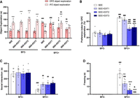
Effect of extinction training. (A) Duration of object exploration. (B) Individual exploration preference index. (C) Time of social interaction. (D) Freezing time. Results are presented as means ± SEM. Holm-Šídák post hoc test. n(SFC−) = 6, n(SFC+) = 5. (##) P < 0.01; (###) P < 0.001 versus SFC−. (++) P < 0.01; (+++) P < 0.001 versus OPZ exploration. (*) P < 0.05; (**) P < 0.01; (***) P < 0.001 versus SEE. ($) P < 0.05; ($$) P < 0.01; ($$$) P < 0.001 versus SEE+EXT1. (OPZ) Outside a proximity zone, (IPZ) inside a proximity zone, (EXT) extinction.
Analysis of the preference for OPZ object exploration (Fig. 5B) using a two-way ANOVA analysis (SFC and SEE test session) revealed significant effects of SFC, SEE test session, and their interaction (F(1,36) = 175.4, P < 0.001; F(3,36) = 18.31, P < 0.001; F(3,36) = 10.01, P < 0.001, respectively). We noticed that the SFC+ group had a significantly higher preference for OPZ object exploration than the SFC− group during SEE (t = 9.26, P < 0.001) and SEE+EXT3 (t = 4.56, P < 0.001). However, within the same SFC+ group, this index significantly decreased during SEE+EXT3 compared to SEE (t = 5.66, P < 0.001).
Two-way ANOVA analysis (SFC and treatment) of social interaction time showed a significant effect of SFC (F(1,36) = 59.1; P < 0.001) (Fig. 5C). However, no significant effects were observed for SEE test session or SFC × SEE test session interaction (F(3,36) = 1.19, P = 0.3274; F(3,36) = 0.5521, P = 0.6501, respectively). Holm-Šídák multiple comparisons revealed that the social interaction time of the SFC− group was greater than that of the SFC+ group during all four test sessions: SEE (t = 4.918, P < 0.01), SEE+EXT1 (t = 3.739, P = 0.0019), SEE+EXT2 (t = 3.464, P = 0.0028), and SEE+EXT3 (t = 3.255, P = 0.0028).
Two-way ANOVA (SFC and SEE test session) analysis showed significant effects of SFC (F(1,36) = 81.38; P < 0.001), SEE test session (F(3,36) = 20.7, P < 0.001), and interaction (F(3,36) = 20.7, P < 0.001) on the freezing time (Fig. 5D). Multiple comparisons revealed that SFC+ mice had significantly higher immobility times compared to SFC− mice during SEE: t = 10.45, P < 0.001 and SEE+EXT1: t = 5.46, P < 0.001. However, when comparing the SFC+ group's freezing times across different sessions, there was a notable decrease during SEE+EXT1 (t = 4.773, P < 0.001), SEE+EXT2 (t = 7.955, P < 0.001), and SEE+EXT3 (t = 10, P < 0.001) compared to SEE. Additionally, SFC+ mice showed reduced freezing times in SEE+EXT2 (t = 3.18, P < 0.01) and SEE+EXT3 (t = 5.22, P < 0.001) compared to SEE+EXT1. Furthermore, there was a decrease in freezing time for the SFC+ group in SEE+EXT3 compared to SEE+EXT2 (t = 2.04, P < 0.05).
Comparison of the therapeutic approaches
Following these results, we carried out a comparative study to evaluate the three therapeutic approaches. This comparison focused on three parameters: preference of OPZ exploration (which more accurately reflects the time spent on both collective and individual exploration of objects), social interaction duration with stimulating mice and freezing duration. This comparison involved four groups of conditioned mice: (a) control mice that received no treatment (CONT), (b) mice during a second exposure to the SEE test (EXT), (c) mice that were given oxytocin (OXT), and (d) mice after HFS.
One-way ANOVA showed a highly significant effect of treatment type on the preference of OPZ exploration (F(3,17) = 17.41, P < 0.001) (Fig. 6A). Furthermore, multiple comparison post hoc tests revealed no significant difference between the CONT and EXT groups (t = 0.1075, P = 0.235). However, compared to the CONT group, a significant decrease was observed in the OXT and HFS groups (t = 6.492, P < 0.001; t = 4.774, P < 0.001, respectively). Interestingly, the EXT mice displayed a higher percentage of the preference of OPZ exploration compared to both the OXT (t = 4.80, P < 0.001) and HFS groups (t = 3.162, P < 0.05). The percentage of preference of OPZ exploration did not differ between the OXT and HFS groups (t = 1.168, P = 0.45).
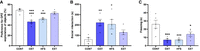
Comparison of the three therapeutic approaches. (A) Individual exploration preference index. (B) Time of social interaction. (C) Freezing time. Results are presented as means ± SEM. Holm-Šídák post hoc test. n(CONT) = 5, n(EXT) = 5, n(OXT) = 6, n(HFS) = 5. (*) P < 0.05; (***) P < 0.001 versus CONT. (+) P < 0.05; (++) P < 0.01; (+++) P < 0.001 versus EXT. (OPZ) Outside a proximity zone, (CONT) control, (EXT) extinction, (Oxt) oxytocin, (HFS) high-frequency stimulation.
On the other hand, a one-way ANOVA analysis revealed a significant effect of treatment type on social interaction time (F(3,17) = 5.991, P = 0.0056) (Fig. 6B). Furthermore, multiple comparisons indicated no significant differences between the CONT and EXT groups in terms of social interaction time (t = 1.59, P = 0.24). However, the OXT and HFS groups showed an increased social interaction time compared to the CONT group (t = 3.66, P < 0.01; t = 3.40, P < 0.05, respectively). Interestingly, although the EXT group had reduced social interaction time, this did not differ from OXT and HFS groups (t = 2.12, P = 0.18; t = 1.81, P = 0.23, respectively). Notably, no significant difference was observed between the OXT and HFS groups (t = 0.19, P = 0.84).
Finally, using a one-way ANOVA, we found that treatment had a significant effect on freezing duration (F(3,17) = 10.25, P < 0.001) (Fig. 6C). The Holm-Šídák post hoc test showed that the EXT, OXT, and HFS groups exhibited significantly a shorter freezing duration compared to the CONT group (t = 2.99, P < 0.05; t = 4.93, P < 0.001; t = 4.72, P < 0.001, respectively). In contrast, no significant difference was observed between the EXT group and the two OXT (t = 1.80, P = 0.244) and HFS (t = 1.72, P = 0.244) groups. Thus, the two OXT and HFS groups showed no significant difference (t = 0.0013, P = 0.99).
Discussion
The primary objective of this study was to assess, in male mice, the efficacy of various treatments in addressing social anxiety induced by SFC. Specifically, we evaluated three therapeutic approaches: pharmacological treatment using oxytocin, electrical brain stimulation targeting the IL cortex, and extinction therapy. These approaches were examined to better understand their potential for mitigating the effects of SFC-induced social anxiety in a controlled experimental setting.
SFA-induced social fear
Numerous animal models, including the social defeat paradigm and exposure to foot shocks, have been employed to investigate the mechanisms of SAD due to its widespread prevalence and severe symptoms. However, these paradigms lack precision in inducing specific behavioral changes, such as generalized anxiety or depression (Denmark et al. 2010; Hollis et al. 2010). In light of these limitations, we adopted the paradigm developed by Toth et al. (2012). This method allows for the elicitation of targeted responses to social fear without introducing additional confounding behavioral changes. As described by Toth et al. (2012), this paradigm is grounded in operant conditioning, in which experimental mice learn to associate social interactions with punishment in the form of electrical shocks, thus inducing fear and avoidance of social stimuli.
In this first part of our study, our aim was to investigate alterations in the social behavior of male mice conditioned to social fear using the SEE test. Our results demonstrate that SFC led to a reduction in IPZ object exploration among conditioned mice compared to control mice. Specifically, conditioned mice showed a tendency to withdraw when unfamiliar mice approached an object. In contrast, their OPZ exploration remained unchanged compared to nonconditioned mice. This suggests that SFC did not alter their exploratory activity, but the presence of stimulus mice nearby interfered with it. This observation is reinforced by the preference index for OPZ exploration, which was higher in the SFC+ group compared to the SFC− group. These findings suggest that SFC promotes an increased preference for OPZ exploration and results in reduced social interaction in conditioned mice. These observations may reflect the social avoidance behaviors seen in individuals with anxiety disorders, where individual exploration is favored while social interaction is limited (American Psychiatric Association 2013). To further explore aspects of social fear, it would be relevant to investigate other components of this behavior. Indeed, the results regarding interaction time further reflect the effectiveness of SFC in inducing SAD. Thus, our conditioned mice exhibited fewer social interactions than nonconditioned mice. These results are consistent with those of Toth et al. (2012). Similarly, in humans, anxiety reduces the motivation to interact with others, leading highly anxious individuals to avoid social interactions even more (Turner 1988). In this regard, the study conducted by Duronto et al. (2005) demonstrates that social interaction avoidance is a reliable indicator of anxiety.
It is important to note that social avoidance can also reflect disinterest in social stimuli, which could bias result interpretation. Therefore, it is crucial to evaluate other fear-related responses, such as immobility (Blanchard et al. 2005). Indeed, data from the SEE test reveal a higher immobility duration in the group subjected to SFC compared to the control group. This suggests that these mice have memorized social interactions associated with electrical shocks during the approach of a conspecific. We posit that this immobility behavior specifically represents avoidance of social stimulus interactions. These observations are consistent with the findings of Toth et al. (2013). In other words, our data confirm that the reduction in social interactions is specifically attributed to social fear and not a lack of interest.
Treatment with acute oxytocin
The pivotal role of oxytocin in social behavior has garnered substantial interest among researchers due to its profound influence on various affiliative behaviors, including mating, parental behavior, and attachment (Bartz and Hollander 2006). Such social involvement suggests that this hormone plays a significant role in the functioning of our “social brain” (Feldman 2012). According to Buchheim et al. (2009), individuals experiencing relational insecurity exhibit an increase in their feelings of attachment and emotional security following oxytocin administration. Similarly, a study by De Oliveira et al. (2012) suggests that oxytocin reduces anticipatory anxiety in healthy subjects when speaking in public.
Using C57BL/6n mice, Zhang et al. (2015) have demonstrated that subcutaneous oxytocin administration (100 μg/kg, 10 mL/kg, administered 30 min before behavioral tests) increased social interaction time and modulated both social and nonsocial behaviors.
Given the above-mentioned effects, we sought to explore the influence of this neuropeptide on social fear. To do this, we induced social fear through SFC in male mice, and, after a 24 h interval, we administered oxytocin (100 µg/kg, 10 mL/kg) subcutaneously. Thirty minutes later, we subjected them to the SEE test.
The results revealed that oxytocin administration in the conditioned group led to a significant increase in IPZ exploration compared to OPZ exploration of objects, in comparison with the conditioned vehicle group. These results suggest that oxytocin may promote collective exploration in conditioned mice. Furthermore, the results of the preference index for OPZ exploration showed a highly significant decrease in this index in the SFC+/OXT group compared to the SFC+/Veh group. Collectively, these findings indicate that oxytocin reduces social fear induced by SFC. Regarding social interaction, the results showed a significant increase in this parameter in the conditioned group treated with oxytocin compared to the conditioned vehicle group. This implies that oxytocin facilitated an increase in social interaction behaviors in conditioned mice. Additionally, we also examined immobility, which was significantly reduced in the conditioned group treated with oxytocin compared to the conditioned group treated with the vehicle. Although this reduction did not reach the level of the nonconditioned groups, it suggests that oxytocin contributed to a reduced fear response in conditioned mice. In summary, based on the analysis of the parameters mentioned above, it appears that oxytocin has an opposing effect on SFC.
In previous studies in rodents, oxytocin has been identified as a neuropeptide that promotes social approach behavior and assists rodents in overcoming social proximity avoidance. This aligns perfectly with our results. Indeed, it has been reported that acute oxytocin treatment increases sociability and social affiliation, extending beyond reproductive-related behaviors (Lukas et al. 2011; Ramos et al. 2013; Holley et al. 2015). Furthermore, mice with mutations in the oxytocin receptor gene exhibit abnormalities in social behavior (Takayanagi et al. 2005). Additionally, according to De Oliveira et al. (2012), both male and female rats and mice respond to oxytocin in the same way as anxiolytics. Just as in animals, oxytocin administration also promotes social interactions in humans. A single intranasal administration of oxytocin has been shown to increase the subjective sensation of attachment security, which is associated with reduced stress reactivity and improved social interaction capacity (Buchheim et al. 2009). Thus, oxytocin diffuses to several brain structures involved in social behavior and fear regulation, including the amygdala (Meyer-Lindenberg et al. 2011). A study in rats has identified a pathway through which oxytocin inhibits amygdala activation, thus modulating its communication with the autonomic nervous system (Huber et al. 2005). This action of oxytocin contributes, in part, to its anxiolytic properties by reducing the hypothalamo-hypophyseal axis reactivity to social stressors (Meyer-Lindenberg et al. 2011). Finally, it has been demonstrated that the amygdala is connected to prefrontal and striatal regions involved in affiliative or avoidance behaviors among individuals of the same species (McDonald 1991). Therefore, the modulation of the activity of these brain structures, as well as others, by oxytocin could play a crucial role in reducing social fear induced by fear conditioning in our study.
Treatment with brain stimulation
The prefrontal cortex plays a crucial role in inhibiting inappropriate responses, particularly the IL region of the medial prefrontal cortex, which is vital for the extinction of conditioned fear (Quirk et al. 2006). In a prior study, the GABAA receptor inhibitor (bicuculline methiodide) was infused into the IL cortex, resulting in an anxiogenic effect (Bi et al. 2013). Furthermore, in the same study, these researchers suggested that imbalances in afferents to pyramidal neurons in the IL cortex could lead to anxiety disorders. Additionally, activation of this region increases motivation, sociability, and reduces stress in rodents (Myers et al. 2019).
Given the above-mentioned effects, we aimed to investigate the efficacy of stimulating this brain structure in reducing social fear responses. To do this, we induced social fear in male mice. Subsequently, the mice underwent two sessions of the SEE test: one before stimulation and one after.
The results demonstrated an increase in social interaction and IPZ object exploration following HFS in both the conditioned and unconditioned groups. We also observed that the value of the OPZ exploration preference index significantly decreased after stimulation in both groups. However, SFC+ mice maintained a higher OPZ index than SFC− mice during the second session of the SEE test. Furthermore, HFS significantly reduced immobility duration in conditioned mice compared to the prestimulation period. In summary, these results suggest that the IL cortex plays a substantial role in the modulation of social anxiety. In line with this last finding, prior research has indicated that microstimulation of the IL cortex reverses the effect of SFC, resulting in decreased freezing (Milad and Quirk 2002). Moreover, the IL cortex influences the hypothalamo-pituitary stress axis response (Radley et al. 2006). This explanation could be extrapolated to our results, indicating a reduction in stress responses and an improvement in social interaction among conditioned mice. On the other hand, the IL cortex projects to numerous sites involved in fear response control, notably the amygdala. More specifically, intercalated neurons receive a significant projection from the IL cortex (McDonald et al. 1996), and stimulation of this connection promotes rapid extinction and inhibition of central amygdala neurons, leading to reduced fear expression (Milad and Quirk 2002).
This study suggests that HFS in the IL cortex may have a potential therapeutic role in alleviating social anxiety and improving social behavior.
Treatment with extinction training
Within the realm of psychotherapy, one approach employed is exposure therapy, which aims to gradually diminish fear by inducing the extinction of fear responses through repeated exposure to aversive conditioned stimuli. This method is applicable, especially in cases of acquired social fear (Fedoroff and Taylor 2001). This therapeutic approach has proven effective in treating SAD in humans (Clark et al. 2003) and in certain animal models of conditioned fear (Toth et al. 2012). Studies suggest that cognitive-behavioral interventions that incorporate exposure techniques yield slightly more favorable results than interventions solely focused on cognitive restructuring (i.e., cognitive therapy focused on anxiety and/or patient's thoughts) (Gould et al. 1997).
In this context, our objective was to explore the effect of exposure therapy on the extinction of social fear using a model of SFC. To achieve this, male mice underwent four extinction sessions with a 48 h interval between each session.
The results confirmed the extinction of social fear based on various measured parameters. Indeed, during the fourth session of the test, a significant increase in IPZ exploration was observed in the conditioned mice compared to their first session, although this level remained lower than that of unconditioned mice. Conversely, for the SFC+ group, the OPZ exploration preference index and immobility duration showed a decrease during the last session compared to the first. Nevertheless, the OPZ exploration preference index and immobility duration remained higher in the SFC+ group compared to the CPS− group. Furthermore, a slight increase in social interaction was observed between the first and fourth session of the test in SFC+ mice. In conclusion, the repetition of the SEE test led to a decrease in conditioned fear.
The obtained results can be attributed to the fact that exposure promotes corrective learning, involving unlearning or deconditioning phobic reactions (Marks and Toben^a 1990). It is worth noting that extinction is a learning process, and the reduction of fear results from the inhibition of the response rather than the complete erasure of the initial fear memory (Garcia 2002; Bouton et al. 2006; Ji and Maren 2007). From a neurobiological perspective, extinction can be considered as a form of learning that does not affect previous plasticity but rather reflects an increase in plasticity in GABAergic inhibitory interneurons. Therefore, the greater the plasticity of GABAergic interneurons, the more they inhibit the central amygdala, and the less fear is expressed (Royer and Paré 2002).
Beyond the amygdala, other neural structures may be involved in extinction, such as the medial prefrontal cortex and the hippocampus, both of which have interconnections with the amygdala (Quirk and Mueller 2008). These structures appear to modulate the inhibition of conditioned responses (Bruchey et al. 2007; Ji and Maren 2007).
This study suggests that repeated exposure tests may have potential therapeutic implications for promoting the extinction of social fear, offering insights into the underlying mechanisms of this process.
Comparison of the three therapeutic approaches
In the final segment of our study, we conducted a comparative assessment of three therapeutic approaches to determine their relative efficacy. Our findings indicate that there is no significant difference in the preference index for OPZ exploration between the CONT group and the EXT group. This suggests that a second exposure to the SEE test does not mitigate the impact of SFC on the behavior of conditioned mice. In contrast, both the OXT and HFS groups exhibited a comparable reduction in OPZ exploration preference, which was significantly different from the CONT and EXT groups. Therefore, it can be inferred that OXT and HFS are more likely to influence the tendency of mice to collectively explore their environment.
Additionally, when examining interaction time, the results show no significant differences between the CONT and EXT groups, indicating that the second exposure to the SEE test and the absence of treatment have a similar effect on social interaction, resulting in reduced interaction time. On the contrary, both the OXT and HFS groups demonstrated a significantly higher interaction time. Notably, the EXT group, while not significantly different, spent less time in social interaction compared to the OXT and HFS groups. These outcomes suggest that both oxytocin administration and HFS treatments are more effective in enhancing social interaction behaviors that were disrupted by SFC, in contrast to the absence of treatment and the second exposure.
Furthermore, the CONT group exhibited significantly longer freezing durations compared to the EXT, OXT, and HFS groups. However, there were no significant differences between the EXT group and the OXT and HFS groups, even though the graphical representation might imply longer freezing durations in the EXT group. This indicates that the second exposure to the SEE test, oxytocin administration, and HFS treatments were equally effective in reducing freezing durations when compared to the absence of treatment.
The obtained results unequivocally demonstrate that our comparative evaluation of three therapeutic approaches in the context of mitigating the effects of SFC in mice provides valuable insights. The findings highlight that both oxytocin administration and HFS treatments show more promise in reversing the effects of SFC on mice. Conversely, it becomes evident, upon close examination of the results, that achieving comparable positive outcomes to those of the OXT and SFC groups would require multiple exposure sessions.
This suggests that all three therapeutic approaches have a positive impact when compared to the absence of treatment, offering potential strategies for addressing the behavioral consequences of SFC in preclinical models.
The choice to conduct behavioral tests, such as the SEE test, 30 min after oxytocin administration was informed by both previous studies and the pharmacokinetic profile of oxytocin. Subcutaneous injections have been shown to result in peak or effective oxytocin activity within a 30 min window, as demonstrated in behavioral tests like the social interaction test (Zhang et al. 2015). This timing aligns with findings from studies on nasal administration, which similarly report that oxytocin reaches its peak concentration ∼30 min post-administration (Neumann et al. 2013). This consistency across different routes of delivery highlights a robust pharmacokinetic profile, making 30 min post-administration the optimal window for assessing oxytocin's effects on behavior.
Materials and Methods
Animals
Experiments were performed on 54 male Swiss mice (10–12 weeks old, weighing 30–35 g) raised in the central animal care facilities of the Cadi Ayyad University, Marrakesh, Morocco. Animals were housed in groups of three to five in transparent Plexiglas cages (26 × 27 × 15 cm) with wood chip bedding under controlled environmental conditions (12:12 light–dark cycle at 22°C ± 2°C) and standard diet and water ad libitum. Behavioral experiments were performed between 9:00 a.m. and 3:00 p.m. All animal procedures were in strict accordance with the ethical standards and guidelines of the European Council Directive (EU2010/63) and the Council Committee of Research Laboratories of the Cadi Ayyad University of Marrakesh, Morocco.
Experimental design
The behavioral schedule is outlined in Figure 2.
Social fear conditioning paradigm
To induce SAD in mice, we used the SFC paradigm established by Toth et al. (2013). The paradigm consisted of the administration of mild electric foot shocks to an experimental mouse while exploring an unfamiliar stimulus mouse, resulting in a specific social fear lasting for at least 15 days. The experiment was designed as follows:
First phase
During this phase, experimental mice were housed individually for three days prior to the SFC phase. Isolation of mice was maintained throughout the subsequent behavioral procedures. Preconditioning isolation is used to enhance the social motivation of mice on the day of SFC (Niesink and Van Ree 1982), whereas postconditioning isolation is maintained to avoid extinction that might occur if mice were placed together in group cages (Cherng et al. 2010).
Second phase
The natural preferential behavior of male mice toward an unfamiliar congener was negatively associated with an aversive stimulus corresponding to an electric shock to the footpads. This shock was delivered by an electro-stimulator (Bioseb, LE10026) connected to the Skinner box, which consisted of a cage (20 × 20 × 25 cm, Bioseb, LE918) with a stainless-steel grid floor.
First, the experimental mouse was placed in the Skinner box previously cleaned with 70% ethanol for a 30 sec habituation period. Subsequently, a small, empty cage (i.e., nonsocial stimulus), electrically isolated and previously cleaned, was placed in a corner of the conditioning box. After 3 min of exploration of the nonsocial stimulus by the experimental mouse, the empty cage was replaced by another identical cage containing a foreign mouse (i.e., social stimulus) and placed in the same corner of the Skinner box. Male social stimulus mice, with identical weights and ages, were acclimatized to stimulus cages for 10 min during the three days before the SFC. The experimental mice explored the entire device for 3 min, and for each direct interaction with the social stimulus, the experimental mouse destined for conditioning (SFC+) received an electric shock (1 sec, 0.7 mA) after 2–3 sec of interaction. To optimize the induction of conditioning to the social stimulus, the SFC+ mice received between two and five electric shocks (Toth et al. 2012). Once the electric shocks were administered, the experimental mice were returned to their cage. The total duration of this conditioning session varies from 3 to 10 min, depending on the number of shocks administered. The same protocol was applied to unconditioned mice (SFC−) without receiving any electric shocks.
Between each conditioning session, the Skinner box and stimulus cages were cleaned with 70% ethanol. Cages used as nonsocial stimuli were different from those used as social stimuli to avoid odor cues from previous subjects. Experimental animals that were not being conditioned remained in a neighboring room to prevent them from hearing vocalizations emitted during SFC.
SEE test
All behavioral tests were recorded using a CCD camera (Sony chip, model: TB-001) placed above the apparatus.
One day after SFC, we assessed the social behavior of mice using the SEE test.
The SEE test allows for a detailed analysis of behavioral changes in mice in a novel enriched environment that mimics a natural setting containing different objects and three freely moving unfamiliar conspecifics for 30 min (Boudjafad et al. 2022). The apparatus for the SEE test consisted of a rectangular, transparent glass arena (40 × 60 × 25 cm) containing clean bedding and eight unfamiliar objects. These objects included three toys, two cubes (5 × 5 × 5 cm), a cylinder (length: 9 cm, diameter: 3.5 cm), and two small white ping-pong balls glued together (diameter: 4 cm). The SEE objects were made from different materials, textures, and colors to promote exploratory activity.
The experimental mice and the stimulus mice were brought into the experimental room 30 min before testing for room acclimatization. Each experimental mouse was tested with a group of three unfamiliar stimulus mice with identical weights that were housed collectively under the same conditions. For each group of experimental mice, two groups of three stimulus mice were used. Between each trial, stimulus mice were transferred into their home cage and replaced by another group. Also, the objects’ disposition was changed to enhance their exploratory activity. Furthermore, the bedding was renewed, and the equipment and objects were cleaned with a 70% ethanol solution after each passage of the experimental mice.
The test started by placing the experimental mouse and the three stimulus mice in the center of the apparatus and allowing them to freely explore the arena for 30 min. The parameters considered were as follows.
Social interaction time
Initiated by the experimental mouse, it represents the total time that this mouse spends exploring a stimulus mouse by making direct contact (including whole-body sniffing, anogenital sniffing, nose-to-nose sniffing, and the time spent following a stimulus mouse while moving in the apparatus). When a stimulus mouse approached the experimental mouse and made direct contact with it, this contact was not scored.
Object exploration time
It represents the time spent exploring an object either outside (OPZ) or inside (IPZ) a proximity zone to conspecifics. Specifically, this measurement was recorded when no stimulus mouse entered a zone of 10 cm (OPZ) or when the head of a stimulus mouse entered the proximity zone (IPZ), respectively. The perimeter of 10 cm was determined after many observations as being approximately the maximum distance of approach, after which the experimental mouse moved away or froze.
Immobility time “freezing time”
This is defined as the complete absence of body movements except for respiratory-related movements (Fanselow 1990). It was measured using a 1 sec time sampling technique.
Index of Preference for OPZ object exploration:

All the behavioral parameters were manually scored by an experienced observer who was blind to the treatment conditions.
Treatment approaches
Experience 1 (drug treatment)
Numerous studies have demonstrated the effect of oxytocin on social behavior. According to Zhang et al. (2015), this neuropeptide would enhance social interaction in mice. Therefore, we tested the effect of acute oxytocin treatment on social fear induced by SFC in the context of the SEE (Fig. 1). To do this, we administered an oxytocin solution (Syntocinon 5U.I./mL, Novartis) subcutaneously to conditioned (SFC+/OXT, n = 6) and unconditioned (SFC−/OXT−, n = 8) male mice (100 µg/kg, 10 mL/kg) (Zhang et al. 2015). The SEE test was conducted 30 min after the injection. Vehicle groups (SFC−/Veh, n = 5; SFC+/Veh, n = 5) received the same amount of saline solution (0.9%).
Experience 2 (HFS)
To test a new therapeutic approach for alleviating symptoms of SAD, we employed HFS in the IL cortex, a brain area strongly implicated in the expression and extinction of fear (Milad et al. 2004). The principle of this technique involves administering trains of electrical pulses at frequencies >50 Hz (Jensen and Durand 2009). Our experimental approach spanned a period of 13 days. For this purpose, the mice were divided into two groups: SFC−/HFS+ (n = 5) and SFC+/HFS+ (n = 5). Each animal underwent the SEE test twice, before (SEE) and after the stimulation (SEE+HFS) (Fig. 1).
The electrodes were made using two PFA-coated tungsten wires, soldered together, and attached to a female MillMax connector. The wires were twisted and electrically insulated at the ends for stimulation delivery. These electrodes were implanted into anesthetized mice using stereotaxic techniques. After anesthesia induction and fixation in a stereotaxic apparatus, the skull was exposed and drilled to create holes for anchoring screws and electrode implantation. The electrode was placed at specific coordinates in the right IL area (AP = 1.8 mm, MD = 0.3 mm, P = 3.0 mm) as referenced to the stereotaxic atlas (Paxinos and Franklin 2001). After securing the electrode with dental cement, mice undergo a 7 day recovery period with close health monitoring.
After 7 days of recovery, the animal was connected to the stimulator (Lab-Trax-4/24T) via a rotary switch on top of the apparatus, allowing it to move freely. Subsequently, the mice were placed in a transparent cylindrical plexiglass box (24 cm in diameter and 20 cm in height) with clean bedding (Fig. 1). After a 3 min habituation phase, each mouse received two trains of 150 pulses (0.4 msec; 250 Hz; 4.5 V) with a 60 sec interval. These HFS parameters were used based on our previous study (Bentefour et al. 2018), which also targeted the IL cortex. After 30 min, the mouse was removed from the apparatus and returned to its home cage. IL HFS was delivered once on day 13 (Fig. 1).
Experience 3 (extinction training)
Repeated exposure is a commonly used method in the treatment of social fear (Pelissolo et al. 2019). In this context, we tested the effect of repeating the SEE test on the social behavior (all the parameters) of conditioned mice.
To do this, two groups of animals were used: one conditioned (SFC+, n = 5), the other unconditioned (SFC−, n = 6). Each group underwent four sessions (SEE1 as baseline and three extinction sessions SEE+EXT1, SEE+EXT2, and SEE+EXT3) of the SEE test with a 2 day interval between each session (Fig. 1).
Statistical analysis of data
Data were statistically analyzed using the GraphPad Prism program, version 8. The Shapiro–Wilk normality test was performed on all data. In cases where the normality test was positive, parametric tests were used. A three-factor ANOVA was used to examine the effect of oxytocin, repeated SEE test, and HFS on object exploration. Furthermore, a two-factor ANOVA was employed to investigate the effect of oxytocin, repeated SEE test, and HFS on individual exploration preference, immobility time, and social interaction time. This analysis also allowed us to assess the effect of SFC on object exploration. Therefore, individual exploration preference, immobility time, and social interaction time were analyzed using t-tests for comparison between the three therapeutic approaches. The Holm-Šídák post hoc test was used for multiple comparisons. Results are expressed as mean ± SEM (standard error of mean), and a significance of P < 0.05 was used throughout the study.
Footnotes
-
Article is online at http://www.learnmem.org/cgi/doi/10.1101/lm.054041.124.
- Received June 20, 2024.
- Accepted March 20, 2025.
This article is distributed exclusively by Cold Spring Harbor Laboratory Press for the first 12 months after the full-issue publication date (see http://learnmem.cshlp.org/site/misc/terms.xhtml). After 12 months, it is available under a Creative Commons License (Attribution-NonCommercial 4.0 International), as described at http://creativecommons.org/licenses/by-nc/4.0/.



