


Elevated corticosterone after fear learning impairs remote auditory memory retrieval and alters brain network connectivity
- Niek Brosens1,
- Sylvie L. Lesuis2,
- Ilse Bassie1,
- Lara Reyes1,
- Priya Gajadien1,
- Paul J. Lucassen1 and
- Harm J. Krugers1
- 1Brain Plasticity Group, Swammerdam Institute for Life Sciences (SILS)-Cognitive and Systems Neuroscience (CNS), University of Amsterdam, Amsterdam 1098 XH, the Netherlands
- 2Program in Neurosciences and Mental Health, Hospital for Sick Children, Toronto, Ontario M5G 1X8, Canada
- Corresponding authors: a.a.d.brosens{at}uva.nl, h.krugers{at}uva.nl
Abstract
Glucocorticoids are potent memory modulators that can modify behavior in an adaptive or maladaptive manner. Elevated glucocorticoid levels after learning promote memory consolidation at recent time points, but their effects on remote time points are not well established. Here we set out to assess whether corticosterone (CORT) given after learning modifies remote fear memory. To that end, mice were exposed to a mild auditory fear conditioning paradigm followed by a single 2 mg/kg CORT injection, and after 28 d, auditory memory was assessed. Neuronal activation was investigated using immunohistochemistry for the immediate early gene c-Fos, and coactivation of brain regions was determined using a correlation matrix analysis. CORT-treated mice displayed significantly less remote auditory memory retrieval. While the net activity of studied brain regions was similar compared with the control condition, CORT-induced remote memory impairment was associated with altered correlated activity between brain regions. Specifically, connectivity of the lateral amygdala with the basal amygdala and the dorsal dentate gyrus was significantly reduced in CORT-treated mice, suggesting disrupted network connectivity that may underlie diminished remote memory retrieval. Elucidating the pathways underlying these effects could help provide mechanistic insight into the effects of stress on memory and possibly provide therapeutic targets for psychopathology.
Stressful experiences can alter cognition by modulating multiple systems involved in learning and memory and their respective brain circuits (Schwabe et al. 2010b; Vogel et al. 2017). The effects of stressful experiences on memory depend on, among other things, the timing of the stressor with respect to learning and the type of learning (Schwabe et al. 2022). The effects of stress on memory are mediated at least in part by glucocorticoid (GC) hormones that are released from the adrenal glands (Roozendaal 2002; Zhou et al. 2010; Kaouane et al. 2012; De Quervain et al. 2017; Bahtiyar et al. 2020). The elevation of GCs and exposure to stress after learning have been reported to enhance consolidation and thereby increase memory in both rodents and humans (Roozendaal 2002; Cahill et al. 2003; Sandi and Pinelo-Nava 2007; Schwabe et al. 2010a; Xiong et al. 2015; Aubry et al. 2016; De Quervain et al. 2017). Stress exposure via GCs can also enhance stimulus–response learning, notably at the cost of spatial learning (Schwabe et al. 2010b).
Studies on the effects of stress on memory consolidation have frequently assessed recent memories, whereas findings on the effects of stress on more remote memory performance have so far been scarce and disparate. Remote memories are often generalized to provide predictive and abiding guidance for adaptive behaviors (Cowan et al. 2021). Episodic memories are initially dependent on the hippocampus, which plays an important role in the transition from short-term into long-term memories that are then consolidated in cortical networks (Kitamura et al. 2017) via its connectivity with subcortical regions (Restivo et al. 2009; Söderlund et al. 2012; Todd et al. 2016; Guo et al. 2018). However, the salient nature of a memory also may determine the network that supports remote memory retrieval. Specifically, the coactivation of the amygdala and hippocampal subregions occurs at remote recall of emotionally salient information but not during remote recall of neutral memories (Silva et al. 2019).
GCs have been suggested to affect the reorganization of neuronal circuits that underlie the transition of recent to remote memories, a process termed systems consolidation (Roozendaal 2002; Roozendaal et al. 2009; Barsegyan et al. 2010). More specifically, GCs may enhance systems consolidation via bidirectional interactions between the prefrontal cortex (PFC) and the amygdala (Roozendaal et al. 2009; Barsegyan et al. 2010; Reis et al. 2016). So far, however, the underlying neurobiological changes in how elevated GCs affect remote memory and whether they alter interactions between brain circuits have remained unclear. Therefore, here we set out to assess the effects of GCs on remote memory performance and neuronal (co)activation in several brain regions relevant for remote memory. To that end, mice were exposed to mild auditory fear conditioning training followed by corticosterone (CORT) administration (Lesuis et al. 2021). Remote memory retrieval was assessed 28 d later upon presentation of the same auditory cue in a novel nonaversive context. Expression of the immediate early gene c-Fos in various brain regions was assessed as a measure of cellular activation and their correlated activation as a measure of network connectivity in the dentate gyrus (DG) of the hippocampus, amygdala, and PFC subregions.
Results
Corticosterone after learning impaired remote auditory memory retrieval
Mice were exposed to an auditory fear conditioning training paradigm followed by systemic 2 mg/kg CORT or vehicle (VEH) administration (Fig. 1A). No difference in freezing levels was observed between the conditioning sessions of the groups that were treated with vehicle and corticosterone, respectively, after training (Fig. 1B). This indicates that learning per se was not different between the two groups. After 28 d, the mice were placed in a novel nonaversive context, and the previously conditioned auditory cue was presented again. Analysis of freezing behavior indicated a significant interaction between time and treatment (F(3.29,46.12) = 5.19, P = 0.003) (Fig. 1C). Post-hoc analysis revealed that this interaction is explained by a significantly lower level of freezing behavior in the CORT-treated mice compared with the VEH-treated mice upon presentation of the auditory cue (P < 0.01).
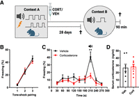
Corticosterone immediately after learning impaired remote auditory memory retrieval. (A) Schematic overview of the remote auditory fear conditioning paradigm. Mice received 2 mg/kg corticosterone (CORT) or vehicle (VEH) immediately after training in context A. After 28 d, remote auditory memory was tested in context B, and the mice were sacrificed 90 min after retrieval. The nonretrieval control group was sacrificed after 28 d to assess the baseline effects of the fear conditioning training paradigm with the CORT/VEH treatment on neuronal activity. Created with BioRender.com. (B) No difference in freezing levels was observed between the conditioning groups that were treated with vehicle and corticosterone, respectively, after training. (C) After 28 d, the mice were placed in a novel nonaversive context, and the previously conditioned auditory cue was presented again. A significant time × treatment interaction was present (F(3.29,46.12) = 5.19, P = 0.003). Post-hoc analysis revealed that this interaction is explained by a significantly higher level of freezing behavior in the VEH-treated mice compared with the CORT-treated mice upon presentation of the auditory cue (P < 0.01). (D) No differences were found in the serum corticosterone levels (in nanograms per milliliter). (**) Post-hoc Tukey effect P < 0.01. Data are presented as mean ± SEM. VEH n = 8, CORT n = 8.
Blood collected upon sacrifice 90 min after retrieval was used to measure plasma CORT levels. No differences were found between the VEH- and CORT-treated mice or in the nonretrieval control group (Supplemental Fig. 1A) in terms of plasma corticosterone levels (t(14) = 0.22, P = 0.83) (Fig. 1D), which were equally low in all experimental groups. Although stress is multifaceted and could be reflected by many parameters, the low plasma corticosterone levels across the groups suggest that mice did not suffer from chronic stress by individual housing. Together, these results show that CORT administration after training—at a dosage that enhances consolidation of recent memories in a generalizing fashion and in a learning-dependent manner (Lesuis et al. 2021)—significantly reduces remote auditory memory retrieval 28 d later.
Corticosterone did not alter the total number of c-Fos-positive cells in the dentate gyrus, amygdala, and prefrontal cortex subregions
Memory retrieval increased the number of c-Fos-positive cells in comparison with the nonretrieval group, where the number of c-Fos-positive cells was generally very low across the brain regions (Supplemental Fig. 1B–J). Memory retrieval induced a significant increase in the number of c-Fos-positive cells (P < 0.01) in comparison with the nonretrieval group in all brain regions except the ventral DG (vDG), where there was a strong trend toward an increase (Fretrieval(1,30) = 3.59, P = 0.06). To assess possible neural correlates of the CORT-induced impairment in remote memory retrieval, we assessed the density of c-Fos-positive cells in the DG, amygdala, and PFC subregions as a measure of neuronal activation at 90 min following the cue (Fig. 2A,B; Denny et al. 2014).
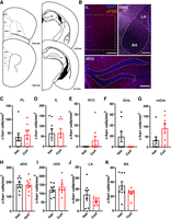
Corticosterone did not alter the total number of c-Fos-positive cells in the dentate gyrus, amygdala, and prefrontal cortex subregions. (A) Schematic diagrams of mouse brain sections depicting the brain regions of interest (Paxinos and Franklin 2004). (Reprinted from Paxinos and Franklin 2004 with permission from Elsevier.) Regions of interest were selected for the dorsal dentate gyrus (dDG; −1.22 to −2.18 mm) and ventral dentate gyrus (vDG; −2.3 to −3.4 mm) of the hippocampus; the lateral/basal amygdala (LA/BA; −0.82 to −1.82 mm); and layer II/III of the prelimbic (PL; 2.58–1.54 mm), medial orbitofrontal (mORB; 2.58–1.98 mm), lateral orbitofrontal (lORB; 2.58–1.98 mm), anterior cingulate cortex (ACC; 2.34–1.34 mm), and infralimbic (IL; 1.98–1.42 mm) areas. (B) Representative pictures of immunofluorescent c-Fos labeling in the IL, AMG, and dDG. No differences were found in the total number of c-Fos-positive cells in the PL (C), IL (D), ACC (E), lORB (F), mORB (G), dDG (H), vDG (I), LA (J), and BA (K). Data are presented as mean ± SEM. VEH n = 8, CORT n = 8. Scale bar = 200 μm.
For the PFC, no differences were found between VEH- and CORT-treated mice in the prelimbic (PL; Z = −0.37, P = 0.71), infralimbic (IL; t(14) = −0.03, P = 0.98), anterior cingulate cortex (ACC; Z = −1.37, P = 0.17), lateral orbitofrontal (lORB; Z = −1.77, P = 0.07), and medial orbitofrontal (mORB; Z = −1.69, P = 0.09) subregions (Fig. 2C–F,G, respectively). Similarly, no differences were found in the numbers of c-Fos-positive cells between the groups in the dorsal DG (dDG; t(14) = 0.18, P = 0.86) and ventral DG (vDG; t(14) = −0.84, P = 0.42) (Fig. 2H,I, respectively). Finally, the number of c-Fos-positive cells was also comparable between both groups in the lateral amygdala (LA; t(14) = 1.46, P = 0.17) and basal amygdala (BA; Z = −1.1, P = 0.27) (Fig. 2J,K, respectively).
To assess whether the CORT treatment may have lastingly affected baseline c-Fos expression regardless of memory retrieval, we included a nonretrieval control. Here, c-Fos density was assessed 28 d after the auditory fear conditioning training paradigm but without active retrieval. The numbers of c-Fos-positive cells of both groups in this control were consistently similar across the brain regions (Supplemental Fig. 1A–J). CORT did not affect the total number of dVenus-positive cells in the DG, amygdala, and prefrontal cortex subregions (Supplemental Fig. 2A–I). The temporal profile of endogenous dVenus expression is subregion-dependent (Eguchi and Yamaguchi 2009; Rao-Ruiz et al. 2019), which may affect the detection of (correlated) activity (for the sake of completeness, these data are included in Supplemental Figs. 2, 3). In sum, CORT given after the FC training had not affected the overall c-Fos and dVenus density in the DG, amygdala, and PFC subregions.
Corticosterone altered correlated activity between the dentate gyrus, amygdala, and prefrontal cortex subregions
The identification of brain regions that covary in their number of c-Fos-positive cells across groups of mice provides insights into the functional connectivity of these regions (Wheeler et al. 2013; Silva et al. 2019). We therefore assessed the covariance of pairs of regions across mice of each group and computed Pearson's R correlation matrices. Due to the high amount of null values of c-Fos-positive cells in the ACC and the lORB, no covariance analysis was performed on these subregions. First, the VEH group displayed positive and strong correlations of the LA with the BA (P < 0.01) and the dDG (P < 0.05) and, to a lesser extent, with the mORB, PL, and IL (P < 0.1) (Fig. 3A). Moreover, positive correlations were present between the mORB with the PL (P < 0.001) and the dDG with the IL (P < 0.05). Finally, the mORB and the PL displayed positive correlations with freezing levels (P < 0.05), and the LA and BA displayed negative correlations with plasma CORT levels (P < 0.05).
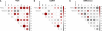
Corticosterone altered correlated activity between the dentate gyrus, amygdala, and prefrontal cortex subregions. (A) Pearson's correlation matrix of interregional covariance (dorsal dentate gyrus [dDG], ventral dentate gyrus [vDG], lateral amygdala [LA], basal amygdala [BA], prelimbic prefrontal cortex [PL], infralimbic prefrontal cortex [IL], and medial orbitofrontal cortex [mORB]) of the VEH group displays multiple positive correlations of the LA with the BA and the dDG and PFC subregions. (B) The Pearson's correlations matrix of the CORT group displays altered interregional coactivation. (C) Heat map of the Δ Pearson's R (CORT–VEH) compared with a bootstrapped population for significance. Axes represent brain regions, colors reflect Pearson's R-values (scale at the right), and labels within squares correspond to P-values. (*) P < 0.05, (**) P < 0.01, (***) P < 0.001.
The CORT group displayed a significant correlation of the mORB with the PL (P < 0.001) (Fig. 3B), similar to the VEH group. However, the positive correlations of the LA with the dDG, BA, mORB, and PL in the VEH group switched to nonsignificant negative correlations in the CORT group, and the positive correlation of LA with the IL in the VEH group was reduced in the CORT group. The positive correlations of the IL with the PL and mORB in the VEH group also switched to nonsignificant negative correlations in the CORT group. Instead, a significant positive correlation was apparent between the dDG and vDG (P < 0.01) in the CORT group (Fig. 3B). We computed ΔR by subtracting the Pearson's R-values of the VEH group from the CORT group (Fig. 3C). Subsequently, to account for the relatively small n number in this study, we performed bootstrapped analysis with the ΔR-values, which allowed for the determination of how the differences between the groups deviated from estimated differences derived from randomly distributed data based on a bootstrapped population that was resampled 10,000 times with replacement (Wheeler et al. 2013). While there were several interregional correlations with a ΔR > −1, the ΔR values of the LA with the BA and the dDG were found to be significant compared with the bootstrapped population (P < 0.05). Correlated activity in terms of dVenus-expressing cells was also differently affected by CORT compared with the VEH group (Supplemental Fig. 3A,B), but bootstrapped analysis did not reveal any significant differences between the groups (Supplemental Fig. 3C). Thus, based on correlated c-Fos activity, the connectivity of the LA with the BA and with the dDG appears to be significantly reduced in CORT-treated mice compared with VEH-treated mice.
Corticosterone altered network connectivity in remote auditory memory
Based on the covariance analysis, network connectivity graphs were generated using positive and negative R-values ≥ 0.5. In this manner, brain regions are represented as nodes that are connected by their respective interregional correlations. Similar to what was observed in the correlation matrices, the LA appears to play a central role in the VEH network, as the node displays five positive correlations with the dDG, BA, IL, PL, and mORB (Fig. 4A). Conversely, the LA does not display any positive interregional correlations in the CORT network (Fig. 4B). The positive correlations of the IL with the PL and mORB in the VEH network are also absent in the CORT network. Instead, a positive interregional correlation for the dDG and vDG is present in the CORT group but absent in the VEH group.
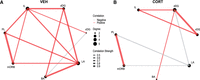
Corticosterone altered network connectivity in remote auditory memory. Network connectivity graph of the VEH (A) and CORT (B) groups based on positive or negative Pearson's R values ≥ 0.5. Positive correlations are in red, and negative correlations are in gray. Connecting line thickness depicts R-value (scale middle), and node size is proportional to the degree of connections.
Discussion
Here, we studied whether stress exposure after learning could modulate remote fear memory. We showed that CORT administration after fear learning significantly reduced remote auditory memory retrieval 28 d later. A similar CORT effect on memory retrieval was already present 14 d after auditory fear conditioning when contextual memory was unaffected (SL Lesuis and HJ Krugers, unpubl.). In parallel, we found at present no effects of CORT on overall neuronal activation in terms of the total number of c-Fos-positive cells in the DG, amygdala, and PFC subregions. Instead, correlation analysis of these brain regions revealed a significant decrease in LA–BA and LA–dDG covariation, indicating an altered functional connectivity between the regions.
Stress can modulate memory and, as such, help guide future behaviors in an adaptive or maladaptive manner (de Kloet and Joëls 2023). Stress can promote the consolidation of emotionally salient information and enhance remote memory retention in humans and rodents (Cahill et al. 2003; Roozendaal et al. 2006; Wichmann et al. 2012; Xiong et al. 2015; Aubry et al. 2016; De Quervain et al. 2017). In fact, CORT levels were shown to be positively correlated with the subsequent strength of memory consolidation (Abercrombie et al. 2003; Zorawski et al. 2006; Smeets et al. 2008), though this has so far mostly been studied in relation to recent memories. Considering that coactivation of dispersed neurons may be more critical than the net activation of each individual brain structure in remote memory (Wheeler et al. 2013; Lisman et al. 2018), the currently observed decrease in coactivation of dispersed neurons as a result of CORT could reflect a disruption of the network(s) underlying remote memory retrieval. In our studies, mice were individually housed throughout the experiment. Although plasma corticosterone levels were not different between experimental groups after retrieval, we cannot exclude that the isolation resulted in a state of chronic stress that may have contributed to the effect of posttraining corticosterone administration.
Functional connectivity per se is known to change with the age of a memory (Tallman et al. 2022). Recent memories are thought to be primarily hippocampus-dependent, whereas remote memory may engage a distinct thalamic–hippocampal–cortical signature (Wheeler et al. 2013; Kitamura et al. 2017). Moreover, while PFC subregions are coactivated in the remote recall of both neutral and emotionally salient memories, coactivation of amygdala and hippocampal subregions occurs in the remote recall of emotionally salient memories (Silva et al. 2019). This is in line with our current findings, as the CORT-induced impairment in remote recall of an emotionally salient memory was associated with a significant decrease in LA–BA and LA–dDG coactivation, whereas PFC coactivation remained largely unaltered. This suggests that specifically the network that is associated with the remote recall of emotionally salient memory is disrupted by the CORT treatment. Moreover, while there is a significant and positive interaction between freezing and the prelimbic and orbitofrontal cortices, the LA does not correlate with freezing levels. This suggests that LA activation does not necessarily reflect memory strength, which is in line with other studies (Morrison et al. 2016) and may indicate the relevance of an interaction of the LA with other cortical areas in the case of the expression of more remote memories.
We showed before (Lesuis et al. 2021) that corticosterone administration failed to alter freezing levels or c-Fos staining after 24 h in nonshock control mice (Wheeler et al. 2013). Hence, corticosterone effects on c-Fos expression, freezing, and neuronal excitability depend on corticosterone in the context of learning. This indicates that any possible alterations in network interactions seen 4 wk after training are independent of the administration of corticosterone given after training alone. Considering that there was a significant coactivation of the dorsal–ventral DG in the CORT but not VEH group, and given that dorsal–ventral hippocampal connectivity is reported to “gate” novelty-induced memory formation (Fredes et al. 2021), the auditory cue could have possibly been perceived as novel, and the altered connectivity might then reflect this particular part of memory formation. The type of memory may also influence the network involved; for example, the retrosplenial cortex forms a functional circuit with the secondary auditory cortex to support in particular auditory memory (Todd et al. 2016). In fact, the secondary auditory cortex drives amygdala activity during remote but not recent memory recall (Cambiaghi et al. 2016). The inclusion of more brain regions, together with a larger sample size in future studies, would provide a more extensive data set for an exhaustive network analysis that would also allow the inclusion of measures of centrality in relation to the above aspects.
GCs can increase cortical dependency and decrease hippocampal dependency of a memory trace (Barsegyan et al. 2010; Reis et al. 2016). At recent time points, this may be associated with memory generalization (Lesuis et al. 2021), a characteristic of cortical-dependent remote memory (Restivo et al. 2009; Söderlund et al. 2012; Todd et al. 2016; Guo et al. 2018). While memory generalization is suggested to depend on memory instability mediated by the PFC (Robertson 2018), this can also render a memory vulnerable to forgetting (Richards and Frankland 2017). As PFC circuits may require more time to mature and strengthen before they can support memory (Kitamura et al. 2017; Lee et al. 2022), an accelerated cortical dependency of a memory trace by CORT may be premature. Consequently, the memory trace would not be retained over time, which may subsequently reduce network connectivity at later time points. In this concept, the “accessibility” of the memory trace may be decreased (Ryan and Frankland 2022), preventing natural recall of emotionally salient events at more remote time points. To assess whether this is a nonspecific or engram-specific effect of CORT, a future approach would be required in which glucocorticoid receptors can be targeted selectively in engram cells. Additionally, future studies are required to further substantiate the current connectivity data by, for example, investigating the projection specificity of labeled neurons based on c-Fos expression.
CORT is proposed to prevent the positive feedback mechanisms by which traumatic memories are constantly retrieved as intrusive memories, a symptom that is, for example, prominent in posttraumatic stress disorder (PTSD) (Schelling et al. 2006). In the clinic, the application of GCs close to trauma exposure may decrease the incidence of PTSD and improve the overall quality of life (Schelling et al. 2001). Our findings thus support this optimal timing for a CORT-mediated prevention of PTSD development. As CORT may enhance memory instability as a gateway to generalization at recent time points, the underdeveloped network cannot retain a memory over time. Although speculative, this may reduce the occurrence of intrusive memories, decreasing the incidence of PTSD.
In sum, we show a significant and lasting remote auditory memory impairment as a result of CORT administration after learning. While net activation of the DG, amygdala, and PFC subregions remained unaltered, the remote memory impairment was associated with an altered coactivation between subregions—in particular, a loss of coactivation between the LA–BA and the LA–dDG. Conversely, an increase in coactivation was observed in the CORT group between the dDG–vDG. Further research is required to establish a more comprehensive analysis of the network responses in relation to stress-induced remote memories and to assess the effects of CORT treatment on engram dynamics in relation to remote memory. The cellular and network actions of corticosterone are mediated via mineralocorticoid and glucocorticoid receptors and also are influenced by the 11beta HSD distribution over the brain (Yau et al. 2011; Wheelan et al. 2018). For future studies, it would be important to investigate the expression of mineralocorticoid and/or glucocorticoid receptors specifically in activated neurons in sensitive brain areas such as the basal and lateral amygdala, (dorsal) dentate gyrus, and prefrontal cortex to get a better understanding of the different sensitivities of the respective brain areas for changes in endogenous plasma corticosterone levels. Elucidating the pathways underlying the effects of CORT on memory performance over time during the consolidation process and retrieval could provide mechanistic insights into the effects of stress on cognition and help provide possible future therapeutic targets for stress-related psychopathologies such as PTSD.
Materials and Methods
Mice and breeding
All animal experiments were conducted under Dutch national law and in compliance with the European Union directive 2010/63/EU. The study design was evaluated and approved by the Animal Welfare Committee of the University of Amsterdam. Mice were housed at a temperature of 20°C–22°C and a humidity of 40%–60% with ad libitum food (standard chow; Special Diets Services 801722 CRM [P]) and water. Mice were kept on a 12:12 h light–dark cycle (lights on at 8:00 a.m.; lights off at 8:00 p.m.). For this study, we used WT and Arc::dVenus mice (kindly provided by Professor Dr. Steven Kushner, Erasmus University Rotterdam) that were backcrossed for >10 generations into C56BL/6J (Eguchi and Yamaguchi 2009). We conducted our studies in Arc::dVenus mice, which enabled the assessment of endogenous experience-dependent changes in Arc expression up until 24 h after fear conditioning training. This approach enabled investigation of the colocalization of retrieval-activated cells with learning-activated cells at this time point (but not at later time points) (Gouty-Colomer et al. 2015; Lesuis et al. 2021). Moreover, the temporal profile of endogenous dVenus expression is subregion-dependent (Eguchi and Yamaguchi 2009; Rao-Ruiz et al. 2019). For these reasons, we do not extensively discuss the Arc::dVenus data (Supplemental Figs. 2, 3) in the ”Results” and “Discussion” sections. Adult male mice (postnatal weeks 10–14) were individually housed in isolated cabinets 7 d prior to the start of the experiments to minimize nonspecific behavioral stimulation. All experiments were performed at the start of the light phase.
Fear conditioning
Mice were placed in a standard square fear conditioning chamber (W × L × H: 30 × 24 × 26 cm) with black walls and a stainless steel grid floor connected to a shock generator (Zhou et al. 2010; Xiong et al. 2015; Lesuis et al. 2017). The chamber was cleaned with 25% EtOH in between trials. Behavior was recorded using an infrared camera (GigE, Basler AG) connected to a computer equipped with Ethovision (version 14, Noldus). Mice were habituated to the conditioning chamber for 180 sec before being subjected to a sequence of three coterminating presentations of an unconditioned stimulus (US: footshock, 2 sec, 0.4 mA) and a conditioned stimulus (CS: auditory cue, 30 sec, 2.8 kHz, 82 dB) with an interstimulus interval of 60 sec. Following the last auditory cue and shock pairing, mice were left in the fear conditioning cage for another 60 sec.
Immediately after training, mice were injected intraperitoneally with 16 mg/mL corticosterone (CORT) (Sigma) dissolved in 99.9% EtOH and diluted 40× in saline (final dose 2 mg/kg; injection volume 5 µL/g bodyweight) or the corresponding vehicle solution (n = 8 per treatment) (Lesuis et al. 2021). Mice were returned to the isolated cabinets and single-housed. After 28 d, CS-evoked freezing was tested in a completely novel context—a circular box (diameter 35 cm, transparent walls, and sawdust floor) cleaned with 1% acidic acid in between trials—in the morning (180-sec baseline, 30-sec tone). Mice from the nonretrieval control group were sacrificed the same morning without exposure to the novel context.
Tissue preparation
Mice were sacrificed by quick decapitation 90 min after fear conditioning testing to capture peak c-Fos expression (Denny et al. 2014). The nonretrieval control group was sacrificed the same morning without undergoing the fear conditioning test phase. Their brains were removed and immersion-fixed in 4% paraformaldehyde in phosphate buffer (0.1 M PB at pH 7.4) for 24 h at 4°C and then stored in 0.01% sodium-azide in 0.1 M PB at 4°C until further processing. Prior to tissue slicing, the fixed hemispheres were cryoprotected overnight in sequential concentrations of 15% and 30% sucrose in 0.1 M PB. Frozen tissue was cut into 40-µm-thick coronal sections in six parallel series using a sliding microtome. The slices were stored in antifreeze solution (30% ethylene glycol, 20% glycerol, 50% 0.05 M phosphate-buffered saline [PBS]) at −20°C until immunohistochemical staining.
Plasma corticosterone measurements
At sacrifice, trunk blood was collected in EDTA-coated tubes (Sarstedt) and centrifuged, and plasma was stored at −20°C for measures of corticosterone levels. A commercially available ELISA kit (IBL International GmbH RE52211) was used to measure plasma corticosterone levels according to the manufacturer's instructions.
Fluorescent immunohistochemistry
Free-floating sections were washed in 0.05 M PBS and incubated with Fab fragments to reduce background staining (1:200; Affinipure Fab fragment goat antimouse IgG, Jackson ImmunoResearch 145879) in 0.05 M PBS for 30 min at room temperature. A washing step of three washes for 10 min in 0.05 M PBS was performed. Subsequently, blocking was performed with 5% bovine serum albumin (BSA) in 0.3% Triton X-100 plus PBS (PBST) for 2 h at room temperature. The slices were then incubated with primary antibodies in blocking mix for 1 h at room temperature followed by an overnight incubation at 4°C. The primary antibodies used were rat anti-c-Fos (1:500; rat anti-c-Fos IgG, SySy 226017), chicken anti-GFP to enhance the endogenous dVenus signal (1:750; chicken anti-GFP, Abcam AB13970), and for sections containing the amygdala, mouse anti-GAD65 to visualize the LA and the BA (1:1000; mouse anti-GAD65, Abcam ab26113). The next day, slices were washed using 0.3% PBST and incubated with secondary antibodies in PBST for 2 h at room temperature. The secondary antibodies used were goat anti-rat (1:500; Alexa fluor 568 goat antirat IgG 2217022), goat antichicken (1:500; Alexa fluor 488 goat antichicken IgG 1899514), and donkey antimouse (1:500; Alexa fluor 647 donkey antimouse IgG 2045337). Slices were then washed in 0.05 M PBS and placed in 0.05 M PB, after which they were mounted on slides and placed on coverslips using DAPI (VectaShield mounting medium with DAPI, Vector Laboratories, Inc. H-1200).
Imaging and quantification
The fluorescent signal of c-Fos-positive cells was imaged using a Nikon DS-Ri2 fluorescent microscope. Images were acquired at 10× magnification and later assigned to corresponding bregma points. ROIs were selected based on the mouse brain atlas (Paxinos and Franklin 2004) using ImageJ analysis software (version 1.52a, Wayne Rasband). For the hippocampus, the ventral dentate gyrus (vDG; bregma −1.22 to −2.18 mm) and dorsal dentate gyrus (dDG; bregma −2.3 to −3.4 mm) were selected. For the amygdala, the lateral and basal amygdala (LA and BA; identified with GAD65 staining [Supplemental Fig. 4A,B], bregma −0.82 to −1.82 mm) were selected. As layer II/III of the PFC is recruited during remote memory retrieval (Jacques et al. 2019), we selected this layer in the prelimbic (PL; bregma 2.58–1.54 mm), medial orbitofrontal (mORB; bregma 2.58–1.98 mm), lateral orbitofrontal (lORB; bregma 2.58–1.98 mm), anterior cingulate cortex (ACC; bregma 2.34–1.34 mm), and infralimbic (IL; bregma 1.98–1.42 mm) areas. Images were converted to 8-bit gray scale to visualize positive cells. A threshold was set for the detection of c-Fos-positive cells. The data are therefore based on the number of c-Fos-positive cells that were detected above a certain threshold (the same for all groups). Experimenters were blinded throughout the analysis, and the absolute values were converted to the number of cells per square millimeter.
Statistical analysis
Data were analyzed using R studio and Graphpad Prism 9 and are expressed as mean ± standard error of the mean (SEM). Data were considered statistically significant when P < 0.05 (two-sided testing). Data points that were outside the 1.5× interquartile range and had methodological or biological deviations were deemed as outliers and excluded from the analysis. Assumptions of parametric analysis were tested using the Shapiro–Wilk normality test and the Levene's test for homogeneity of variance. Subsequently, appropriate parametric or nonparametric statistic tests were performed. The general approach was as follows: For behavioral data with repeated measures, a mixed model ANOVA was used in which time was a within unit and condition was a between unit. Post-hoc Tukey tests were performed for pairwise comparisons. For VEH × CORT comparisons, independent t-tests (or nonparametric Wilcoxon rank-sum tests) were performed. Pearson's R correlations were calculated for the correlation matrices, and the correlation coefficients were tested against critical values on a two-tailed distribution (Leal Santos et al. 2021). Significant ΔR-values were determined upon comparison with the 95% quartile of a bootstrapped data set (n = 10,000) (Wheeler et al. 2013).
Acknowledgments
We thank Jan van den Blaauwen for carrying out the ELISA measurements of the plasma CORT levels, and Andrea Muñoz Zamora (Trinity College Dublin) and Judith Lim (University of Amsterdam) for help and advice on the coactivation analysis. N.B. is supported by Alzheimer Nederland. P.J.L. and H.J.K. are supported by the Memorabel Dementia Program of ZonMW (Mechanisms of Dementia [MODEM]), Alzheimer Nederland, and the Center for Urban Mental Health.
Footnotes
-
[Supplemental material is available for this article.]
-
Article is online at http://www.learnmem.org/cgi/doi/10.1101/lm.053836.123.
- Received June 15, 2023.
- Accepted June 23, 2023.
This article is distributed exclusively by Cold Spring Harbor Laboratory Press for the first 12 months after the full-issue publication date (see http://learnmem.cshlp.org/site/misc/terms.xhtml). After 12 months, it is available under a Creative Commons License (Attribution-NonCommercial 4.0 International), as described at http://creativecommons.org/licenses/by-nc/4.0/.



