


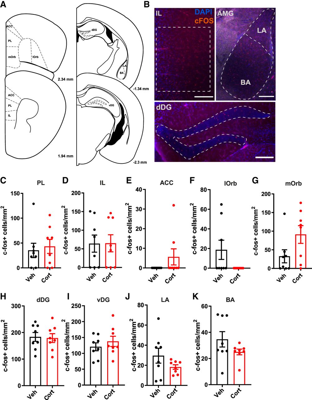
Corticosterone did not alter the total number of c-Fos-positive cells in the dentate gyrus, amygdala, and prefrontal cortex subregions. (A) Schematic diagrams of mouse brain sections depicting the brain regions of interest (Paxinos and Franklin 2004). (Reprinted from Paxinos and Franklin 2004 with permission from Elsevier.) Regions of interest were selected for the dorsal dentate gyrus (dDG; −1.22 to −2.18 mm) and ventral dentate gyrus (vDG; −2.3 to −3.4 mm) of the hippocampus; the lateral/basal amygdala (LA/BA; −0.82 to −1.82 mm); and layer II/III of the prelimbic (PL; 2.58–1.54 mm), medial orbitofrontal (mORB; 2.58–1.98 mm), lateral orbitofrontal (lORB; 2.58–1.98 mm), anterior cingulate cortex (ACC; 2.34–1.34 mm), and infralimbic (IL; 1.98–1.42 mm) areas. (B) Representative pictures of immunofluorescent c-Fos labeling in the IL, AMG, and dDG. No differences were found in the total number of c-Fos-positive cells in the PL (C), IL (D), ACC (E), lORB (F), mORB (G), dDG (H), vDG (I), LA (J), and BA (K). Data are presented as mean ± SEM. VEH n = 8, CORT n = 8. Scale bar = 200 μm.



