


Decreases in H2A monoubiquitination in the amygdala constrain fear memory formation
- 1School of Animal Sciences, Virginia Polytechnic Institute and State University, Blacksburg, Virginia 24061, USA
- 2School of Neuroscience, Virginia Polytechnic Institute and State University, Blacksburg, Virginia 24061, USA
- Corresponding author: tjjarome{at}vt.edu
Abstract
Evidence suggests a role for monoubiquitination of histone H2B, a regulator of increased gene transcription, in memory formation. However, whether monoubiquitination of histone H2A (H2Aubi), a transcriptional repressor, is involved in memory formation has not been explored. We found global and gene-specific decreases in H2Aubi in the amygdala following fear conditioning. H2Aubi decreased at Pten, an inhibitor of PI3K–AKT–mTOR signaling, which occurred concurrently with increases in PTEN expression. CRISPR–dCas9 mediated upregulation of the H2Aubi ligase, Ring1b, in the amygdala enhanced contextual memory. These results suggest that decreases in transcriptionally repressive H2Aubi in the amygdala functions to constrain fear memory strength.
Over the last decade, strong evidence has highlighted a critical role of chromatin modifications in regulating gene transcription underlying memory formation (Kwapis et al. 2018). Epigenetic changes like methylation can occur directly on DNA or via various posttranslational modifications on the N-terminal tails of the four core histone proteins (H2A, H2B, H3, and H4), which package DNA into nucleosomes. While acetylation and methylation have been the primary focus of most studies, other modifications, such as ubiquitination, are gaining attention for their regulatory roles in gene expression and chromatin dynamics (Shilatifard 2006; Jarome et al. 2021; Yang et al. 2022; Liu et al. 2023; Wu et al. 2023).
The ubiquitin-proteasome system (UPS) is traditionally known for degrading short-lived proteins and has also been extensively implicated in synaptic plasticity (Ehlers 2003; Dong et al. 2008; Djakovic et al. 2012; Hegde 2017; Patrick et al. 2023). Dysregulation of the UPS has been associated with the pathophysiology of numerous neurodegenerative and psychiatric disorders, further emphasizing its importance in maintaining neuronal function and plasticity (Rubio et al. 2013; Zheng et al. 2016; Cheon et al. 2019). While protein degradation is the canonical function of the UPS, proteins can also be marked by ubiquitin for other nonproteolytic functions. One such modification is histone H2B monoubiquitination at lysine 120, a critical regulator of active gene transcription that has been shown to be necessary for synaptic plasticity and memory formation in the hippocampus through the regulation of histone methylation (Jarome et al. 2021; Navabpour et al. 2024). Another form of monoubiquitination occurs on histone H2A at lysine 119 (H2Aubi) which serves as a repressor of gene transcription (Meas and Mao 2015) where it acts primarily through the Polycomb Repressive Complex 1 (PRC1) (Gutierrez et al. 2012). This modification is critical for silencing specific genes during development in single-cell organisms and has been linked to the regulation of genes involved in stress responses and DNA repair (Oss-Ronen et al. 2022). Furthermore, studies in addiction have demonstrated that chronic cocaine use increases H2Aubi in the paraventricular thalamus, a process regulated by scaffold protein melanoma-associated antigen D1 (Maged1) and USP7, with genetic variations in these proteins being associated with altered susceptibility to cocaine addiction and related symptoms in humans (Cheron et al. 2023). However, H2Aubi has never been examined in the brain in the context of memory formation, leaving its potential role in this process unknown. Here, we tested the hypothesis that H2Aubi is a critical regulator of fear memory formation in the amygdala.
Refer to the Supplemental Material for expanded details on methodology. Experiments used 8- to 9-week-old male and female Sprague Dawley rats. All molecular experiments (western blotting, etc.) used mixed-sex groups while the final behavioral experiment used only male rats to limit variability in the fear response as female rats can sometimes use darting in place of freezing behavior (Gruene et al. 2015). Animals were handled for 4 days before undergoing the contextual fear conditioning procedure, which was performed as previously described by our group (Farrell et al. 2023). Both hemispheres of the basolateral amygdala (BLA) were collected, separated, and used for histone extraction or chromatin immunoprecipitation (ChIP) procedures; hemispheres were counterbalanced across these analyses to account for any possible laterality effects. Total phosphatase and tensin homolog (PTEN) protein levels were assessed using a whole cell lysate. In the behavioral experiment, animals received intracranial infusions of CRISPR–dCas9 plasmids into the BLA. These animals underwent behavioral training 4 weeks later and were tested for memory retention the following day. One week after the contextual fear memory test, an elevated plus maze test was performed to assess anxiety-like behavior.
We initially examined changes in global H2Aubi levels in bulk histone extracts from the amygdala of male and female rats 1 h after contextual fear conditioning (Fig. 1A), as this time point is when many studies have reported significant changes in epigenetic mechanisms following learning (Maddox and Schafe 2011; Gupta-Agarwal et al. 2014; Jarome et al. 2015, 2018, 2021; Halder et al. 2016; Butler et al. 2019; Farrell et al. 2022; Navabpour et al. 2024). Further, we chose to use contextual fear conditioning as our behavioral model, as the only prior studies on histone ubiquitination in the brain (H2Bubi) and the majority of epigenetic literature examining memory formation have used this paradigm. Unexpectedly, we observed a significant decrease in H2Aubi levels following fear conditioning (t18 = 2.848, P = 0.0107) (Fig. 1B). As this was at a global level, we next examined whether H2Aubi levels change at a gene-specific level in the amygdala following fear conditioning. We chose to focus on the PTEN, a protein phosphatase that regulates the activation of mTOR, which acts as a critical regulator of fear memory formation in the amygdala (Parsons et al. 2006). PTEN has been shown to be epigenetically regulated in the hippocampus following fear memory retrieval through the Polycomb Repressive Complex 2 (PRC2) (Jarome et al. 2018) that typically functions in conjunction with PRC1. As H2Aubi is a major component of PRC1 and is known to associate with PRC2-mediated epigenetic modifications (Ohtomo et al. 2023), it is likely that H2Aubi can target similar genes as those of PRC2, such as Pten. We tested whether H2Aubi levels were altered at Pten DNA regions during fear memory formation by using ChIP. Congruent with the global analysis, we observed a significant reduction in H2Aubi occupancy in the Exon 1 coding region of Pten (t14 = 6.811, P < 0.0001) (Fig. 2A), but no change in the promoter region after fear conditioning (t17 = 0.9773, P = 0.342), suggesting the loss of transcriptional repression of Pten during memory formation. Consistent with this, we observed an increase in PTEN protein expression in the amygdala 1 h after fear conditioning (t21 = 2.329, P = 0.0299) (Fig. 2B). Together, these data suggest that fear conditioning results in a reduction of H2Aubi at Pten, which correlates with increased PTEN levels during the memory consolidation process.
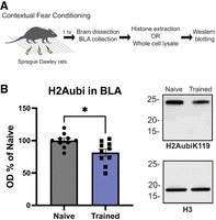
Global H2Aubi levels decrease in the amygdala following fear conditioning. (A) Experimental design. Rats were trained to contextual fear conditioning, and the BLA was dissected 1 h later. (B) Western blot analysis of H2Aubi levels in BLA bulk histone extractions of young male and female adult rats (n = 10 per group, mixed sex, 8-week-old). H2Aubi levels decreased in the fear-conditioned group relative to the naive animals. Representative images of the H2Aubi on the top and H3 (loading control) on the bottom. (*) P < 0.05 from Naive.
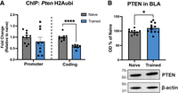
H2Aubi levels decrease at Pten following fear conditioning. (A) ChIP analysis revealed that fear conditioning resulted in decreased H2Aubi occupancy in the coding region of Pten (n = 8–9 per group, mixed sex, 8-week-old), but not in the promoter region (n = 9–10 per group, mixed sex) in the amygdala. (B) Fear-conditioned animals (n = 12, mixed sex, 8-week-old) had increased PTEN protein expression compared to the naive group (n = 11, mixed sex, 8-week-old). Western blot representative images of PTEN in the middle and β-actin (loading control) on the bottom. (*) P < 0.05 from Naive. (****) P < 0.0001 from Naive.
We next tested whether reductions of H2Aubi are critical for contextual fear memory in the amygdala. We designed CRISPR guide RNAs (gRNAs) against Ring1a and Ring1b, which are both necessary for H2Aubi conjugation through their combined actions and serve as critical components of the PRC1 (Fig. 3A). We first tested our gRNA in vitro using B35 rat neuroblastoma cells transfected with either gRNA + dCas9-VPR (a transcriptional activator) or gRNA alone (control). We observed significant increases in Ring1b (t14 = 3.385, P = 0.0044) (Fig. 3B) but only moderate changes in Ring1a expression (t13 = 1.596, P = 0.1344) (Fig. 3B) in cells transfected with gRNA + dCas9-VPR. Considering this, we used the Ring1b gRNA for subsequent experiments in vivo. Animals received gRNA alone (control) or gRNA + dCas9-VPR into the amygdala (Fig. 3A). After 4 weeks to allow plasmid expression (Farrell et al. 2024) and successful upregulation of H2Aubi levels in the amygdala (t14 = 2.149, P = 0.0496) (Fig. 3C), animals were trained and tested to contextual fear conditioning. During training, no significant differences were observed between treatment groups (F(1, 11) = 0.4497, P = 0.5163) (Fig. 3D), nor was there a Time × Treatment interaction (F(4, 44) = 1.390, P = 0.2531). However, we observed a significant effect for Time (F(2.388, 26.27) = 12.98, P < 0.0001), showing performance improved over time regardless of the treatment conditions. During testing, the gRNA + dCas9-VPR group had significantly enhanced memory compared to control animals (Kolmogorov–Smirnov test, D11 = 0.7143, P = 0.0385) (Fig. 3E). Shock reactivity analysis revealed that the gRNA + dCas9-VPR treatment did not affect sensory processing as there was not a significant effect for Treatment (F(1, 11) = 0.2968, P = 0.5968) (Fig. 3F) or Shock (F(3, 33) = 2.307, P = 0.0947), and there was not a significant Shock × Treatment interaction (F(4, 44) = 1.170, P = 0.3361). Further, we examined whether H2Aubi manipulation in the amygdala resulted in changes in anxiety-like behavior by conducting an elevated plus maze test. However, we did not observe a significant impact of the CRISPR manipulation for time spent in the open arms (t14 = 0.2160, P = 0.8321) (Fig. 3G) or closed arms (t14 = 0.2160, P = 0.8321), suggesting that the upregulation of the H2Aubi ligase Ring1b did not broadly affect normal amygdala functioning.
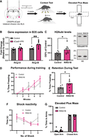
CRISPR–dCas9 mediated upregulation of H2Aubi ligase Ring1b in the amygdala enhances contextual fear memory. (A) Schematic of experimental design for CRISPR–dCas9 infusion in the amygdala followed by contextual fear conditioning and elevated plus maze. (B) The gRNA plasmid targeting Ring1a and Ring1b alone (control) or with dCas9-VPR transcriptional activator were transfected into rat B35 neuroblastoma cells and collected 48 h later. RT-qPCR analysis revealed an increase in Ring1b, but not Ring1a. Subsequent in vivo experiments used the Ring1b gRNA. (C) Western blot analysis revealed that upregulation of Ring1b in the amygdala increased H2Aubi levels 4 weeks later. H2Aubi was normalized to H3. The representative image is H2Aubi (top) and H3 (bottom). (D) During training, there was no effect of treatment on performance. (E) During testing, the gRNA + dCas9-VPR group had increased memory retention compared to the control group. (F) Shock reactivity analysis showed no effect for treatment. (G) During the elevated plus maze test, we found no difference in time spent in the open or closed arms (n = 6–7 per group, males only, 8- to 9-week-old). (*) P < 0.05 from Control. (**) P < 0.01 from Control.
The regulation of gene transcription through various histone modifications has been strongly linked to long-term memory formation (Kwapis et al. 2018). Monoubiquitination of H2A and H2B, though less prevalent than other modifications, plays a significant role in chromatin dynamics and transcriptional regulation (Meas and Mao 2015). Our team has recently revealed that monoubiquitination of H2B is a crucial regulator of the transcriptome induced in the dorsal hippocampus following contextual fear conditioning (Navabpour et al. 2024). However, the role of H2Aubi in memory formation has not yet been explored.
Here, we provide significant insight into the role of H2Aubi in the amygdala during contextual fear memory formation. We observed that fear conditioning leads to a reduction in H2Aubi level in the amygdala, with a specific decrease in the coding region of Pten. The reduction in H2Aubi at the coding region of the Pten likely facilitates the release of transcriptional repression, promoting increases in PTEN levels following fear conditioning. Interestingly, our prior work indicated that H2Aubi levels did not significantly change in the hippocampus following contextual fear conditioning (Jarome et al. 2021), suggesting that, unlike H2B ubiquitination, H2Aubi may have a specialized role in the amygdala. While it is unclear why H2Aubi may have a region-specific function, it is interesting to speculate that this could be attributed to the amygdala's role in processing emotionally salient stimuli and its need for distinct epigenetic regulatory pathways (Alexandra Kredlow et al. 2021). For example, while the epigenome has been far less studied in the amygdala than in the hippocampus, the current data suggest that there may be a stronger heterochromatin state in the amygdala following fear conditioning (Gupta-Agarwal et al. 2014). Considering our data that H2Aubi decrease in the amygdala following fear conditioning, it is possible that fear memory formation may also require a loss of heterochromatin, which fits with data showing a critical role for histone acetylation in this region (Maddox et al. 2013). The region-specific role of H2Aubi may also be due to the differences in the availability of ubiquitination machinery in the amygdala, particularly ubiquitin ligases critical for conjugating ubiquitin to H2A. Future studies should focus on identifying why this region-specific role for H2Aubi in fear memory formation exists.
PTEN is a negative regulator of PI3K–AKT–mTOR signaling and plays a key role in multiple molecular processes including apoptosis, metabolism, cell proliferation, survival, and synaptic plasticity (Manning and Cantley 2007; Jurado et al. 2010; Knafo et al. 2016; Ahmad et al. 2023; Glaviano et al. 2023; Tian et al. 2023). A prior study has demonstrated that the epigenetic regulation of Pten via Enhancer of Zeste Homolog 2 (EZH2) is pivotal in modulating mTOR signaling during memory reconsolidation following retrieval (Jarome et al. 2018). Moreover, impairments in the PI3K/Akt/mTOR pathway have been implicated in Alzheimer's disease like neurodegeneration in Down syndrome, and the loss of PTEN expression in the brain is associated with learning deficits, epilepsy, and autism spectrum disorder. Additionally, previous research reported that PTEN negatively regulates synaptic activity and structural plasticity, with its downregulation being associated with enhanced synaptic plasticity and memory performance (Sperow et al. 2012). The increase in PTEN expression observed along with H2Aubi reduction may reflect a temporary need to fine-tune cellular signaling during memory consolidation, which may occur through control over mTOR activation. This suggests that maintaining a balanced level of PTEN activity is critical for achieving optimal cognitive function (Tilot et al. 2015) and that H2Aubi acts as a regulatory “brake” on transcription, which is reduced during memory formation. Future studies will aim to directly test the importance of H2Aubi-mediated regulation of Pten expression to mTOR activation in the amygdala during fear memory formation.
Based on the above observation, we then investigated whether enhancing H2Aubi levels in the amygdala can modulate fear memory. Using the CRISPR–dCas9 system targeting Ring1b, we found that the upregulation of H2Aubi significantly enhanced fear memory without impacting sensory functioning and anxiety-like behavior. It is interesting to speculate that H2Aubi upregulation influenced memory via repression of Pten, though we were unable to directly test this in the present study. Future studies will aim to investigate the causal relationship between increased H2Aubi levels and regulation of fear memory strength through PTEN expression.
Our finding demonstrates a link between H2Aubi, PTEN, and fear memory formation in the amygdala. However, the broader impact of H2Aubi on the other target genes in the amygdala has not been explored. Given the wide influence of histone modification on the transcriptome, there may be additional downstream effects of H2Aubi modulation that contribute to memory enhancement that were not investigated in this study. A future direction could also examine H2Aubi dynamics using auditory-cued fear memory, a more amygdala-centric task, to further isolate and clarify the role of H2Aubi in amygdala-dependent learning. Additionally, due to the persistent nature of the CRISPR–dCas9 system, the role of H2Aubi in different stages of memory processing (acquisition, consolidation, and retrieval) could not be delineated in this study. Despite this, our study presents the first evidence of a critical role for H2Aubi in fear memory formation in the amygdala.
Acknowledgments
This work was supported by the National Institutes of Health (NIH) grants MH122414, MH131587, AG081851, AG071523, and AG079292 to T.J.J. All experiments were approved by the Institutional Animal Care and Use Committee at the Virginia Polytechnic Institute and State University (protocol #23-249) and conducted within the ethical guidelines of the National Institutes of Health (NIH).
Author contributions: Y.B.: conceptualization, investigation, formal analysis, writing—original draft. H.V.: investigation. N.P.: investigation. M.T.: investigation. T.J.J.: writing—review and editing, supervision, funding acquisition, conceptualization.
Footnotes
-
[Supplemental material is available for this article.]
-
Article is online at http://www.learnmem.org/cgi/doi/10.1101/lm.054092.125.
- Received January 17, 2025.
- Accepted February 26, 2025.
This article is distributed exclusively by Cold Spring Harbor Laboratory Press for the first 12 months after the full-issue publication date (see http://learnmem.cshlp.org/site/misc/terms.xhtml). After 12 months, it is available under a Creative Commons License (Attribution-NonCommercial 4.0 International), as described at http://creativecommons.org/licenses/by-nc/4.0/.



