


Inhibition of prefrontal glutamatergic neuron activity during the recovery period following chronic stress disrupts fear memory in male rats: potential role of the infralimbic cortex
- Jessica M. Judd,
- Dylan N. Peay,
- Jinah L. Kim,
- Elliot A. Smith,
- Megan E. Donnay,
- Joel Miller,
- Jean-Paul Klein,
- Erin K. Nagy,
- Amanda M. Acuña,
- M. Foster Olive and
- Cheryl D. Conrad
- Corresponding author: conradc{at}asu.edu
Abstract
Chronic stress typically leads to deficits in fear extinction. However, when a delay occurs from the end of chronic stress and the start of fear conditioning (a “recovery”), rats show improved context-cue discrimination, compared to recently stressed rats or nonstressed rats. The infralimbic cortex (IL) is important for fear extinction and undergoes neuronal remodeling after chronic stress ends, which could drive improved context-cue discrimination. Here, glutamatergic IL neurons of Sprague-Dawley male rats were targeted for inhibition using inhibitory designer receptors exclusively activated by designer drugs (DREADDs) and daily injections of clozapine N-oxide (CNO) during a 21-day recovery period from chronic stress. Histological verification confirmed DREADDs in the IL with some spread to nearby medial prefrontal cortex (PFC) regions. CNO administration was then discontinued before fear conditioning started and behavioral testing thereafter so that behavioral assessments occurred without neuronal inhibition. Fear conditioning involved presenting male rats with three tone-foot shock pairings on 1 day, which was followed by 2 days of 15 tone-alone extinction sessions. Daily and repeated inhibition of mainly IL neurons during the 21-day recovery period did not disrupt fear learning or fear extinction in all groups (controls, stressed rats without a recovery, and stressed rats with a recovery). However, chronically stressed rats given a recovery and with DREADD activation showed impaired spontaneous recovery, indicating a failure to form a tone-foot shock association. The findings show that daily inhibition of mainly IL neurons prior to fear conditioning and extinction depends upon the changes that occur during the recovery period following the end of chronic stress.
The formation of fear memories is necessary for long-term survival, but maladaptive memories can be detrimental (Boissy 2004). Some individuals exposed to trauma may express overly robust and intrusive memories (Milad et al. 2008; American Psychiatric Association 2013), which can manifest as posttraumatic stress disorder (PTSD). About 6%–9% of the general U.S. population and nearly 40% of those exposed to extreme trauma develop PTSD (Hoge and Warner 2014; Sareen 2014). Unfortunately, current therapies for PTSD have limited effectiveness (Bradley et al. 2005; Steckler and Risbrough 2012; Difede et al. 2014; VanElzakker et al. 2014). Thus, additional research on mechanisms underlying PTSD is necessary to identify novel targets for intervention.
Chronic stress is a risk factor for the development of PTSD (Breslau et al. 1999; Sareen 2014) and can be used to prime rodents toward a PTSD-like phenotype (Daskalakis et al. 2013). For both PTSD patients and chronically stressed rodents, the acquisition of fear memories is rapid and overtly robust (Conrad et al. 1999; Cordero et al. 2003; Yehuda and LeDoux 2007; Norrholm et al. 2011; American Psychiatric Association 2013; Hoffman et al. 2015; Sep et al. 2023). When fear memories are formed, PTSD patients often fail to show complete remission following treatment with extinction-based therapies, that is, the repeated presentation of a fearful stimulus in a safe environment (Blechert et al. 2007; Milad et al. 2009; Norrholm et al. 2011; Sep et al. 2023). Similarly, chronically stressed rodents commonly show impaired extinction learning (Rau et al. 2005; Izquierdo et al. 2006; Hoffman et al. 2014). When extinction occurs within a single session, chronically stressed rodents show impaired recall of the prior extinction experience (Hoffman et al. 2010; Wilber et al. 2011). In addition, both PTSD patients and chronically stressed rodents demonstrate heightened anxiety (Vyas et al. 2004; Mikics et al. 2008; Grillon et al. 2009), which can become anxiety generalization, where a safe environment or stimuli unassociated with the trauma now triggers the aversive response (Radulovic et al. 1998; Blechert et al. 2007; Hoffman et al. 2014; Judd et al. 2020). Consequently, chronic stress combined with fear conditioning appears to parallel many of the characteristics of PTSD (Bryant 2017) and can aid in parsing out the biological mechanisms underlying PTSD.
Recent research from our laboratory and others has investigated how the detrimental cognitive effects that occur following chronic stress can be time-dependent, in some cases. Numerous studies show that chronic stress leads to impairments in spatial memory (Luine et al. 1994; Ghiglieri et al. 1997; Bowman et al. 2002; Abidin et al. 2004; Kleen et al. 2006; Song et al. 2006; Ortiz et al. 2015), which requires the hippocampus (Moser et al. 1995). However, when weeks pass between the end of the chronic stress manipulation and the start of spatial memory testing, spatial ability improves and can be better than that of unstressed control rats (Luine et al. 1994; Sousa et al. 2000; Radley et al. 2005; Goldwater et al. 2009; Bloss et al. 2010; Hoffman et al. 2011; Bian et al. 2012; Ortiz et al. 2015; Conrad et al. 2017). We recently investigated whether a temporal component between the end of chronic stress and the start of behavioral testing also impacts fear extinction learning (Judd et al. 2020). We observed that fear responses during extinction were lower in chronically stressed rats provided with a delay after stress ended and before the start of a fear conditioning paradigm compared to recently stressed or control rats (Judd et al. 2020). This finding suggests that the impact of chronic stress on fear extinction might be time-dependent, with fear extinction performance having the potential to improve in the weeks following the end of chronic stress. Consequently, chronically stressed rats that are cognitively tested after a delay following the end of chronic stress may represent a distinct phenotype from chronically stressed rats tested soon after stress exposure or even from nonstressed control rats.
Important structures involved in fear extinction are the prelimbic cortex (PL) and infralimbic cortex (IL) of the medial prefrontal cortex (mPFC). Inhibition of the PL impairs the expression of conditioned fear responses to contextual and auditory fear signals (Sotres-Bayon and Quirk 2010). In lesion studies, permanent destruction of the IL impairs fear extinction memories, but the acquisition of the fear memory itself is unperturbed (Milad and Quirk 2002). Temporarily inhibiting the IL using pharmacological methods during fear extinction leads to impairments in fear extinction learning (Sierra-Mercado et al. 2010) and retrieval (Mueller et al. 2010), which demonstrates that even transient inhibition of the IL before fear extinction can lead to impairments that last through the recall of fear extinction, despite the IL being fully functional at recall. The IL-mediated extinction process is a form of new learning that requires IL protein synthesis (Hugues et al. 2004; Santini 2004; Burgos-Robles et al. 2007) and IL neuronal activity (Milad and Quirk 2002; Burgos-Robles et al. 2007; Wilber et al. 2011). The IL essentially acts as a break on the emotional responses (Knapska and Maren 2009; Bennett et al. 2016; Bloodgood et al. 2018) to allow safe cues to be differentiated from unsafe cues.
Chronic stress negatively impacts the PL and IL, both functionally and structurally, with a temporal component. Under chronic stress conditions, the PL becomes functionally inhibited (McKlveen et al. 2016) and the PL neurons express dendritic hypotrophy (Moench and Wellman 2017). When chronic stress ends and a recovery period ensues, the PL dendritic arbors remodel and resemble unstressed PL neurons (Moench and Wellman 2017). Similarly, chronic stress decreases the activity of IL neurons (Goldwater et al. 2009; Wilber et al. 2011), produces IL dendritic hypotrophy (Izquierdo et al. 2006; Cerqueira et al. 2007; Goldwater et al. 2009), and IL dendritic spine reductions (Moench and Wellman 2014). Unlike the PL, neurons in the IL show a different phenotype with recovery from chronic stress. The dendritic arbors of IL neurons become more complex and show increased neuronal activity following recovery from chronic stress (Bloss et al. 2010) and are accompanied by decreased neuronal activity (Radley et al. 2005; Goldwater et al. 2009; Wilber et al. 2011), but with more dendritic complexity proximal to the soma compared to nonstressed controls (Goldwater et al. 2009). As for behaviors, chronic stress impairs IL-dependent fear extinction and the recall of fear extinction (Baran et al. 2009; Holmes and Wellman 2009; Oualian and Gisquet-Verrier 2010). However, how a recovery from chronic stress may alter fear extinction is unknown. Comparing the immediate effects of chronic stress with the effects following a recovery from chronic stress is important due to the structural changes that occur in the brain, which may result in unique behavioral phenotypes that have not yet been investigated.
In the present study, the goal was to interfere with processes that take place in the IL following the end of chronic stress to understand the role that such processes might play in improving fear and safety cue discrimination. IL glutamatergic neurons were inactivated daily for 3 weeks following the end of chronic stress and then uninhibited when behavioral testing started. Functional inactivation of the IL was achieved by using designer receptors exclusively activated by designer drugs (DREADDs). The goal of this timing was to try and block any potential alterations that may take place in the IL during the period between the end of chronic stress and the start of fear conditioning. Importantly, the IL was unmanipulated during fear conditioning and extinction, which allowed us to focus on the role of changes in the IL during the recovery period from when stress ended through when fear conditioning started. Finally, we used an extended acclimation paradigm before the start of fear conditioning so that the rats would show similar acquisition of fear conditioning (Hoffman et al. 2014, 2015; Jacobs et al. 2010) and thereby allow for assessment during fear extinction. We hypothesized that the changes in the IL during the recovery period following chronic stress drive optimal fear extinction learning. We predicted that the inactivation of the IL following the end of chronic stress would lead to higher fear responses during fear extinction in chronically stressed rats that underwent daily IL inhibition during recovery compared to chronically stressed rats that did not receive this manipulation.
Results
Experimental design, time line, and placement
A summary of the experimental design using the independent variables for stress group (CON, STR-IMM, STR-R3) and virus type (enhanced green fluorescent protein [EGFP], hM4Di) is shown, as well as the time line for the manipulations relative to behavioral testing (Fig. 1A). After behavioral testing finished, the brains were processed to visualize fluorescence from the virus and sections matched using a rat brain atlas (Paxinos and Watson 2013). Brains from most animals showed that the IL was successfully targeted, but some virus spread into nearby regions, such as the PL (Fig. 1B). Representative images of virus expression are also illustrated (Fig. 1C). Rats were included in the subsequent analyses when fluorescent placement was bilateral and within the IL or a combination of being within the IL with some spread into the PL. Rats were excluded from analysis when fluorescence was not observed, expressed unilaterally in the IL, or expressed in the PL only. Using these criteria, data from 41 out of 69 rats were included (for a 59% success rate). Additionally, one CON-active was excluded for low engagement during training. This resulted in the final number of rats per group: (CON-EGFP = 5; CON-hM4Di = 5; STR-IMM-EGFP = 9; STR-IMM-hM4Di = 6; STR-R3-EGFP = 11; STR-R3-hM4Di = 5).
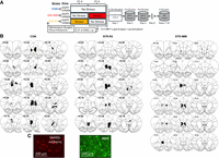
Experimental time line and images. (A) Time line. Rats were assigned to one of three stress groups. Rats in the stress immediate group were chronically stressed and underwent fear conditioning within days after restraint ended (STR-IMM). Rats in the stress-recovery group were chronically stressed and then had 3 weeks without restraint before the start of fear conditioning (STR-R3). The control group did not undergo a stressor manipulation (CON). During the 3-week break for STR-R3, which overlapped with the stressor manipulation for STR-IMM, all rats (CON, STR-IMM, STR-R3) were given daily injections of CNO (1 mg/kg BW, i.p.) for chronic activation of the DREADDs, which were introduced in the IL region of the mPFC via viral infusions before the start of any manipulation. Six days before the start of fear conditioning, rats were acclimated to contexts A and B for 6 days for 10 min per day, alternating between contexts. On the day after the end of the stress manipulation for the STR-IMM cohort and 3 weeks after the end of the stress manipulation for the STR-R3 cohort, CNO administration stopped, and then all rats were fear conditioned in context A with three tone and foot shock pairings. On the subsequent 2 days, rats were placed into context B and were presented with 15 tone-only presentations (no foot shock) for extinction training for a total of 30 tone presentations over 2 days. The day after the second extinction training ended, anxiety-like behavior was measured on the EPM. All rats underwent spontaneous recovery (SR) 4–7 days after the end of extinction training. (CS) Conditioned stimulus (tone), (US) unconditioned stimulus (foot shock). Context A = Ctxt A is denoted by the white box. Context B = Ctxt B is denoted by gray cylinders. (B) Viral expression placement in rats with hM4Di virus. Viral placement was primarily in the IL, but 81% had spread into the PL. Rats were included in subsequent analyses when placement was bilateral and in the IL only, or when placement was bilateral and in the IL with spread to the PL. Please note that the Paxinos and Watson stereotaxic coordinate markers are overlaid on the image to help illustrate brain regions and reflect the histological axis of A/P: +2.7; M/L: +0.5, −0.65; D/V: −5.2 from bregma (Paxinos and Watson 2013). (C) Magnified images. A magnified image showing neurons expressing the inhibitory virus AAV8-CamKIIα-hM4Di-mCherry (left) and the control (CON) virus AAV8-CamKIIα-EGFP (right).
Fear conditioning training
Summary
All groups displayed low and minimal freezing behavior to tone at the start of the fear conditioning training session. As trials progressed, all groups increased freezing to tone and context. However, stress group differences were observed over the course of the session with STR-IMM freezing more to the context than STR-R3.
Freezing to tone during training
The first tone trial of the fear conditioning session was analyzed for any initial differences in response to tone before the introduction of a foot shock (the fear conditioning paradigm is shown as a reference in Fig. 2A). A 3 × 2 analysis of variance (ANOVA) for the stress group (CON, STR-IMM, STR-R3) and virus type (EGFP, hM4Di) for freezing to tone during trial 1 revealed a significant main effect of virus (F(2,19) = 8.038, P = 0.0106) and significant stress by virus interaction (F(2,19) = 5.175, P = 0.0161). The STR-IMM-EGFP rats froze more than STR-IMM-hM4Di (P = 0.0449) and CON-hM4Di (P = 0.0449) rats (Fig. 2B). Consequently, STR-IMM enhanced freezing to tone despite acclimation to the chambers to reduce the potential for a priori differences to fear conditioning training.
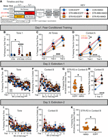
Fear conditioning and extinction. (A) Time line and key are illustrated to the groups, stress manipulations and fear conditioning timing. (B) For freezing to the first tone, the STR-IMM-EGFP group froze more than STR-IMM-hM4Di and CON-EGFP. (C) Freezing to tone increased from trial 2 to trial 3, with similar levels of freezing to tone on trial 3. No statistical group differences were detected. (D) Freezing to context A increased across the session, and the rats in the STR-R3 (EGFP and hM4Di) group froze less to context than the rats in the STR-IMM (EGFP and hM4Di) group. (E) During the first extinction session of 15 CS-only trials, all groups decreased their freezing to tone (E) and context B (F) across the session. (G) For the STR-R3 group, freezing to context B was higher across the extinction session for rats in the EGFP condition than for rats in the hM4Di condition. (H) Analysis of trials 2 and 3, after the tone had been introduced to context B, revealed that freezing to context initially increased, before decreasing later in the session (as seen in F). (I) During the second extinction session of 15 CS-only trials, freezing decreased to tone (I) and context (J). (K) Within the STR-R3 group, rats in the hM4Di condition froze less to context during trials 2 and 3 than those in the EGFP condition. (CON) Blue circles, (STR-IMM) red triangles, (STR-R3) orange squares. EGFP are shown in full color for the respective symbols, and hM4Di are shown by symbols that are blocked in half by color and black. Data are reported as means ± SEM. (*) P < 0.05, (***) P < 0.001, (****) P < 0.0001.
Freezing across tone trials 2 and 3 was assessed to determine behavior during fear conditioning acquisition, after the introduction of the foot shock. A repeated-measures ANOVA for stress group (CON, STR-IMM, STR-R3) and virus type (EGFP, hM4Di) by conditioning trial (2, 3) showed there was a significant effect of trial (F(1,435) = 25.360, P < 0.001), with an increase in freezing from trial 2 to trial 3, as would be expected (Fig. 2C). To confirm that all groups had acquired similarly by the end of the session, trial 3 was analyzed individually. A 3 × 2 ANOVA for the stress group (CON, STR-IMM, STR-R3) and virus type (EGFP, hM4Di) for the third tone did not reveal significant differences between groups, suggesting that all groups went into extinction with similar levels of fear response to the tone. No other significant effects or interactions were observed.
Freezing to context A
Freezing to the context was measured before the introduction of either tone or foot shock, which indicates that acclimation to this context was similar across groups. A 3 × 2 ANOVA for the stress group (CON, STR-IMM, STR-R3) and virus type (EGFP, hM4Di) for context trial 1 showed that all groups froze similar and low and that there were no differences based on stress group, virus type, and no stress by virus interaction (Fig. 2D).
Freezing across context trials 2 and 3 during training was assessed to determine how freezing to context A changed across acquisition trials, after the introduction of the tone and the foot shock. A repeated-measures ANOVA for the stress group (CON, STR-IMM, STR-R3) and virus type (EGFP, hM4Di) by conditioning trial (2, 3) showed that for all groups, there was a significant effect of trial (F(1,35) = 36.981, P < 0.001), with an overall increase in freezing as trials progressed (more freezing to trial 3 than trial 2 (P < 0.001). Additionally, a significant main effect of stress (F(2,35) = 3.489, P = 0.042) showed that STR-R3 froze less than STR-IMM (P = 0.044, Fig. 2D). There were no other significant interactions or group differences.
Trial 3 was analyzed separately to assess whether group differences existed by the end of the training session and a 3 × 2 ANOVA for the stress group (CON, STR-IMM, STR-R3) and virus type (EGFP, hM4Di) for context trial 3 showed no significant effects or interactions (Fig. 2D).
Extinction-1 session (EXT-1)
Summary
Groups froze similarly to the first tone and context B with differences emerging early in extinction on trials 2 and 3 before extinction learning occurs, with STR-IMM freezing more to context than CON. Moreover, STR-R3-hM4Di froze less to context B than did STR-R3-EGFP throughout the extinction-1 session.
Freezing to tone during EXT-1
Freezing to the first tone was assessed alone because it was the first time that the shock-associated tone was presented in this context (context B) and the rats had not experienced the tone without a foot shock before the first extinction trial. No significant differences were found, as revealed by an ANOVA for the stress group (CON, STR-IMM, STR-R3) and virus type (EGFP, hM4Di) for tone trial 1 during EXT-1 (Fig. 2E).
In our prior work on the effect of chronic stress on fear extinction, differences in freezing responses were observed in trials 2 and 3 of extinction training (Judd et al. 2020). Consequently, freezing to tone during trials 2 and 3 was assessed by a repeated measures ANOVA for stress group (CON, STR-IMM, STR-R3) and virus type (GFP, hM4Di) across trials 2 and 3. Freezing to tone increased from trial 2 to trial 3 (F(1,35) = 25.360, P < 0.001), but no significant effects for stress group, virus type, or interactions were found (Fig. 2E).
To determine whether extinction occurred over the 15 trials, the subsequent analysis used the average freezing over three trials for a total of five bins and supported that less freezing to tone occurred as bins progressed, with no significant effects from stress group, virus type, or interactions (Fig. 2E). A repeated-measures ANOVA for stress group (CON, STR-IMM, STR-R3) and virus type (EGFP, hM4Di) by extinction bin (1, 2, 3, 4, 5) showed a significant effect of bin (F(4,140) = 8.820, P < 0.001), with no other significant effects. Freezing to tone decreased as bins progressed for all groups (Fig. 2E).
Freezing to context B during EXT-1
To understand whether the rats froze differently before the introduction of the shock-associated tone, freezing to context B was measured before the presentation of the first tone and showed no differences among groups and that freezing to context B was low at the start of the extinction-1 session 3 × 2 ANOVA for stress group (CON, STR-IMM, STR-R3) and virus type (EGFP, hM4Di) for context trial 1 (Fig. 2F). To assess whether freezing to context B would show extinction as trials progressed during the first extinction-1 session, freezing to context B was averaged over three trials/bin and analyzed across the five bins in the first extinction. A repeated-measures ANOVA for stress group (CON, STR-IMM, STR-R3) and virus type (EGFP, hM4Di) by extinction bin (1, 2, 3, 4, 5) showed a significant effect across bins (F(4,140) = 5.799, P < 0.001), with freezing during bin 4 being less than during bin 2 (P = 0.005) and bin 1 (P = 0.017, Fig. 2F). In addition, a significant interaction between stress group and virus type was found (F(1,35) = 4.329, P = 0.021), with no other significant effects. Post-hoc analysis revealed that across all trials of the extinction-1 session, STR-R3-hM4Di rats froze less to context B than their STR-R3-EGFP counterparts (P = 0.028, Fig. 2G).
Freezing to context B during trials 2 and 3 for the extinction-1 session was analyzed as an assessment of how rats were responding to context B after the introduction of the shock-associated tone, but before extinction learning occurs following multiple trials (Fig. 2H). A repeated-measures ANOVA for stress group (CON, STR-IMM, STR-R3) and virus type (EGFP, hM4Di) by trial (2, 3) for freezing to Context B showed that freezing to context increased from trial 2 to trial 3 (F(1,35) = 36.981, P < 0.001). A significant main effect of the stress group (F(1,35) = 3.489, P = 0.042) and significant stress by trial interaction was found (F(2,35) = 3.714, P = 0.034), with no other significant effects. Post-hoc tests of the stress group by trial interaction showed that for trial 2, STR-IMM froze more than CON (P = 0.037), which is consistent with our previous findings (Judd et al. 2020).
Extinction-2 session (EXT-2)
Summary
Freezing to tone and context continued to decrease across the extinction session. Moreover, for freezing to context, STR-R3 continued to show lower freezing in the hM4Di group than in the EGFP group.
Freezing to tone trials during EXT-2
To assess whether freezing to tone would extinguish across trials during the second extinction session, a repeated-measures ANOVA for the stress group (CON, STR-IMM, STR-R3) and virus type (EGFP, hM4Di) by extinction bin showed a significant effect of bin (F(4,160) = 32.398, P < 0.001) with no other significant effects. Freezing to tone decreased as bins progressed for all groups (Fig. 2I). Since prior work showed effects early during extinction sessions (Judd et al. 2020), trials 2 and 3 were examined separately. A repeated-measures ANOVA for stress group (CON, STR-IMM, STR-R3) and virus type (EGFP, hM4Di) by trial (2, 3) revealed that freezing to tone decreased from trial 2 to trial 3 (F(1,35) = 5.903, P = 0.020), but there were no stress or viral effects.
Freezing to context B trials during EXT-2
To assess whether freezing to context B decreased across extinction 2, freezing to context B during three trials was binned into averages and analyzed, showing decreased freezing to context B during extinction 2 (Fig. 2J). A repeated-measures ANOVA for stress group (CON, STR-IMM, STR-R3) and virus type (EGFP, hM4Di) by extinction bin averaged freezing to three trials (1, 2, 3, 4, 5) did not reveal differences in freezing across bins; freezing to context was low across extinction 2.
Since prior work revealed differences early during the extinction sessions (Judd et al. 2020), trials 2 and 3 were investigated separately. A repeated-measures ANOVA for stress group (CON, STR-IMM, STR-R3) and virus type (EGFP, hM4Di) by trial (2,3) was performed to assess freezing to context B after the reintroduction of the tone, but before many extinction trials were given on this day. Freezing to context decreased across these trials (F(1,35) = 9.413, P = 0.004). Additionally, a significant interaction of stress group by virus type was found (F(1,35) = 4.240, P = 0.022), with no other significant effects. STR-R3 rats with the hM4Di virus froze less than their EGFP counterparts (Fig. 2K).
Spontaneous recovery
Freezing to all three tones during spontaneous recovery was analyzed to determine whether freezing to tone reflected associated processes and whether differences across groups existed. A repeated-measures ANOVA for stress group (CON, STR-IMM, STR-R3) and virus type (EGFP, hM4Di) across all three tones (trial 1, 2, 3) revealed a significant effect of trial (F(2,70) = 15.247, P < 0.001) whereby freezing to tones decreased as trials progressed, and a significant stress group by virus type interaction (F(2,35) = 4.283, P = 0.022). Post-hoc comparisons revealed that STR-R3 with hM4Di froze significantly less than their counterparts STR-R3 with EGFP (P = 0.013, Fig. 3A).
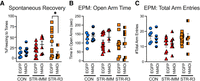
Freezing during spontaneous recovery and performance on the EPM. (A) Spontaneous recovery to tone was assessed 4–7 days after extinction. Group differences emerged when examining the sum of freezing time to all three tones. STR-R3-hM4Di rats froze significantly less and at chance levels compared to STR-R3-EGFP rats. This low freezing to tone indicated that STR-R3-hM4Di rats failed to show spontaneous recovery and as such, did not form a tone-foot shock association. No significant group differences were found in (B) time spent in open arms or (C) number of total arm entries. Data are reported as means ± SEM. (*) P < 0.05.
Elevated plus maze (EPM)
Differences in anxiety-like behavior on the EPM were not found. A two-way ANOVA for the stress group (CON, STR-IMM, STR-R3) and virus type (EGFP, hM4Di) was performed for time in open arms, total arm entries, and the anxiety index and no significant main effects nor an interaction was observed. Together, this suggests that both anxiety-like behavior and overall exploration were similar regardless of stress or virus condition (Fig. 3B,C).
Body weights
Body weight (BW) increased over the course of the experiment and weight gain was attenuated during chronic stress manipulation (Fig. 4). A repeated-measures ANOVA for stress group (CON, STR-IMM, STR-R3) and virus type (EGFP, hM4Di) by week (1, 4, 5, 6, 7, 8, 9, 10, 11) revealed a significant effect of week (F(8,264) = 347.722, P < 0.001). Note that weekly weights were not taken on weeks 2 and 3 due to surgical procedures at that time. Stressors began for STR-R3 at week 3 and ended at week 7, then began for STR-IMM at week 7 and ended at week 10. Across the weeks, rats gained more weight by week 11 than week 1 (P < 0.001). A significant main effect of stress group (F(2,33) = 10.124, P < 0.001), and a significant stress by week interaction (F(16,264) = 17.267, P < 0.001) were found. In summary, the weeks that the stressor was implemented, the stress groups lost weight initially, followed by weight gain, albeit less robustly than CON. For STR-R3, BW dipped from week 4 to week 5 and stayed lower than both CON and STR-IMM (ranging from P < 0.05 to P < 0.001). Then, during weeks 7–10 when the stressor stopped for STR-R3 and began for STR-IMM, the STR-IMM rats lost weight initially and then stabilized but were lower than CON during those weeks (ranging from P < 0.05 to P < 0.001) and were like STR-R3. Importantly, viral type had no impact on weight gain.
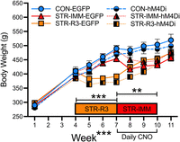
BW measures taken throughout the duration of the experiment. Body weight increased throughout the experiment in all treatment groups. CON gained weight across the duration of the experiment and was unaffected by virus type. For STR-R3, body weight gain was attenuated during restraint compared to STR-IMM and CON and was unaffected by virus type. For STR-IMM, body weight gain was attenuated during restraint compared to CON and was unaffected by virus type. Data are reported as means ± SEM. (**) P < 0.01, (***) P < 0.0001.
Discussion
In the current study, glutamatergic neurons in the IL were targeted for inhibition using DREADDs during the 21 days before the start of fear conditioning, which corresponded to the recovery period for chronically stressed rats given a recovery (STR-R3). A day prior to the start of fear conditioning, the DREADD manipulation was stopped so that these neurons would be theoretically functional by the start of fear conditioning. Contrary to our prediction, long-term neuronal inhibition during the recovery period failed to enhance freezing to tone and context during extinction in chronically stressed rats with a recovery period (STR-R3-hM4Di). Instead, repeated daily inhibition that was to target the IL in STR-R3-hM4Di led to less freezing to the context during extinction compared to their counterparts that did not receive daily, long-term inhibition (i.e., vs. STR-R3-GRP). This reduced freezing to context during extinction was likely attributed to the failure to form a tone-foot shock association of the original fear memory because the STR-R3-hM4Di group failed to show spontaneous recovery to tone. These results indicated that the daily and repeated inhibition of mainly IL neurons on fear conditioning and extinction depends upon whether chronic stress immediately preceded fear conditioning (STR-IMM), was followed by a recovery period prior to the start of fear conditioning (STR-R3), and the experience of chronic stress itself (CON).
The main target of the DREADD infusions was the IL region of the mPFC, but some rats included in our data had viral spread that extended into neighboring regions, including the PL (see Fig. 1B). In our study, 13 of the 16 (81%) rats that received the hM4Di virus had viral expression in both the IL and in portions of the PL. The concern is that the PL and IL have opposing actions in the expression of conditioned fear (Perusini and Fanselow 2015; Bennett et al. 2016). For the PL, temporary inactivation suppresses fear expression, without affecting the acquisition of a fear memory (Corcoran and Quirk 2007). Consequently, one would expect less freezing during fear training or conditioning, but the memory of the tone-foot shock association would persist. Since the spontaneous recovery data revealed that the original tone-foot shock association was not formed in the STR-R3-hM4Di group, potential suppression of the PL is unlikely, as it does not explain our results. For the other two conditions (chronically stressed rats tested a day after stress has ended and the nonstressed controls), the IL (and possible PL) inactivation process did not suppress the fear response during conditioning or extinction when compared to their respective noninhibited controls (hM4Di vs. EGFP, respectively). Consequently, the putative spread of the virus into the PL and other nearby regions was unlikely to have produced the results of the present study.
The IL is important when new learning occurs that contradicts previously learned information. For example, the IL helps form new extinction memory, but not in the formation of the original cued-foot shock fear memory (Milad and Quirk 2002; Mueller et al. 2010; Sierra-Mercado et al. 2010). However, the current study seemed to contradict this by suggesting that the original memory failed to form. One explanation for these seemingly differing views is that the IL appears to be involved whenever flexible learning is required, such as when a new strategy is needed in a familiar situation (Oualian and Gisquet-Verrier 2010; Barker et al. 2013; Mukherjee and Caroni 2018). Furthermore, the IL facilitates actions that would be appropriate for a given situation, and so temporary inactivation of this region can impair both appropriate avoidance and appetitive operant behaviors (Capuzzo and Floresco 2020). Given that the rats were extensively acclimated to fear conditioning context before the start of fear conditioning, they would have learned that this context was safe, which would have changed with the introduction of a foot shock during fear conditioning. This requires flexible learning and suggests that rats with mainly IL inactivation (STR-R3-hM4Di) had suffered impaired flexible learning that prevented them from expressing an adaptive response to a novel threat in a familiar environment when assessed a week later.
The present study supports the growing evidence that chronically stressed rats provided with recovery periods (STR-R3) are different from chronically stressed rats that are fear conditioned a day after stress ends (STR-IMM) and from nonstressed controls (CON). During the presentation of the first tone in fear conditioning, the STR-IMM-GFP rats froze significantly more to the context than did CON-EGFP, while the STR-R3-EGFP froze minimally and similarly to the CON-EGFP group (Fig. 2B). During the training process, STR-IMM (both EGFP and hM4Di) froze significantly more to context A than did STR-R3 (both EGFP and hM4Di, Fig. 2D), demonstrating that the immediate effects of chronic stress had different effects that likely included generalized freezing. This adds to prior studies on hippocampal-dependent spatial memory in rats that were cognitively assessed immediately following the end of chronic stress (Luine et al. 1994; Ghiglieri et al. 1997; Bowman et al. 2002; Abidin et al. 2004; Kleen et al. 2006; Song et al. 2006; Ortiz et al. 2015), but not in chronically stressed rats that were given a recovery period (Luine et al. 1994; Hoffman et al. 2011; Bian et al. 2012; Ortiz et al. 2015; Conrad et al. 2017; Peay et al. 2020). These findings add to the literature that a recovery period following the end of chronic stress has a critical window of effectiveness.
Heightened anxiety-like behavior is often another result following chronic stress and is typically measured through analysis of the exploration of the open arms on the EPM (Strekalova et al. 2004; Vyas et al. 2004; Mitra et al. 2005; Bondi et al. 2008). However, we found no differences across stress or hM4Di patterns for the anxiety index, as measured by the time spent in the open arms and the total number of arm entries on the EPM. The EPM taps into both innate anxiety-like behaviors and exploratory behaviors (Pellow et al. 1985). Typically, rats find exploration of novel areas rewarding and a lack of novelty exploration can be an indicator of a lack of motivation or anhedonia-like behavior (Strekalova et al. 2004; Rygula et al. 2005). Considering the lack of significant differences for both the anxiety-like measures and the exploratory behaviors, this suggests that motivation level and anhedonia-like behavior were similar in all groups (Rygula et al. 2005; Conrad 2006; Huynh et al. 2011). Additionally, the anxiety index considers the total number of entries into all arms of the EPM and did not reveal group differences in motivation or locomotion in the present study.
This experiment was innovative in the use of a chemogenetic approach to study chronic stress effects on fear conditioning, but with some limitations. We administered clozapine N-oxide (CNO) via daily injections over 3 weeks and ended CNO treatment a day before fear conditioning began. However, chemogenetic manipulations are typically performed to influence behavior when DREADDS are concurrently activated (Mahler et al. 2014; Roth 2016; Smith et al. 2016; Whissell et al. 2016; Rapanelli et al. 2017). Nonetheless, chemogenetic activation over weeks has produced long-term changes in the manipulated cell populations, even after chemogenetic activation has ended (Basude et al. 2012; Poyraz et al. 2016; Vetere et al. 2017; Campbell and Marchant 2018). As such, long-term changes could have continued within the mPFC of our study, although additional explanation and further investigation are needed. Another limitation is that ligands to activate DREADDs are typically administered in drinking water to produce a continuous source of the DREADD-activating ligand (Basude et al. 2012; Poyraz et al. 2016; Roth 2016; Smith et al. 2016). In the present study, CNO was administered through daily injections and as such, could have provided an opportunity for the mPFC to be uninhibited. However, a prior study showed that CNO can continue to have behavioral effects for up to 9 h following systemic injections (Alexander et al. 2009), and another found that daily CNO injections produced similar outcomes on behavior as with CNO in drinking water (Basude et al. 2012). Consequently, the existing evidence suggests that the daily and repeated activation of DDREADs via injection in our study was likely effective. Another limitation is that CAMKII promoters might not be as specific to glutamatergic neurons as originally thought and may possibly influence GABAergic neurons as well (Veres et al. 2023). As this could have impacted both glutamatergic and GABAergic neurons in the present study, future studies should tease apart the contribution of these neuronal populations to the observed results.
The current research contributes to and expands upon the existing literature on the role of the IL in fear extinction following chronic stress. The failure to form a fear memory when the majority of IL neurons were inhibited during the recovery period from chronic stress, suggests that the combination of chronic stress-induced changes to the IL and the inhibition of the majority of IL neurons during recovery is particularly disruptive to forming fear memories. The DREADD activation protocol did not disrupt the fear learning process; fear learning itself was similar across all groups. While the current study used male subjects only, it is notable that females express PTSD at higher rates than males (Christiansen and Berke 2020; Iqbal et al. 2024). We chose to focus on males because our past work on chronic stress in both sexes revealed that males expressed impaired recall of fear extinction, which depends upon the IL, whereas females expressed facilitated fear extinction (Baran et al. 2009). Understanding these sex differences are important but target different approaches and was beyond the scope of this investigation. In summary, this work shows that disrupting the majority of neurons in the IL during the recovery process following chronic stress is important in the formation of a fear memory.
Materials and Methods
Subjects
Male Sprague-Dawley rats (Charles River Laboratories) weighing 225–250 g upon arrival were housed with two or three conspecifics in standard laboratory cages (21–22°C). Except where noted below, animals were allowed food and water ad libitum. Animals were housed on a reverse 12:12 light cycle, with lights off at 7 A.M. All procedures occurred during the dark phase of the light cycle and were performed following the Guide for the Care and Use of Laboratory Animals and the approval of the Arizona State University Institutional Animal Care and Use Committee.
DREADD viral infusion surgery
A week after arrival, all rats underwent stereotaxic surgery to target the IL of the mPFC under aseptic sterile conditions. Rats were anesthetized with a ketamine/xylazine/acepromazine cocktail (ketamine = 95 mg/kg BW, xylazine = 5 mg/kg BW, acepromazine = 1 mg/kg BW) in sterile 0.9% sodium chloride (0.2 mL/100 gm BW, i.p.). Once anesthetized, rats were given 1 mg/kg BW of meloxicam (i.p.) and 0.03 mg/kg BW buprenorphine (s.c.). Ketamine boosters were given as needed during surgery (0.25 mL, i.p.). Ophthalmic ointment was applied over the eye to prevent eye dryness. Rats were secured in a stereotaxic apparatus (David Knopf Instruments). Rats received a local anesthetic (s.c.) injection of bupivacaine (2.5 mg/mL) at the incision site to further manage pain. The rats’ heads were next scrubbed thrice with alternating betadine (Perdue Products) and isopropyl alcohol (Vi-Jon, Inc.). A drape cloth was placed over the surgical area. An incision on the skin was made along the midline from around bregma to λ and the head leveled to zero. Dedicated Hamilton microsyringes with a blunt tip (5 μL; National Scientific Company) were used for infusions. Bilateral infusions of one of the viral vectors (see below for details) were given in each hemisphere at 0.5 μL per side. The targeted coordinates to the IL of the mPFC from bregma and skull surface were A/P: +2.7; M/L: +0.5, −0.65; D/V: −5.2 (Paxinos and Watson 2013). The infusion duration was 2–3 min, allowing the virus to spread, and then syringes were left in place for at least another 2 min after infusion was completed. Following infusion, the injector was removed, and the head incision was closed using coated vicryl sutures (Ethicon). The area was cleaned with sterile water and then swabbed with triple antibiotic cream. Rats were placed in a heated, empty cage until they awoke. Rats were individually housed for at least 3 days or until the scalp incisions healed. During postoperative care, rats received meloxicam and buprenorphine on postsurgery days 1 and 2. In some cases, rats had to be resutured during the postoperative period, before being re-pair housed. All rats had at least a week of recovery before the start of chronic stress. There was a 96% survival rate for surgery (nprior to surgery = 72, nsurvived surgery = 69).
Designer receptors exclusively activated by designer drugs (DREADDs)
The following viral vectors were purchased from Addgene: hM4Di inhibitory DREADD virus AAV8-CamKIIα-hM4Di-mCherry (Addgene #50477) and control virus AAV8-CamKII〈-EGFP (Addgene #50476). Vectors incorporating CaMKIIα (calcium/calmodulin-dependent protein kinase IIα) as the promoter were chosen to allow for selective expression in cortical glutamatergic excitatory neurons (Liu and Jones 1996). Virus titers were 3 × 10¹² vg/mL. CNO was obtained from the NIH Chemical Synthesis and Drug Supply Program (Batch 14073-1) and used to activate DREADDs via daily injection at a dose of (1 mg/kg BW, i.p.). The dose was chosen based on work that shows that at low doses CNO is less likely to reverse metabolize to clozapine and have potential confounding effects (MacLaren et al. 2016; Gomez et al. 2017).
Chronic stress procedure
Rats were chronically stressed by restraint for 6 h/day for 21 days. Our previous work demonstrated that these restraint parameters are the minimum required duration for restraint stress to produce behavioral and structural changes (McLaughlin et al. 2007) and were consistent with our past work on fear conditioning (Conrad et al. 1999, 2001). Restraint took place between 9 A.M. and 3 P.M. and occurred in the animal's home cage. Sound-attenuating chambers were used to isolate animals undergoing restraint from animals not undergoing restraint. To keep conditions similar across stress treatments, the control group had food and water removed during restraint hours and were handled at the start of each day. Animals were initially restrained using a wire mesh tube (6.4 cm DIA × 26.7 cm L) that had the ends sealed using grip guard sealer (Flynn and Enslow) to keep the wire ends coated. Rats were upgraded to a larger restrainer (7.6 cm DIA × 29.2 cm L) as they grew. BWs were recorded weekly.
Groups and time line
Rats were assigned one of three stress groups. Rats in the stress immediate group were chronically stressed and underwent fear conditioning and extinction immediately (i.e., within days) after restraint ended (STR-IMM). Rats in the stress-recovery group were chronically stressed and then had a 3-week break or recovery period without restraint before the start of fear conditioning and extinction (STR-R3). The control group did not undergo stress manipulation before fear conditioning and extinction (CON). During the 3-week break for STR-R3, which overlapped with the stressor manipulation for STR-IMM, all rats (CON, STR-IMM, STR-R3) were given daily injections of CNO to activate the DREADDs in those with the hM4Di inhibitory DREADD virus. Those with the control virus (abbreviated EGFP for its reporter) also received CNO to rule out any possible confounding effects of CNO actions (Gomez et al. 2017; Mahler and Aston-Jones 2018). The independent variables of stress groups (CON, STR-IMM, STR-R3) and viral types (EGFP, hM4Di) created a 3 × 2 design, as shown in the time line (Fig. 1A).
Behavioral testing
Fear conditioning
Fear conditioning apparatus
Rat test cages were square and made of metal and plastic (30.5 cm W × 25.4 cm D × 30.5 cm H: Coulbourn Instruments, E10-18TC or H10-11R-TC) and were modified so that the top metal panel was replaced with clear Plexiglas for video recording. Both arenas were housed within a sound-attenuating cabinet. Tones (75 dB steady tone, 20 sec) were delivered through a speaker (Coulbourn H12-01R) mounted on the inside of the sound-attenuating cabinet and were produced by a frequency generator (Coulbourn E12-01 or H12-07). An animal shock generator (Coulbourn H13-15) administered mild foot shocks (0.5 mA, 1 sec) through a shock floor (Coulbourn E10-18RF or H10-11RTC-NSF), with current equally distributed between parallel metal bars. Illumination was provided throughout testing by LED light bulbs mounted to the ceiling of the isolation cubicles.
All stimuli were controlled using Graphic State software (v 4.0 GS4-UP) via a computer connected to a linc system (Coulbourn H02-08) that controlled the stimuli output via an USB interface (Coulbourn U90-11H). Infrared lights (Coulbourn H27-91R) were positioned to be observed by the video and were programmed to denote the context and tone. The infrared lights could not be visually detected unless viewed on video.
Environments for fear conditioning procedures
Two different contexts were used for training and testing within the same room. In context A, the testing cages were square metal and plastic and had a metal floor of parallel rods (Coulbourn H10-11R-TC-SF), silver side panels (Coulbourn H90-00R-M-KT01), and black and white striped panels on the clear plastic back wall. The sound-attenuating cabinet contained a 40-watt equivalent LED bulb (450 Lumens) and a white LED computer fan (Thermaltake, CL-F020-PL12WT-A or Coulbourn ACT-130). The cleaning solution used after each rat was an all-purpose, grapefruit-scented cleaner (Method, Johnson & Son Inc.) and the room lighting of the overall holding room was white light. Experimenters wore a yellow wrap gown and black gloves. Rats were transported from the colony room to the testing room by hand-carrying the rats in their home cages. For context B, the testing cages were round, blue plastic buckets (37 cm H × 30.5 cm DIA, Lowes). A 3-watt, Red LED bulb (91 Lumens) was used as illumination in the isolation cubical. A 35.6 cm, computer fan with red LED light (Thermaltake, TT-1425) provided white noise/ventilation in the cubicle. The cleaning solution used after each rat was 70% isopropyl alcohol (Vi-Jon, Inc.). Experimenters wore a white laboratory coat and blue gloves. The rats were transported from the colony room to the testing room in their home cages on a cart and the room lighting of the holding room was red light.
Procedure
Rats were acclimated to both contexts A and B before the start of fear conditioning to prevent context generalization and to increase the likelihood that the groups would acquire fear conditioning similarly so that fear extinction could be studied (Hoffman et al. 2014). On the last 6 days of the stress manipulation, rats were exposed to the contexts for 10 min, alternating between them for three exposures per environment. On the day after the end of the stress manipulation for the STR-IMM cohort, all rats were fear-conditioned in context A with three tone-foot shock pairings. On the subsequent 2 days, rats were placed into context B and were presented with 15 tone-only presentations. The day after extinction training ended, anxiety-like behavior was measured on the elevated plus maze (EPM). All rats then underwent spontaneous recovery by being placed back into context B and getting three tone-only presentations. For cohort 1, this occurred a week after extinction training. For cohort 2, it took place 4–6 days after extinction training to allow spacing for sacrifice 90 min after behavioral assessment (Fig. 1).
Behavioral quantification
All behavior was digitally recorded on GoPro Hero 3 cameras (GoPro, Inc.) for offline analysis. Video from the cameras was monitored using a Quad Splitter Processor (Evertech), which allowed four videos to be viewed on one monitor. Behavior was manually scored offline by trained observers. Freezing was defined as the lack of all movement, except those associated with respiration (Blanchard and Blanchard 1969). Freezing to tone was defined as any freezing that took place during the 20 sec tone presentation and freezing to context was defined as any freezing that took place in the 20 sec immediately before the presentation of the tone. A fear conditioning difference score was calculated to assist in understanding how much of the freezing to the tone was due to associative processes over a more generalized, nonassociative freezing response that may occur in the absence of the discrete cue. This was calculated as the amount of freezing to tone minus the amount of freezing to context 20 sec before the tone (similar to Majchrzak et al. 2006). Inter- and intra-rater reliability met or exceeded 95.0%.
Elevated plus maze (EPM)
The EPM was used to assess anxiety responses in rats by examining the conflict between exploration of novel environments and innate fear of heights and preference toward dark, enclosed areas (Walf and Frye 2007) and is commonly used by our team (Huynh et al. 2011; Nishimura et al. 2017). The EPM was a raised platform (50 cm off ground) with two opposing open arms (50 cm long × 10 cm wide) and two opposing closed arms (50 × 10 × 30 cm tall walls) that connected to a central area. A camera (GoPro Hero 3, GoPro, Inc.) mounted on the ceiling recorded behavior for offline quantification.
Rats were transported in their home cage with their littermate from the animal colony room to a novel testing room. Littermates were tested one at a time and could not see the apparatus while awaiting their turn from their home cage. At the start of a trial, a rat was placed in the center of the EPM and facing a closed arm. Rats were then given 5 min to explore. After the trial ended, the rats were returned to their home cage. Any debris was wiped off the EPM and it was cleaned with an all-purpose, lavender-scented cleaner (Method, Johnson & Son Inc.).
EMP behavioral quantification
The video recordings of the EPM were scored for entries into the open arms, entries into the closed arms, total entries, and
time spent in open arms. Time spent in closed arms was calculated as the total trial time minus time in the open arms. An
anxiety index was calculated by using the following formula:

Tissue collection and verification of virus placement
Due to the number of rats involved in these studies, behavioral testing occurred using two rat cohorts with all treatment groups represented in each cohort. After behavioral testing concluded, rats were anesthetized with an overdose of sodium pentobarbital, i.p. (100 mg/kg BW, Virbac) before cardiac perfusion. Rats were perfused with 0.1M phosphate-buffered saline followed by 4% paraformaldehyde in 0.1M PBS. After perfusion, brains were removed and postfixed in 4% PFA. Forty-eight hours before sectioning, brains were transferred to a 30% sucrose solution (in 0.1M PBS) for cryoprotection.
Brains were prepared to visualize the virus placement. Brains were sectioned (60 μm, −28°C) using a cryostat (Leica) and mounted onto 2% gelatin subbed glass slides. Fluorescence was protected with Vectashield (Vector Laboratories), and the slides were cover slipped and sealed with clear nail polish. Fluorescence and bilateral placement in the IL were confirmed using a Leica MZ FLIII Stereozoom microscope equipped with a digital camera interfaced with a computer. The spread of the virus was visualized under green light excitation for the inhibitory DREADD (mCherry reporter) or blue light excitation for the EGFP reporter for the control virus. The placement was determined from corresponding coronal sections from a rat brain atlas (Paxinos and Watson 2013) and illustrated in Figure 1.
Statistical analysis
Results for a single time point were analyzed using two-way ANOVA with stress group and viral type as independent factors in Prism GraphPad (v10.3.1). For analyses over multiple time points, a repeated-measures ANOVA was performed using stress group and viral type as the two independent factors. After excluding rats based on viral placement, sample sizes were unequal. In an experimental design with “subject dropout,” correcting for unequal variances was necessary to allow statistical programs to handle the inequality (Fidell and Tabachnick 2007). Consequently, data were transformed using x + 1 (Fidell and Tabachnick 2007). Data analysis was performed using SPSS (v24). Data are represented as means ± SEM, using P-values < 0.05 as being statistically significant.
Acknowledgments
Funding was provided by Arizona State University Knowledge Enterprise. We valued the expertise of the Arizona State University Department of Animal Care and Technologies and contributions from Susan Neill-Eastwood. The authors acknowledge the help from Carol Chen, Camila De Avila Dal'Bo, J. Bryce Ortiz, Cindy Reynolds, Meenal Srivastava, Rujuta Takalkar, Gillian Thornton, Kara Ugarte, and Christopher Willis.
Footnotes
-
Article is online at http://www.learnmem.org/cgi/doi/10.1101/lm.053957.124.
- Received May 31, 2024.
- Accepted November 14, 2024.
This article is distributed exclusively by Cold Spring Harbor Laboratory Press for the first 12 months after the full-issue publication date (see http://learnmem.cshlp.org/site/misc/terms.xhtml). After 12 months, it is available under a Creative Commons License (Attribution-NonCommercial 4.0 International), as described at http://creativecommons.org/licenses/by-nc/4.0/.



