


Social and nonsocial environmental loss have differential effects on ventral hippocampus-dependent behavior and inhibitory synaptic markers in adult male mice
- Corresponding author: goulde{at}princeton.edu
Abstract
In humans, psychological loss, whether social or nonsocial, can lead to clinical depression, anxiety disorders, and social memory impairments. Researchers have modeled combined social and nonsocial loss in rodents by transitioning them from social, enriched environments (EE) to individual housing, affecting behaviors related to avoidance, stress coping, and cognitive function. However, it remains unclear if these effects are driven by social or nonsocial loss. We examined the effects of nonsocial loss by housing adult male mice in EE before moving them to standard cages, where they were pair-housed, and compared this to mice experiencing complete social loss. Continuous EE reduced social investigation time while leaving social memory intact, also decreasing avoidance behavior. Nonsocial loss restored social investigation and avoidance behavior to control levels, while social loss impaired social memory and increased avoidance. In rodents, social memory and avoidance require ventral hippocampus (vHIP) neuronal oscillations, which involve parvalbumin-positive (PV+) inhibitory interneurons. We found decreased vHIP PV intensity in the social loss group, with no differences in the nonsocial loss group. Most PV+ cells are surrounded by perineuronal nets (PNNs) concentrating GABAA receptors in their lattice-like holes. Social loss decreased GABAA-δ expression, a subunit associated with extrasynaptic receptors, across PNN+ soma and in PNN holes, while nonsocial loss reduced gephyrin in these regions. These findings suggest social and nonsocial losses differentially affect vHIP function and behavior, with social loss having a more pronounced impact through mechanisms involving PV+ interneurons, PNN structure, and neurotransmitter receptor expression.
Psychological loss can be a precipitating event for the development of clinical depression and anxiety disorders (Zisook and Shuchter 1991a,b; Kaplow et al. 2010; Sikorski et al. 2014). These losses can be mostly social, such as the death of a loved one or a relationship ending, or mostly nonsocial, like eviction or job loss. Both social and nonsocial psychological loss have been linked to the development of anxiety disorders and major depressive disorder (Monroe et al. 1999; Sikorski et al. 2014; Hoke and Boen 2021; van Eersel et al. 2021), which are leading causes of disability (Brenes et al. 2008). Anxiety disorders and clinical depression have been linked to social memory deficits and behavioral inhibition (White et al. 2011). Researchers have modeled combined social and nonsocial loss in rodents by transitioning them from social, enriched environments (EE) to individual, standard housing (Smith et al. 2017; Smail et al. 2023). This paradigm changes behaviors associated with memory function, avoidance, and stress coping. However, it is not clear whether these effects are driven primarily by social or nonsocial loss. Understanding how social and nonsocial loss affect brain regions and cellular substrates associated with social memory and behavioral inhibition may suggest targets for future therapeutic interventions.
In humans, the anterior hippocampus has been linked to memory encoding and anxiety (Strange et al. 1999). The rodent ventral hippocampus (vHIP), specifically ventral CA1 (vCA1), is considered to be functionally analogous to the primate anterior hippocampus, with links to social memory (Okuyama et al. 2016; Cope et al. 2023) and avoidance behavior (Adhikari et al. 2010; Murthy et al. 2019; Padilla-Coreano et al. 2019; Laham et al. 2022). The vCA1 is also a stress-sensitive region, showing molecular, cellular, and circuit-level alterations in response to both early-life stress (Murthy et al. 2019; Laham et al. 2022) and adult stress (Ivens et al. 2019; Albrecht et al. 2022). Neuronal oscillations of multiple frequencies have been linked to various behaviors associated with vCA1. Specifically, social recognition, as well as retrieval of stress-related memories, have been associated with sharp wave ripples (SWRs) in the vCA1 (Rao et al. 2019; Tao et al. 2022; Kuga et al. 2023), while avoidance behavior has been associated with power in the theta frequency range (4–12 Hz) (Adhikari et al. 2010; Padilla-Coreano et al. 2019).
Parvalbumin-positive (PV+) interneurons have been shown to contribute to neuronal oscillations, including theta, gamma, and SWRs (Stark et al. 2014; Amilhon et al. 2015; Antonoudiou et al. 2020), by coordinating the rhythmic inhibition and release from inhibition of excitatory neurons (Klausberger 2009; Gulyás and Freund 2015). In the vCA1, the majority of PV+ interneurons are surrounded by perineuronal nets (PNNs), which are specialized extracellular matrix structures (Murthy et al. 2019; Laham et al. 2022). Although PNNs have been associated with restricted plasticity both during development and in pathological states (Takesian and Hensch 2013; Cope et al. 2022), their function in the adult healthy brain is incompletely understood. PNNs typically surround the cell body and proximal dendrites of neurons, and have a lattice-like structure. This results in regions where the neuron has limited access to the extracellular space because of PNN coverage, and regions where the neuron has greater access to the extracellular space, namely in the perforations or holes of the PNN. Some evidence suggests that PNNs regulate neuronal activity by concentrating neurotransmitter receptors at their holes (Wingert and Sorg 2021; Tewari et al. 2024), thus allowing for a greater postsynaptic response to neurotransmitter release. Since the majority of synapses on the cell body are inhibitory, access of GABAergic receptors to GABA may be modulated by the presence and configuration of PNNs surrounding PV+ interneurons.
Gamma-aminobutyric acid A (GABAA) receptors, which are fast-acting ionotropic receptors, are present on PV+ interneurons, where they participate in perisomatic inhibition. Inhibitory inputs to PV+ interneurons originate from long-range GABAergic neurons of the medial septum as well as from inhibitory interneurons, including nearby PV+ cells (Unal et al. 2015). GABAA receptors consist of multiple subunits, which can influence their location in the neuronal membrane. For example, GABAA receptors with delta (δ) subunits are more often located extrasynaptically, while those lacking this subunit are more often located at synapses (Farrant and Nusser 2005). This distinction has functional implications because activation of extrasynaptic GABAA receptors contributes to tonic inhibition, while activation of synaptic GABAA receptors contributes to phasic inhibition. Optimal hippocampal function seems to require a balance between tonic and phasic inhibition, while too much or too little of either type produces dysfunction (Glykys and Mody 2006). PNNs may affect this balance by preventing extrasynaptic receptors from accessing ambient GABA or by altering the distribution or concentration of receptors in their holes. However, no previous studies have examined whether changes in vCA1 PNN holes, or GABAA synaptic and extrasynaptic receptors are associated with behavioral changes after different types of experiential loss.
To further characterize the influence of experiential loss on vCA1 behavioral function and PNNs and their relationship to extrasynaptic and synaptic GABAA receptors, we examined the effects of both social and nonsocial loss in male mice. First, to examine only nonsocial loss, groups of mice were housed in EE for 4 weeks before moving them to standard cages, where they were pair-housed for 2 weeks. These results were then compared to mice that experienced only social loss, as they were transferred from standard group housing to individual housing on the same timescale. We found that social, but not nonsocial, loss impaired social memory. In addition, we found that social, but not nonsocial, loss increased avoidance behavior. Mice from all housing groups (EE, enrichment removal [ER], and social isolation [SI]) exhibited a reduction in the area of PNNs occupied by PNN holes compared to controls. SI mice had less GABAA-δ and more gephyrin, a GABAA receptor synaptic scaffolding protein, in the holes, while EE and ER mice showed the reverse pattern of changes in these markers compared to controls. These findings show that in adulthood, changes in environmental living conditions can alter the expression of vCA1 GABAA receptor markers as well as PNN configurations, raising the possibility that SI impairs vCA1 behavioral function by reducing the expression of GABAA extrasynaptic receptors in PNN holes.
Results
Social and nonsocial loss effects on vCA1 behavior
To determine the impact of social and nonsocial loss (Fig. 1) on social memory, we conducted the direct social interaction test with an intertrial interval of 1 h between novel and familiar trials (Fig. 2A). EE and ER groups had comparable discrimination indices to controls (Fig. 2B; Table 1). In contrast, SI mice show a significantly lower discrimination index when compared to control and EE mice (Fig. 2B; Table 1). No significant differences were observed between ER and SI mice for the discrimination index, possibly due to the fact that there was greater variability in the ER group compared to control and EE groups (Fig. 2B; Table 1). We also observed large differences in overall social investigation times, as well as in investigation times of novel and familiar mice, across groups (Fig. 2C–E; Table 1), which did not parallel changes in discrimination abilities. EE mice had the lowest social investigation times during both novel and familiar trials, despite maintaining the ability to distinguish between a novel and a familiar mouse. ER mice exhibited intermediate social investigation times during both novel and familiar trials between control and EE mice. Despite diminished social discrimination abilities, SI mice had overall social investigation times that were comparable to controls, with no significant differences in investigation times during novel or familiar trials compared to controls. These latter findings suggest that diminished social discrimination in SI mice was not the result of reduced motivation to investigation or rapid habituation during social exposure, but rather to diminished social recognition. Next, we investigated the impact of social loss (SI) and nonsocial loss (ER) on avoidance behavior using the EPM (Fig. 2F). We found that compared to controls, EE mice spent more time on the open arms, suggesting reduced avoidance behavior (Fig. 2G; Table 1). SI mice showed the opposite effect with less time in the open arms compared to controls, and ER mice had open arm times that were intermediate between SI and EE, although these values were only significantly different from EE (Fig. 2G; Table 1). The difference between SI and the control group was not related to an overall difference in the amount of time spent in open and closed arms (as opposed to the center). This analysis, however, showed that EE and ER mice spent significantly more time in the arms than controls (Fig. 2H; Table 1). Overall, these findings show that while ER mice differ on some vCA1-dependent behavioral measures from controls or EE mice, the largest behavioral differences were observed in the SI group, which showed impaired social discrimination and increased avoidance behavior.
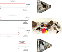
Timelines and environmental living conditions. Experimental timeline for control housing, EE, ER, and SI.
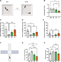
Social, but not nonsocial, loss impairs social recognition and increases avoidance behavior. (A) Schematic of direct social interaction test. (B) SI mice have a significantly lower discrimination index compared to CON, while no differences are observed in ER mice compared to controls. (C) Overall social investigation times across all groups. EE mice exhibited the lowest social investigation times yet retained the ability to discriminate between novel and familiar mice. ER mice showed intermediate investigation times positioned between CON and EE groups, while SI mice showed investigation times comparable to CON. (D) Novel social investigation times across all groups. EE and ER mice showed lower novel interaction times compared to CON, while SI mice showed novel investigation times comparable to CON. (E) Familiar social investigation times across all groups. EE mice exhibit lowest familiar investigation times, while ER and SI mice are comparable to CON. (F) Schematic of EPM test. (G) Time spent in the open arms is significantly higher in the EE group compared to the CON group, while the SI group spent significantly less time in the open arms compared to the CON group. No significant differences were observed between the ER and CON groups. (H) EE and ER mice spent more time in the arms (open + closed) compared to CON. No significant differences were observed between CON and SI mice. Data are presented as mean ± SEM (CON n = 12, EE n = 6, ER n = 6, SI n = 10). Significance is denoted as adjusted P-values after FDR post hoc tests: (*) P < 0.05, (**) P < 0.01, (***) P < 0.001. See Table 1 for complete statistics. (CON) Control, (EE) environmental enrichment, (ER) enrichment removal, (SI) social isolation. Images in A and F were created using BioRender.com and are published under a CC BY-NC-ND 4.0.
Statistical analysis
Social and nonsocial loss effects on vCA1 PNNs and GABAA receptor measures
Previous studies have shown that developmental and adult stress alter PNN intensity in several brain regions, including the vCA1 (Laham and Gould 2022; Laham et al. 2022), so we measured PNN intensity around PV+ cells in the vCA1 using the plant lectin Wisteria floribunda agglutinin (WFA) (Fig. 3A). Unexpectedly, we found no overall differences in PNN intensity in both the social loss (SI) and nonsocial loss (ER) groups when compared to controls. (Fig. 3C; Table 1). We further evaluated PV intensity of PV+/WFA+ cells and found a decrease in overall PV intensity in the SI group compared to controls, while no differences in intensity were noted between the ER and control group (Fig. 3B; Table 1). Since one of the functions attributed to PNNs is to influence neurotransmitter receptor distribution through their concentration in PNN holes, we next carried out a high-resolution confocal analysis of vCA1 PNNs (Fig. 3D) and found that although there were no changes in the density of holes (mean ± SEM-CON: 0.2494 ± 0.006288; EE: 0.2733 ± 0.00774; ER: 0.2501 ± 0.00714; SI: 0.257 ± 0.007332; P > 0.1 for all comparisons), the percent area of PNN-enwrapped cell body occupied by holes was reduced in all groups, including EE, ER, and SI compared to controls (Fig. 3E; Table 1). These results arise from a reduction in hole area, and strongly suggest that housing experience alters the size of holes in PNNs. These findings suggest that vCA1 PNNs in EE, ER, and SI mice undergo changes in PNN hole size that are similar in direction (decreased) but differ in magnitude, with the greatest decreases observed in EE mice.
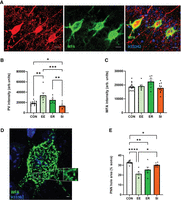
Social and nonsocial loss decreases the percentage of PNN occupied by holes in the vCA1. (A) Confocal image of PV+/WFA+ cells in vCA1. (B) EE mice show a higher PV intensity compared to CON, while SI mice have significantly lower PV intensity compared to CON mice. No differences were observed between ER and CON groups. (C) No differences in WFA intensity were observed across groups. (D) Confocal image of WFA+ cell in vCA1. (E) Decrease in percentage of WFA area covered by holes is observed in all groups compared to CON. Data are presented as mean ± SEM (CON n = 16, EE n = 6, ER n = 6, SI n = 9–10). Significance is denoted as adjusted P-values after FDR post hoc tests: (*) P < 0.05, (**) P < 0.01, (***) P < 0.001, (****) P < 0.0001. See Table 1 for complete statistics. (CON) Control, (EE) environmental enrichment, (ER) enrichment removal, (SI) social isolation, (PNN) perineuronal net, (PV) parvalbumin, (WFA) Wisteria floribunda agglutinin, (H33342) Hoechst 33342. Scale bars, 20 μm (A), 5 μm (D).
Because GABAA receptors have been linked to vCA1 behavioral function (Rezayof et al. 2007; Ardi et al. 2019), we next looked at the intensity and distribution of the GABAA receptor scaffolding protein gephyrin (Fig. 4A), which is known to anchor GABAA receptors to synapses (Kneussel et al. 1999). We found that overall gephyrin intensity was reduced across soma of PNN+ cells in EE and ER, but not SI mice (Fig. 4B; Table 1). This difference was greatest in EE mice, where gephyrin labeling was about 50% of controls (Fig. 4B; Table 1). Since gephyrin is a marker of synaptic GABAA receptors, which contribute to phasic firing, the large gephyrin decrease in EE mice might contribute to reduced avoidance behavior observed in our EPM analysis. We then examined gephyrin labeling in PNN holes and found results that were very similar to overall gephyrin intensity for EE and ER groups (reduced gephyrin intensity in holes) with an increase in the SI group (Fig. 4C; Table 1). We next investigated the intensity and distribution of the GABAA-δ subunit, a common subunit of GABAA extrasynaptic receptors (Fig. 4D), which contribute to tonic inhibition (Petrini et al. 2004), on vCA1 PNN+ cell bodies. We found no differences between controls and EE or ER groups, but a decrease in GABAA-δ intensity on cell bodies of the SI group (Fig. 4E; Table 1). This decrease in overall GABAA-δ expression in vCA1 PNN+ cells in SI mice was paralleled by the results of a separate analysis within PNN holes, suggesting that fewer GABAA-δ receptors are exposed to the extracellular space (Fig. 4F; Table 1). In contrast, both EE and ER groups had elevated GABAA-δ in PNN holes as well as across the cell body (Fig. 4F; Table 1). These findings suggest environmental conditions exert differential plasticity of GABAA receptor markers in terms of expression.
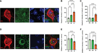
Social and nonsocial loss alter expression of GABAA receptor markers and alter their concentration within PNN holes in the vCA1. (A) Confocal image of WFA+ Gephyrin+ cell in vCA1. (B) Higher gephyrin intensity represented as percent control is observed in the SI group compared to EE and ER group. (C) Higher gephyrin intensity in holes, represented as percent control, was observed in the SI group compared to EE and ER. (D) Confocal image of WFA+ GABAA-δ+ cell in vCA1. (E) Overall GABAA-δ intensity, when considered as percent control, showed no differences between the EE and ER groups but a difference in the SI group compared to both the EE and ER groups. (F) Lower GABAA-δ intensity in PNN holes was observed in the SI group compared to EE and ER. No difference in GABAA-δ intensity in PNN holes was observed between the EE and ER groups. Data are mean ± SEM (EE n = 6, ER n = 6, SI n = 10). Significance is denoted as P-values: (*) P < 0.05, (**) P < 0.01, (***) P < 0.001, (****) P < 0.0001. See Table 1 for complete statistics. (EE) Environmental enrichment, (ER) enrichment removal, (SI) social isolation, (GABAA-δ) gamma-aminobutyric acid receptor subunit δ, (WFA) Wisteria floribunda agglutinin, (H33342) Hoechst 33342. Scale bars, 5 μm (A,D).
Discussion
The results of this study show that for behaviors linked to the vCA1, including social recognition and avoidance, social loss exerts a much larger effect than nonsocial loss on adult male mice. Mice subjected to ER with continued social (pair) housing did not show any significant changes in these behavioral measures, while mice subjected to SI had impaired social recognition and increased avoidance behavior. In addition, mice that were housed in EE for the entire duration of the experiment showed reduced avoidance behavior and overall diminished social investigation times despite intact social recognition. Analysis of PV+ PNN+ cells in the vCA1 showed significantly diminished PV intensity in social loss mice with no significant change in the nonsocial loss group. PNN analyses revealed similar profiles across experimental groups with social loss and nonsocial loss groups showing reduced soma areas occupied by PNN holes with no change in PNN intensity or PNN hole density. In contrast, GABAA receptor markers showed differential plasticity depending on the environmental manipulation. Mice subjected to social loss had a reduction in GABAA-δ expression across the entire cell body and a corresponding decrease in GABAA-δ expression in PNN holes. Mice subjected to nonsocial loss showed a completely different set of results with a reduction of gephyrin expression across the cell body, as well as reduced gephyrin in PNN holes. Mice that remained in the EE through the entire experiment had elevated PV intensity, but otherwise looked similar to nonsocial loss mice, with the former group having larger effects, suggesting that the nonsocial loss group may be continuing to exhibit lasting effects of enrichment.
Social and nonsocial loss effects on ventral hippocampal behavioral function
Our results add to a growing body of literature indicating that the vHIP of adult mice is sensitive to experience. In particular, studies have shown that the vCA1 is sensitive to stress both during development and in adulthood (Sood et al. 2014; Floriou-Servou et al. 2018; Fee et al. 2020; Ma et al. 2021; Laham et al. 2022). Our results showing that SI increases avoidance behavior and impairs social recognition function are consistent with previous studies that measured effects of SI for longer durations of time or beginning just after weaning or during adolescence (Kwak et al. 2009; Li et al. 2017; Deng et al. 2019; Zhang et al. 2021; Guimarães et al. 2023).We found robust behavioral changes after just 2 weeks of SI, which suggests that ventral hippocampal function is especially sensitive to social deprivation. Our results differ from a previous study that showed reduced instead of increased avoidance behavior, as well as no effect on social recognition, in mice subjected to SI (Sukegawa et al. 2022). The reasons for these discrepant results are unknown, but it is likely relevant that different strains of mice were used (BALB/c in Sukegawa and C57 in our study). Since BALB/c mice are known to be less social than C57 mice (Sankoorikal et al. 2006), negative impacts of SI might be less on the former strain.
Our results also show that some aspects of ventral hippocampal behavioral function are sensitive to EE living with reduced avoidance behavior being a notable outcome. This finding is consistent with several other studies showing that environmental enrichment reduces behavioral inhibition on several tasks, including the elevated plus maze (EPM), light–dark test, and zero maze (Benaroya-Milshtein et al. 2004; Rogers et al. 2017; Singhal et al. 2019). We found no significant changes in avoidance behavior with ER; this measure was restored to control-like levels within 2 weeks. Taken together with our SI results, these findings suggest that nonsocial loss reverses beneficial effects of enrichment but at least at the time points and with the assays we examined, there are no further negative effects. In contrast, social loss has a substantial effect on both avoidance behavior and social recognition independent of any prior enrichment. Since the enrichment loss mice went from larger group to pair housing, they experienced partial social loss. Given that the enrichment loss group was very similar on almost all measures to the enrichment group, these findings collectively suggest that social loss only becomes impactful when it is complete, that is, SI.
In addition to emphasizing the robustness of SI effects on behavioral function, these findings also suggest a potential dissociation between behaviors linked to the vCA1, because although SI altered avoidance and social recognition, EE living affected only avoidance while leaving social recognition largely intact (although we did observe differences in overall social investigation times). The vCA1 region provides a major output of the hippocampus integrating information from several afferents to support multiple behavioral functions. For example, inputs from the dorsal CA2 (dCA2) to the vCA1 are critical for social recognition (Meira et al. 2018), while those from the basolateral amygdala (BLA) to the vCA1 contribute to avoidance behavior (Yang and Wang 2017). Thus, exposure to environmental enrichment may affect avoidance behavior by targeting the BLA-vCA1 circuit more specifically, while SI may alter both BLA-vCA1 and dCA2-vCA1 circuits. The specific stimuli related to each environmental condition that drive differential effects on vCA1 circuitry and behavior remain unknown.
Parvalbumin-positive interneuron involvement in ventral hippocampal function
Previous studies suggest that PV+ interneurons play important roles in ventral hippocampal behavioral function, particularly in social recognition and avoidance behavior. Inhibition of vCA1 PV+ interneurons has been shown to impair social recognition and alter avoidance behavior (Deng et al. 2019; Volitaki et al. 2024). It is likely that PV+ interneurons influence these behaviors through their effects on coordinated network activity; these cells have been shown to play a role in high-frequency oscillations (gamma and SWRs) as well as low-frequency oscillations (theta) (Wulff et al. 2009; Schlingloff et al. 2014). Social recognition has been causally linked to both gamma and SWRs (Aoki et al. 2017; Brown et al. 2020; Cope et al. 2023), while avoidance behavior has been causally linked to theta rhythm in the vCA1 (Padilla et al. 2019). Thus, it seems likely that environmental manipulations influence these two behavioral functions by modulating oscillations at different frequencies, although PV+ interneurons are likely involved in both behavioral effects.
Developmental and adult stress have been reported to alter neuronal oscillations across multiple frequencies in the vHIP, including SWR, gamma, and theta (Çalışkan and Stork 2019; Murthy et al. 2019; Laham et al. 2022; Kuga et al. 2023). Along with these functional changes, developmental and adult stress have been shown to reduce PV+ cell number as well as PV intensity (Czéh et al. 2015; Murthy et al. 2019), with some evidence suggesting that reduced PV+ cell number occurs because of reduced PV expression, not an overall loss of PV+ cells (Murthy et al. 2019). Our findings that social loss in adulthood also reduces PV intensity are consistent with this finding, as well as with findings in rats showing that SI reduces the number of PV+ cells in the vCA1 (Deng et al. 2019). Although it remains unknown whether the PV intensity decrease results from or is a cause of changes in network activity, previous studies have shown that experimentally induced theta bursts reduce PV expression in other brain regions (Mix et al. 2010, 2015). The effects of social loss on vCA1 neuronal oscillations and a potential relationship with PV expression remain to be investigated. We also noted that vCA1 PV expression was increased in mice living in EE, an effect that may be related to reduced avoidance behavior in this group. However, no significant differences were observed in PV expression or avoidance behavior between control and nonsocial loss mice, suggesting that by the two-week time point after ER, these effects of enrichment have dissipated.
Role for PNNs in neurotransmitter receptor exposure and ventral hippocampal function
The majority of PV+ interneurons in the vCA1 are surrounded by PNNs (Murthy et al. 2019; Laham et al. 2022). Several functions have been attributed to PNNs, including that they suppress plasticity by enhancing inhibitory input through control of neurotransmitter receptor distribution across the surface of the cell (Corvetti and Rossi 2005; Tewari et al. 2024). Although PNNs have been associated with developmental events that represent the closure of critical periods (Takesian and Hensch 2013), it is known that they remain sensitive to experience in adulthood. Studies have shown that stress and EE living can alter PNN intensity in the hippocampus (Carstens et al. 2016; Foggetti et al. 2019; Laham and Gould 2022). Unexpectedly, we observed no differences in PNN intensity in the vCA1 between controls and any of our environmental manipulations. PNNs are not homogeneous structures, with differences in biochemical composition and structure noted across cell types and brain regions (Laham and Gould 2022). The holes embedded in the lattice-like structure of the PNNs appear to be areas where neurotransmitter receptors concentrate and some evidence suggests that PNNs regulate the lateral movement of receptors on the membrane (Sorg et al. 2016; Tewari et al. 2024). We observed that despite finding no differences in PNN hole density, all environmental manipulations we examined (EE, ER, and SI) had proportionately lower soma surface area occupied by PNN holes. This decrease was greatest in the EE group and least in the SI group, with ER in between, again suggesting the possibility that ER represents the dissipation of EE effects, as opposed to added stress. Differences in the percentage of the soma with PNN holes suggest a plastic response to environmental manipulation that may alter the distribution of afferent inputs across the soma and correspondingly change the availability of neurotransmitter receptors to the extracellular space. Because many afferents synapsing on the cell body are inhibitory, changes in PNN holes may have a large impact on GABAergic receptors, most of which are GABAA receptors.
Studies have shown that GABAA receptors, which are Cl- channel receptors, can be synaptic or extrasynaptic (Farrant and Nusser 2005). Synaptic GABAA receptors are associated with the anchoring protein gephyrin (Petrini et al. 2014), and their activation is known to contribute to phasic inhibition of the postsynaptic cell (Stell and Mody 2002; Farrant and Nusser 2005). Phasic inhibition of hippocampal PV+ interneurons produces neuronal oscillations, with different frequencies of inhibitory release contributing to different behavioral outcomes. In contrast, extrasynaptic GABAA receptors are associated with tonic inhibition of postsynaptic cells, which can oppose phasic inhibition by disrupting the intensity of large-scale rhythmic inhibition (Farrant and Nusser 2005; Brickley and Mody 2012). GABAA receptors with δ subunits are most often located extrasynaptically but can also contribute to synaptic GABAA receptors (Bogdanov et al. 2006). Their location on the membrane, i.e., whether they are adjacent to GABA-releasing afferents and near gephyrin-associated GABAA receptors, plays a major role in determining whether GABAA-δ receptors are synaptic or extrasynaptic. Thus, changes in vCA1 PNN holes and the concentration of GABAA receptors therein have the potential to alter the balance of phasic and tonic inhibition and affect behavioral function.
We found that SI reduced the overall expression of GABAA-δ across the soma and had a similar diminishing effect on GABAA-δ located in PNN holes. Taken together with the reduction in percent soma occupied by PNN holes, these findings suggest that SI may disrupt social memory and increase avoidance behavior by reducing the amount of GABAA-δ receptors exposed to the extracellular space, potentially diminishing extrasynaptic GABAA receptor activation and related tonic inhibition. EE living also alters the expression of GABAA receptor markers, with reductions in gephyrin across the soma as well as within PNN holes. These changes may result in less synaptic GABAA receptor activation and a corresponding reduction in phasic inhibition, potentially contributing to reduced avoidance behavior in this group. As with the other measures we considered in this study, the ER group shared similar features with the EE group but in some cases showed measures that were closer to controls.
Limitations and future directions
While the current study indicates that social loss is more impactful on vCA1-mediated behavior and PV+ interneurons than nonsocial loss, it remains possible that if combined, larger or different effects beyond what was observed with social or nonsocial loss individually would be seen. Some previous studies have examined combined nonsocial and social loss, without comparing to social loss alone, and found large impacts on behavior that may arise from synergistic effects of social and nonsocial loss (Smith et al. 2017; Smail et al. 2023). Future studies should investigate this possibility more completely.
Our findings show parallels between behavioral changes and changes in PV+ interneurons, and the relationship between PNN holes and GABAA receptor markers. However, we have yet to explore causal links among these findings, which would likely require the development of new tools. However, since previous studies have shown that PV+ interneurons, PNNs, and GABAA receptors (synaptic and extrasynaptic) are sensitive to experience and contribute to hippocampal function (Czéh et al. 2015; Deng et al. 2019; Murthy et al. 2019; Volitaki et al. 2024), our findings support a functional contribution of vCA1 brain changes to social loss-induced alterations in social memory and avoidance behavior. These connections should be more closely explored in the future.
An additional limitation of our study is the exclusive use of male mice for these comparisons. It is well known that sex differences exist in many stress effects; however, findings from rodent studies have been mixed with some showing greater vulnerability in females (Adamec et al. 2006; Farrell et al. 2016; Goodwill et al. 2019) and others showing greater vulnerability in males (Adkins et al. 2022; Furman et al. 2022). Enrichment loss studies have been done in both sexes of rats although not in the same experiment where direct comparisons can be made (Smith et al. 2017; Morano et al. 2019; Smail et al. 2023). Although these studies focused on fear-related and stress-coping behaviors (Smith et al. 2017; Morano et al. 2019; Smail et al. 2023) and did not use the same behavioral measures we used, they raise the possibility of larger effects of enrichment loss in females. Future studies designed to directly investigate potential sex differences of social and nonsocial loss on ventral hippocampal structure and function will be illuminating.
Concluding remarks
Our study highlights the differential impacts of social and nonsocial loss on ventral hippocampal (vCA1) function and associated behaviors. We observed that social, but not nonsocial, loss has detrimental effects on social recognition and avoidance behavior, likely mediated through alterations in PV+ interneurons and GABAA receptor distribution. Specifically, social loss leads to a significant reduction in GABAA-δ expression across the soma as well as within PNN holes, suggesting a possible shift toward synaptic localization and phasic inhibition. In contrast, nonsocial loss results in reduction of gephyrin expression across the PNN+ cell body, reduced gephyrin in PNN holes, and increased GABAA-δ in PNN holes similar to what is observed with continuous EE living. Our findings underscore the sensitivity of the vCA1 to environmental manipulations and highlight that distinct neural differences result from social and nonsocial experiences. These insights contribute to a broader understanding of how different forms of environmental stress influence hippocampal function and behavior, with potential implications for therapeutic strategies targeting neuropsychiatric disorders associated with social and nonsocial loss. Future studies should explore the precise circuitry and molecular pathways involved in these differential responses to further elucidate the mechanisms driving these complex behaviors.
Materials and Methods
Animals
Animal procedures were approved by the Princeton University Institutional Animal Care and Use Committee and followed the National Research Council Guide for the Care and Use of Laboratory Animals (2011). Adult male C57BL/6J mice were obtained from the Jackson Laboratory and were used when they were 7 to 8 weeks old (n = 6–16/group). During the study, all mice had ad lib access to food and water. Mice were housed under a reverse 12/12 h light–dark cycle and were tested in the dark. The humidity of the room was ∼50%.
Environmental manipulations
Standard housing consisted of two to five mice housed in Optimice cages. SI housing consisted of one mouse per cage in Optimice cages. Enrichment groups were housed six to a group in containers that were 34 inch length × 18 inch width × 12 inch height with multiple air holes on the lid. The containers each included several rodent pet toys and objects, such as wooden textured huts, climbing structures, hollow balls, small arch shelters, plastic rings, and nestlets (Fig. 1). Cages were cleaned and, to maintain a high level of novelty, supplied with a different set of objects every week. Ad lib access to standard rodent chow was provided in bowls and ad lib water access was provided through water bottles hung on the outside of the containers at a reachable level to the mice.
Mice were divided into four groups: control housing (CON, n = 16), continuous EE (n = 6), ER (n = 6) as a model for nonsocial loss, and SI (n = 10) as a model for social loss (Fig. 1). The control mice were group-housed in standard cages for the total duration of the study. The EE and ER groups were housed in EE 24 h/day. The EE mice were housed in the EE for a total duration of 6 weeks. The ER mice were housed in the EE for 4 weeks and then transferred to control housing, where they were pair-housed for 2 weeks. The SI mice were socially isolated and placed in standard cages for a duration of 2 weeks (Fig. 1).
Behavioral analysis
Social recognition testing
The direct social interaction test was used as previously described (Cope et al. 2023) to assess social memory/novelty preference in all groups. The DSIT was conducted in a 12 × 12 in plexiglass arena with black walls and a clear base under low light (10–15 lux). Prior to the test beginning, mice were acclimated to the behavior testing room for at least 30 min, and then also habituated to the testing box for 5 min prior to the first social stimulus trial. The test consisted of two 5 min trials separated by an intertrial interval of 1 h. In trial 1 (novel), mice were paired with a novel age- and sex-matched stimulus mouse from a different litter and allowed to interact freely for 5 min. After the intertrial interval during which the mice were returned to the home cage, trial 2 (familiar) was conducted with the same test mouse and stimulus mouse pairing for 5 min. The test mouse and the stimulus mouse were positioned in the center of the arena, facing each other with a 1 cm gap to ensure detection. Both trials were recorded from above using a single camera. A trained experimenter, blind to the treatment group, manually scored the time the test mouse spent investigating the stimulus mouse. Social investigation was defined as the test mouse directing its snout toward the stimulus mouse's anogenital region or body within 1 cm, following, or initiating allogrooming. Strict criteria were applied consistently across all experiments to ensure the behavior measured reflected active investigation by the test mouse, rather than passive interaction prompted by the stimulus mouse. As mice generally prefer novel social stimuli over familiar ones, a reduction in investigation time from trial 1 to trial 2 suggests social recognition or memory. The arena was thoroughly cleaned with 70% ethanol between each mouse and trial. The discrimination index was calculated as follows: Discrimination index = ([Novel – Familiar]/ [Novel + Familiar]). The total investigation time was calculated as follows: Total investigation time = Novel investigation time + Familiar investigation time
Elevated plus maze testing
To measure avoidance behavior, the EPM was used as previously described (Laham et al. 2022), where increased time not in the open arms (in the closed arms and center) is considered to be evidence of avoidance behavior. Mice were placed on an EPM that consisted of an elevated (50 cm) plus-shaped track with two arms that were enclosed with high walls (30 cm) and two open arms that had no walls. The open arms were sprayed with water and illuminated to 600 lux (Laham et al. 2022). All arms were 50 cm in length. On the day of testing, each mouse was placed into the center of the maze and their behavior was videotaped for 5 min. The arena was thoroughly cleaned with 70% ethanol between each test. The number of entries into the open and closed arms and time spent in the open arms, closed arms, and center was measured for each mouse from video recordings using ANY-Maze (version 7.34).
Perineuronal net, parvalbumin interneuron, and inhibitory synapse labeling
Mice were deeply anesthetized with Euthasol (Virbac) and were transcardially perfused with cold 4% paraformaldehyde (PFA). Extracted brains were postfixed for 48 h in 4% PFA at 4°C, followed by an additional 48 h in 30% sucrose at 4°C for cryoprotection before being frozen in cryostat embedding medium at −80°C. Hippocampal coronal sections (40 µm) were collected using a cryostat (Leica). Sections were blocked for 1½ h at room temperature in a PBS solution that contained 0.3% Triton X-100 and 3% normal donkey serum. Sections were then incubated overnight while shaking at 4°C in the blocking solution that contained combinations of the following primary reagents: mouse anti-parvalbumin (PV, 1:500, Synaptic Systems, Cat # 195004), mouse anti-Gephyrin (1:500, Synaptic Systems, Cat # 147011), rabbit anti-gamma-aminobutyric acid delta (GABA-A δ, 1:250, Synaptic Systems, Cat # 868A-GDN), and the plant lectin Wisteria floribunda agglutinin (WFA, 1:1000, Sigma-Aldrich, Cat # L1516-2MG). For GABAA-δ immunohistochemistry, sections were subjected to an antigen retrieval protocol that involved incubation in sodium citrate and citric acid buffer for 30 min at room temperature and 30 min at 80°C prior to blocking solution incubation. For all primary antibody reactions, washed sections were then incubated for 1½ h at room temperature in secondary antibody solutions that contained combinations of the following secondaries: donkey anti-mouse Alexa Fluor 488 or 568 (1:500, Invitrogen), streptavidin Alexa Fluor 488 or 568 (1:1000, Invitrogen), donkey anti-rabbit Alexa Fluor (1:250, Invitrogen). Washed sections were then counterstained with Hoechst 33342 (1:5000, Molecular Probes), mounted onto slides, and coverslipped with Vectashield (Vector Laboratories). Slides were coded until the completion of data analysis.
Confocal imaging
High-resolution z-stack images of the vCA1 were taken on a Leica SP8 confocal using LAS X software version 35.6 and a 40× oil objective. For each analysis, two brain sections were examined, with five cells analyzed per section.
Optical intensity measurements
Collected z-stack images of PV+/WFA+, GABAA-δ+/WFA+, and gephyrin+/WFA+ double labeled cells were analyzed for optical intensity in Image J (NIH, version 1.54f). Prior to measuring optical intensity, the background was subtracted (rolling ball radius = 50 pixels). Using the ROI function, a perimeter was drawn around five individual WFA+ cells in the vCA1 per section, including the cell body and proximal dendrites. For each WFA+ cell, the maximum intensity value was calculated by identifying the optical section with the highest mean gray value across the cell and multiplying this value by the cell's area. The maximum intensity values of PV, WFA, GABAA-δ, and gephyrin were then averaged for each section. Finally, the averages from the two sections (per brain) were averaged to obtain a single intensity value for each mouse.
PNN hole measurements
To analyze PNNs and identify holes within them, we utilized a custom ImageJ macro script focused on WFA+ cells. The experimenter specified the range of stacks to analyze, ensuring that only the external top or bottom portion of the net was included. An initial threshold was applied using ImageJ's default auto-thresholding method. Using the ROI (region of interest) function, a perimeter was drawn around each WFA+ cell–cell body, excluding proximal dendrites. Within the selected ROI, the “Analyze Particles” function quantified holes in the PNNs. An overlay mask was generated to visually represent the identified holes, and a summary of the particle analysis was recorded, including the count of holes, total area occupied by holes, average hole size, and the total area of the cell body. The number of holes was then expressed as density by dividing by the cross-sectional area of the cell body (number of PNN holes per um2).
Optical intensity measurement of gephyrin and GABAA-δ in the PNN holes
To analyze the intensity of gephyrin or GABAA-δ in the PNN holes, we utilized a custom ImageJ macro script. For the quantification of gephyrin or GABAA-δ signals within the identified PNN holes (15–150 holes/cell), a user-defined ROI was drawn around the area of interest in the gephyrin or GABAA-δ channel. The gephyrin or GABAA-δ intensity was measured in each identified PNN hole by selecting the corresponding PNN hole ROIs in the Gephyrin channel. For each cell, the average intensity of the signal within all identified holes was calculated. The average intensities from five cells per section were then computed. Finally, the averages from two sections per brain were averaged to obtain a single representative intensity value for each brain.
Statistical analysis
Data sets that met the criteria for parametric statistics were analyzed using one-way ANOVA followed by Sídák's post hoc tests. Data sets that did not meet requirements for parametric statistics were analyzed with Kruskal–Wallis tests followed by false discovery rate (FDR) post hoc tests to yield adjusted P-values. For colabeling PNN hole analyses, data were compared as percent control. Statistical analyses were performed using GraphPad Prism (version 10.1.2) and can be found in Table 1.
Acknowledgments
This work was supported by the National Institutes of Health, NIMH R01 MH117459, to E.G. The authors thank Austin Carpenter for technical assistance and Biorender for assistance with the figure schematics.
Footnotes
-
Article is online at http://www.learnmem.org/cgi/doi/10.1101/lm.053968.124.
- Received June 28, 2024.
- Accepted October 1, 2024.
This article is distributed exclusively by Cold Spring Harbor Laboratory Press for the first 12 months after the full-issue publication date (see http://learnmem.cshlp.org/site/misc/terms.xhtml). After 12 months, it is available under a Creative Commons License (Attribution-NonCommercial 4.0 International), as described at http://creativecommons.org/licenses/by-nc/4.0/.



