


Deletion of PTEN produces deficits in conditioned fear and increases fragile X mental retardation protein
- 1Department of Psychology and Neuroscience, Baylor University, Waco, Texas 76798, USA
- 2Institute of Biomedical Studies, Baylor University, Waco, Texas 76798, USA
Abstract
The phosphatase and tensin homolog detected on chromosome 10 (PTEN) gene product modulates activation of the phosphatidylinositol 3-kinase (PI3K)/AKT pathway. The PI3K pathway has been found to be involved in the regulation of the fragile X mental retardation protein, which is important for long-term depression and in the formation of new memories. We used delayed fear conditioning and trace fear conditioning to determine learning and memory deficits in neuron subset-specific Pten (NS-Pten) conditional knockout (KO) mice. We found that NS-Pten KO mice had deficits in contextual learning and trace conditioning, but did not have deficits in the ability to learn a conditioned stimulus. Furthermore, we found increased levels in the total and phosphorylated forms of the fragile X mental retardation protein (FMRP) in the hippocampus of NS-Pten KO mice.
The phosphatidylinositol 3-kinase (PI3K)/AKT pathway has a large influence on cell growth, cell proliferation, and cell survival (Leevers et al. 1999). When PI3K is activated it phosphorylates phosphatidylinositol 4,5-bisphosphate (PIP2) to become phosphatidylinositol 3,4,5-trisphosphate (PIP3). The phosphatase and tensin homolog detected on chromosome 10 (PTEN) gene product is a lipid phosphatase that converts PIP3 to PIP2 and inhibits downstream activation of the PI3K pathway (Maehama and Dixon 1998). Therefore, deletion of the Pten gene results in hyperactivation of the PI3K signaling pathway, which then leads to increased activation of the downstream effectors such as AKT and the mammalian target of rapamycin (mTOR) pathway.
Mutation in Pten is associated with different syndromes: Cowden syndrome, Proteus syndrome and Proteus-like conditions, Bannayan–Riley–Ruvalcaba syndrome, juvenile polyposis syndrome, and Lhermitte–Duclos disease (Waite and Eng 2002; Buxbaum et al. 2007; Busa et al. 2013). Even though the most commonly observed outcome in these syndromes are tumors, deletion of Pten can significantly alter neurological functioning. Individuals with germline mutations in Pten have a higher rate of autism spectrum disorder, macrocephaly, and cognitive deficits (Goffin et al. 2001; Waite and Eng 2002; Butler et al. 2005; Endersby and Baker 2008).
Deletion of Pten in mice is lethal during early development (Di Cristofano et al. 1998). For this reason several different conditional mouse knockout lines have been created. Mice with a neuron-specific enolase (Nse) promoter-driven cre for the PTEN gene are associated with neuronal and dendritic hypertrophy (Backman et al. 2001; Kwon et al. 2001), deficits in long-term potentiation (Fraser et al. 2008), and long-term depression (Wang et al. 2006; Jurado et al. 2010). In addition, mice with deletion of Pten have social behavior deficits, altered anxiety, and deficits in spatial learning (Kwon et al. 2006; Sperow et al. 2012).
Even though mice with deletion of Pten show deficits in learning and memory, the neural circuitry that is altered and the neural mechanisms that underlie learning deficits are unknown. There has recently been significant investigation into the role of the PI3K/AKT/mTOR pathway in fragile X. Fragile X is the most common cause of mental retardation and is due to a trinucleotide repeat (CGG) expansion in the X-linked FMR1 gene for the protein fragile X mental retardation protein (FMRP) (Verkerk et al. 1991). One consequence of the loss of FMRP is aberrant enhancement of long-term depression (LTD) at synapses in the hippocampus (Bear et al. 2004). This enhancement in LTD is believed to be a strong contributor to the mental retardation seen in individuals with fragile X. There are several reports that demonstrate that elimination of FMRP results in dysregulation of the mTOR pathway (Sharma et al. 2010; Bhattacharya et al. 2012). However, it is less clear whether deregulation of the PI3K/AKT/mTOR pathway results in aberrant levels of FMRP, which could then impact learning and memory. In these studies we used different paradigms of Pavlovian fear conditioning to examine the neural circuitry that is altered when the Pten gene is deleted in neurons. Furthermore, we examined whether deletion of Pten affects the level of total and phosphorylated FMRP.
Neuron subset-specific Pten (NS-Pten) conditional mice have been previously described as GFAP-Cre; PtenloxP/loxP (Backman et al. 2001; Kwon et al. 2001). They are on a FVB-based mixed background strain and have been bred for more than 10 generations. We bred NS-PtenloxP/+ heterozygote parents to produce NS-Pten+/+ wild-type (WT), NS-PtenloxP/+ heterozygous (HT), and PtenloxP/loxP knockouts (KO). These mice were generated and housed at Baylor University at an ambient temperature of 22°C, with a 14-h light and 10-h dark (20:00 to 6:00 h) diurnal cycle. The mice were given ad libitum access to food and water. All procedures to the mice were in compliance with the National Institutes of Health Guidelines for the Care and Use of Laboratory Animals and the animal protocol was approved by Baylor University Animal Care and Use Committee.
We used Pavlovian fear conditioning to examine whether the NS-Pten KO mice have learning and memory deficits. In this type of conditioning an animal learns to associate an initially neutral stimulus (NS) with an aversive unconditioned stimulus (US). For the first set of experiments we used a delay conditioning protocol in which the NS and US were presented in a temporally contiguous manner. The protocol we used allows for learning to occur rapidly so the NS becomes a conditioned stimulus (CS) within two trials. Through this protocol we examined whether deletion of Pten results in amygdala-dependent memory deficits and/or hippocampal deficits, since this conditioning uses both amygdala and hippocampal-dependent processes (Fanselow and LeDoux 1999). Contextual conditioning is hypothesized to be primarily hippocampal-dependent, while auditory cue conditioning is hypothesized to be primarily amygdala-dependent (Kim and Fanselow 1992; Phillips and LeDoux 1992).
The animal's associative learning in a conditioned fear task was measured using previously described methods (Lugo et al. 2012). Detailed methods for delay fear conditioning can be found in the Supplemental Material. We examined contextual memory and cued fear conditioning 24 h after conditioning. There was a significant difference in freezing in contextual conditioning (F(2,28) = 3.44, P < 0.05) (Fig. 1A) and Tukey post hoc tests revealed that NS-Pten knockout mice spent less time freezing than the heterozygous mice, P < 0.05. A separate independent-measures t-test was used to compare NS-Pten WT and KO mice and confirmed that the KO mice had impaired memory compared to the WT mice (t(1,19) = 2.6, P < 0.05).
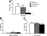
Loss of PTEN is associated with contextual memory deficits after delayed fear memory. (A) Graph shows the mean (±SEM) percent time freezing during the contextual memory test 24 h after training. Graphs show the mean (±SEM) percent freezing during the new context condition (B) and the conditioned stimulus (tone) condition (C). n = 11, 10, and 10 for WT, HT, and KO, respectively. Significant group difference: (*) P < 0.05.
The cued fear test was conducted 2 h later (see Supplemental Material). There was no difference in percent time freezing in the new context condition (F(2,28) = 0.72, P = 0.49) (Fig. 1B) and in the CS condition when the tone was presented in a new context for 3 min (F(2,28) = 0.80, P = 0.46) (Fig. 1C). These results demonstrate that the NS-Pten KO mice have contextual memory deficits compared to WT without deficits in tone conditioning and show similar freezing to a new context.
Next, we examined the hippocampal-dependent memory deficits through the use of trace fear conditioning. In trace fear conditioning the NS and US are separated by a period of time with no stimuli, known as the trace period. For this test we used 20 sec for the trace period. Although delayed fear conditioning is thought to rely on the amygdala, trace fear conditioning is thought to rely on the dorsal hippocampus and to be amygdala-independent (Raybuck and Lattal 2011).
The trace conditioning protocol was performed as previously described (Wiltgen et al. 2005). A different cohort of WT, HT, and KO mice was used in this test. The testing chamber was as described in the delayed conditioning methods (Supplemental Material). On the training day, mice were placed in the test chamber and allowed to explore for 3 min. A 20-sec tone (80 dB, 2700 Hz) was presented and then, after a 20-sec delay, a mild shock was administered (2 sec, 0.5 mA). A 200-sec intertrial interval separated five conditioning trails.
Twenty-four hours later the mice were placed in a new context for the trace conditioning test. The new context was created as described in the delayed fear conditioning methods (Supplemental Material). During the tone test there was a 2-min baseline (BL) period followed by three 20-sec tone presentations. There was a 220-sec ITI between each tone presentation. There was a significant decrease in the percent time freezing for the NS-Pten KO mice compared to that for WT during the presentation of the tone (F(2,60) = 5.67, P < 0.01), during the 20-sec interval after the tone was presented (F(2,60) = 4.06, P < 0.05), and in the intertrial interval between trials (F(2,60) = 3.36, P < 0.05) (Fig. 2A). In the three conditions the Tukey post hoc tests revealed that the NS-Pten KO mice froze less than the WT mice, P < 0.05. There was no difference in the BL condition between the groups (F(2,60) = 0.21, P > 0.8).
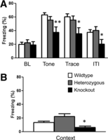
Mutation in PTEN results in trace memory deficits and deficits in contextual memory. (A) Graph shows the mean (±SEM) percent time freezing during the trace memory testing 24 h after training. (B) Graph shows the mean (±SEM) percent time freezing during the context test. n = 31, 20, 12 for WT, HT, and KO, respectively. Significant group difference: (**) P < 0.01, (*) P < 0.05).
Contextual conditioning was conducted 24 h later. In this phase the mice were placed back into the original training context and their freezing behavior was recorded over the 8-min trial. NS-Pten KO mice froze significantly less in a contextual fear conditioning test than the WT and HT mice (K = 6.136, P < 0.05) (Fig. 2B). We used the Kruskal–Wallis test, since the data violated the homogeneity of variance assumption for an ANOVA test.
In order to examine the neural basis of the hippocampal-dependent learning deficits we evaluated protein levels of FMRP, phosphorylated FMRP, total AKT, phosphorylated AKT, total S6, phosphorylated S6, and S6K1 in a separate cohort of NS-Pten mice. Adult mice were decapitated, brains removed, and rapidly placed into ice-cold phosphate-buffered saline. The hippocampi, cortex, and amygdala were removed, frozen on dry ice, and stored at −80°C until use. Total homogenate and crude synaptosomal preparations from samples were prepared using previously described methods (Lugo et al. 2008).
There was a significant increase in FMRP protein levels in the NS-Pten KO mice (F(2,33) = 9.8, P < 0.001) (Fig. 3A), where Tukey post hoc tests revealed a significant increase in KO compared to the WT mice. There was a significant increase in the ratio of phosphorylated FMRP/total FMRP in the KO mice (F(2,33) = 11.1, P = 0.001) (Fig. 3B). Tukey post hoc tests revealed a significant increase in the KO compared to WT and HT mice, P < 0.05. We confirmed that deletion of Pten results in hyperactivation of the PI3K/AKT/mTOR pathway, since there was a significant increase in the ratio of phosphorylated AKT/total AKT (F(2,15) = 19.5, P < 0.001) (Fig. 3D), an increase in total S6 (F(2,15) = 4.9, P < 0.05) (Fig. 3E), the ratio of phosphorylated S6/total S6 in the NS-Pten KO mice compared to the WT and HT mice (F(2,15) = 6.7, P < 0.05) (Fig. 3F), and increase in S6K1 (F(2,15) = 6.3, P < 0.05) (Fig. 3G).
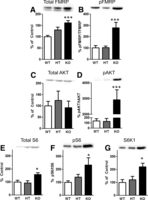
Mutation in PTEN results in elevated levels of fragile X mental retardation protein (FMRP) and phosphorylated FMRP in hippocampus NS-Pten knockout (KO) mice. Western blotting was performed in hippocampal samples from NS-Pten wild-type (WT), heterozygous (HT), and KO mice. Representative immunoblots from hippocampal proteins are shown above each graph. (A) Graph shows the mean (±SEM) for total FMRP in wild-type (WT), heterozygous (HT), and knockout mice (KO). (B) Graph shows the mean (±SEM) for phosphorylated FMRP levels in WT, HT, and KO. (C) Graph shows the mean (±SEM) for total AKT in WT, HT, and KO. (D) Graph shows the mean (±SEM) for phosphorylated AKT in WT, HT, and KO. (E) Graph shows the mean (±SEM) for total S6 in WT, HT, and KO. (F) Graph shows the mean (±SEM) for phosphorylated S6 in WT, HT, and KO. (G) Graph shows the mean (±SEM) for S6K1 in WT, HT, and KO. The primary antibodies used were as follows: FMRP, ribosomal S6 protein, P-S6 (S240/244), AKT, P-AKT (S473), and S6K1 (1:500, Cell Signaling Technology), phosphorylated FMRP-S499 (1:500, Abcam), and actin (1:1000, Sigma Chemical Co.). Total homogenate samples were used for total and phosphorylated AKT, total and phosphorylated S6, and S6K1. Synaptosomal preparations were used for total and phosphorylated FMRP. All primary antibodies were incubated with the membranes overnight at 4°C, followed by incubation with a 1:20,000 goat HRP tagged anti-rabbit IgG secondary antibody (Cell Signaling Technology). Immunoreactive bands were visualized by ECL Prime (GE Healthcare), images were captured on the Proteinsimple FluroChem E & M imager (Santa Clara), and immunoreactivity was quantified using Proteinsimple software (AlphaView SA). The band for each sample was normalized to its actin level. Optical densities of phospho-proteins were normalized to the levels of total protein from the same sample and are depicted as a ratio of phospho-to-total protein level. n = 12 per group for the total and phosphorylated FMRP/total western blots; n = 6 per group for total AKT, phosphorylated AKT, total S6, phosphorylated S6, and S6K1. Significant group difference: (*) P < 0.05, (***) P < 0.001.
The NS-Pten KO mice exhibit hippocampal-dependent learning and memory deficits as demonstrated by impairment in contextual learning in the delay fear conditioning test (Fig. 1A) and by impairment in trace fear conditioning (Fig. 2A,B). However, NS-Pten KO mice show normal learning in the amygdala-dependent tone conditioning test (Fig. 1C). The behavioral results are in line with the molecular results which found no difference in total or phosphorylated FMRP in amygdala (Supplemental Fig. S2) or in cortex tissue (Supplemental Fig. S1). Furthermore, NS-Pten heterozygous mice do not have memory deficits compared to WT mice and have similar levels of total and phosphorylated FMRP. Backman et al. (2001) reported that deletion of Pten in the dentate gyrus of NS-Pten KO mice is ∼35%, so heterozygous deletion of Pten in this conditional knockout would not be expected to impact hippocampus-dependent learning.
FMRP negatively regulates translation of target mRNAs (Laggerbauer et al. 2001; Li et al. 2001) and has a particular influence on dendrites and synapses. Mutations in the FMR1 gene that encodes FMRP are associated with exaggerated LTD in rodent hippocampus (Huber et al. 2002). In addition, phosphorylated FMRP is necessary for FMRP to function as a negative repressor of translation and subsequently to influence synaptic connectivity (Coffee et al. 2012). Therefore, we speculate that elevated FMRP levels in the hippocampus of NS-Pten KO mice may have a functional impact on plasticity since phosphorylated FMRP levels were also significantly increased. It is possible that the elevated phosphorylated FMRP levels are having an additive or possibly a synergistic effect on baseline total FMRP levels. The increased FMRP levels in NS-Pten KO mice in our studies may be responsible for the diminished LTD in Pten knockout mice previously reported (Sperow et al. 2012). Furthermore, a recent study using a similar conditional knockout of Pten as in the current study found impaired mGluR-LTD in the dentate gyrus (Takeuchi et al. 2013). The increase in total and phosphorylated FMRP that we observed in our study is likely contributing to the diminished LTD found in conditional Pten knockout mice.
The type of cre mouse knockout used in the current study has been found to have deletion of Pten in a subset of differentiated neurons in the hippocampus, cortex, and cerebellum (Kwon et al. 2006). The increase in phosphorylated AKT only occurs in neurons with little or no change in glia (Backman et al. 2001; Kwon et al. 2001). Therefore there is little or no impact on behavior due to glia cells. The absence of Pten results in greater PIP3, which then results in greater levels of phosphorylated AKT, and subsequently results in hyperactivation of the mammalian target of rapamycin (mTOR) pathway. The PI3K/AKT/mTOR pathway has been demonstrated to be involved in translation control and in long-lasting synaptic plasticity (Hoeffer and Klann 2010). Furthermore, genetic deletions of regulators of the pathway at the level of TSC1/TSC2 result in learning and memory deficits (Kwon et al. 2006; Ehninger et al. 2008). Hyperactivation of the downstream mTOR pathway appears to be limited to the hippocampus since we did not observe increased levels of total S6, phosphorylated S6, and S6K1 in the cortex (Supplemental Fig. S1) or in the amygdala (Supplemental Fig. S2).
Even though there is significant evidence to suggest that alteration of the pathway results in learning and memory deficits and deficits in LTP and LTD, the mechanism that underlies these changes is not known. One consideration is that deletion of PTEN may produce many effects. Deletion of Pten disrupts neuronal migration, disrupts neuronal proliferation, and results in hypertrophy of the neuronal somata and processes (Backman et al. 2001; Kwon et al. 2001; Kazdoba et al. 2012). Therefore, deletion of Pten may impact plasticity and learning directly or through secondary mechanisms. However, a recent paper used a Pten knockout mouse that expresses Cre under the control of a CamKIIα promoter (Sperow et al. 2012). The use of this promoter bypasses many of the developmental impacts of Pten deletions since the deletion occurs after postnatal day 14. The neurons in these mice do not show hypertrophy of the neuronal somata or other processes, do not have gross abnormalities in the hippocampus, and have normal basal synaptic transmission at the CA3–CA1 synapses. However, the NS-Pten KO mice continue to have deficits in LTP, LTD, and spatial learning. Furthermore, another study found that alterations in synaptic plasticity, such as in alterations in LTP and LTD, occur before morphological defects in a conditional knockout mouse of Pten are detected (Takeuchi et al. 2013).
There have recently been several studies that have examined the changes in translational control during learning. One study used a pharmacological and genetic approach to determine that mTORC1 has a direct role in memory consolidation that is dependent upon protein synthesis (Stoica et al. 2011). Similar approaches could be used to determine the influence of inhibiting the mTORC1 pathway in the Pten conditional knockout mice. Future studies could examine whether additional changes in total and phosphorylated FMRP occur during memory acquisition and retrieval in Pten conditional knockout mice. Such studies will be able to provide insights into the role of the PI3K/AKT/mTOR pathway in regulating FMRP and the role in FMRP in mediating synaptic plasticity.
Acknowledgments
This study was supported from a Baylor University Research Council grant and a research grant from the Epilepsy Foundation.
Footnotes
-
↵3 Corresponding author
E-mail Joaquin_lugo{at}baylor.edu
-
[Supplemental material is available for this article.]
- Received August 14, 2013.
- Accepted September 9, 2013.
This article is distributed exclusively by Cold Spring Harbor Laboratory Press for the first 12 months after the full-issue publication date (see http://learnmem.cshlp.org/site/misc/terms.xhtml). After 12 months, it is available under a Creative Commons License (Attribution-NonCommercial 3.0 Unported), as described at http://creativecommons.org/licenses/by-nc/3.0/.



