


Individual contribution of PDE4B and PDE4D subfamilies to the prevention of object location memory impairments induced by sleep deprivation
- Hongyu Zhao1,
- Arjan Blokland1,
- Jos Prickaerts2,
- Robbert Havekes3,
- Elroy L. Meijer3 and
- Pim R.A. Heckman1
- 1Department of Neuropsychology and Psychopharmacology, Faculty of Psychology and Neuroscience, Maastricht University, Maastricht 6229 ER, The Netherlands
- 2Peitho Translational, Maastricht 6229 ER, The Netherlands
- 3Neurobiology Expert Group, Groningen Institute for Evolutionary Life Sciences (GELIFES), University of Groningen, 9700 AB Groningen, The Netherlands
- Corresponding author: p.heckman{at}maastrichtuniversity.nl
Abstract
Insufficient sleep compromises the cAMP signaling pathway in the hippocampus, negatively impacting hippocampus-dependent memory. In the current study, we explored whether selective PDE4B or selective PDE4D inhibition can improve hippocampus-dependent spatial memory in the object location test (OLT) in mice after sleep deprivation (SD). The results demonstrated that SD impaired the OLT performance, and both A-33 and zatolmilast protected against the negative consequences of SD when administered at the start and middle of the SD period. These findings suggest that both PDE4B and PDE4D subfamilies contribute to the beneficial effect of PDE4 inhibition against SD-induced memory consolidation impairment.
In contemporary societies, inadequate sleep is a prevalent issue. Sleep loss exhibits a close association with our daily routines, exerting a substantial influence on various advanced cognitive functions, particularly declarative memory (Chuah and Chee 2008; Killgore 2010; Lim and Dinges 2010). Earlier work has demonstrated the pivotal involvement of sleep in the maintenance and consolidation of these memories, with sleep deprivation (SD) resulting in a negative impact on hippocampal memory processes (Cousins and Fernández 2019). SD exerts these memory deficits, in part, by disrupting hippocampal cyclic adenosine monophosphate (cAMP) signaling (Vecsey et al. 2009; Havekes et al. 2014, 2016b).
PDE4 is an enzyme that degrades cAMP, thereby regulating its intracellular levels (Paes et al. 2021). Thus, PDE4 upregulation after sleep loss accelerates cAMP breakdown, reducing its availability, and impairing the signaling pathway crucial for synaptic plasticity and memory formation (Vecsey et al. 2009; Alhaider et al. 2011; Luo et al. 2013). Accordingly, studies have shown that treatment with a PDE4 inhibitor can mitigate memory deficits after SD.
The PDE4 family includes four subfamilies: PDE4A, PDE4B, PDE4C, and PDE4D (Paes et al. 2021). PDE4C is not expressed in the brain, and there are no selective PDE4A inhibitors. The effects of PDE4B inhibition have been related to anxiolytic, antidepressant, and procognitive action (McGirr et al. 2016; Zhang et al. 2017b; Shi et al. 2019; Zhao et al. 2024), and the effect of PDE4D inhibition appears to be more closely associated with memory enhancement (Zhang et al. 2017b, 2018; Jino et al. 2024). However, the individual contribution of PDE4B and PDE4D to the memory improving effects in after SD is not well studied (Zhao et al. 2024). Based on the studies conducted with PDE4B and PDE4D inhibitors, we hypothesized that PDE4D inhibition should be more effective in preventing SD-induced spatial memory deficits than PDE4B inhibition.
Twenty-two male C57BL/6J mice, aged 8 weeks (24–27.6 g), were obtained from Charles River. Afterward, a 2-week habituation phase was conducted. During this period, the mice were familiarized with handling, oral gavage administration, and allowed to explore an empty circular arena (diameter 48 cm, height 40 cm) five times. The vivarium maintained a constant room temperature (22 ± 1°C) and humidity (55% ± 10%), coupled with a standardized 12 h light and dark cycle (lights on from 7 a.m. to 7 p.m.). Mice had ad libitum access to standard chow diet and water. All experiments were in accordance with Guidance on the operation of the Animals (Scientific Procedures) Act 1986 and associated guidelines, EU Directive 2010/63.
OLT, a spatial memory paradigm dependent on the hippocampus, is based on the mice's inherent preference for spatial novelty (Ennaceur and Meliani 1992; Ennaceur et al. 1997). The experimental testing of animals, objects, and equipment was similar to an earlier study (Zhao et al. 2024). Briefly, four pairs of objects were used (aluminum cubes, white plastic cubes, metal rectangular prisms, yellow iron pillars). Mice were allowed to explore the objects for 10 min in the first trial (T1). In the second trial (T2), the time the mice interacted with the objects in the original and displaced position was recorded. Test sessions were separated by 48 h to prevent carry-over effects of A33 (half-life 3.8–4.5 h) and zatolmilast (10–12 h). The discrimination index d2 was calculated as follows: d2 = ([time spent to moved object in T2] − [time spent to stationary object in T2])/(total exploration time in T2). The design and OLT testing procedures are shown in Figure 1A and B.
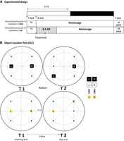
General design. (A) Experimental design. Mice underwent two sleep conditions (SD and NSD), along with three drug administrations (vehicle, zatolmilast, and A-33). All the drugs were administrated immediately after T1 (at the start of SD), and then again 3 h post-T1 (midway during SD). T1 and T2 occurred consecutively at 7 a.m. with SD commencing immediately after T1 at 9 a.m. (B) Object location task (OLT). In T1, one pair out of four objects (see the four pairs on the right) was placed at the midline of the arena. In T2, one object from the same pair was moved to a different, predetermined position based on a location randomization scheme. T1 and T2 had a 24 h interval. (SD) Sleep deprivation, (NSD) non-sleep deprivation, (T1) learning trial, (T2) test trial, (L) left, (R) right.
A 6 h SD was induced in mice directly following T1, using the “gentle stimulation method” (Meerlo et al. 2001; Hagewoud et al. 2010; Vecsey et al. 2013).
The animals were subjected to three treatment conditions: vehicle, PDE4B inhibitor A-33 (3 mg/kg; IC50: 15 nM; Sigma-Aldrich), and PDE4D inhibitor zatolmilast (0.1 mg/kg IC50: 7.8 nM; Med Chem Express via Bio-Connect B.V.) were prepared as described earlier (Zhao et al. 2024). A 48 h washout period was applied between testing days to ensure proper drug elimination as based on PK estimations (see Titus et al. 2016; Gurney et al. 2019).
We conducted a two separate 2 × 2 analyses with the two factors—Sleep (NSD vs. SD) and Drug (vehicle vs. A-33 or vehicle vs. zatolmilast)—using a general linear model (GLM) with repeated measures ANOVA in SPSS (n = 22 per condition). Of note, the pooled vehicle groups (NSD and SD) were used for the analysis of A33 and zatolmilast. Additionally, we analyzed total exploration times between T1 and T2 using paired sample t-tests.
The NSD animals had a better discrimination performance than the SD mice [t(21) = 3.23, P = 0.004, Fig. 2]. In addition, the d2 index for the SD animals in the vehicle condition did not significantly differ from zero [t(21) = −0.86], N.S., indicating that they did not discriminate between the position of the objects. The exploration times of the animals in the two trials did not differ between the NSD and SD groups [T1 NSD-VEH vs. T1 SD-VEH, t(21) = 0.80, N.S.; T2 NSD-VEH vs. T2 SD-VEH; t(21) = 0.61, N.S.].
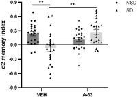
Effects of A-33 and sleep conditions on OLT performance in mice. The pooled data of the vehicle groups are the same as presented in Figure 3. (SD) Sleep deprivation, (NSD) non-sleep deprivation, (OLT) object location task, (VEH) vehicle. Data represent mean (+SEM). (**) P < 0.01
For the A-33 treatment, a significant SD × Treatment interaction was found [F(1, 21) = 10.718, P = 0.004; see Fig. 2]. Direct comparison of the conditions showed that A-33 treatment reversed the SD-induced impairment [t(21) = −3.48, P = 0.002], but there was a weak statistical support that A-33 impaired discrimination performance in the NSD condition [t(21) = 1.81, P = 0.084]. Treatment with A-33 did not affect the exploration times of the NSD and the SD animals in the two trials [T1 SD-VEH vs. T1 SD-A-33; t(21) = −0.39, N.S.; T2 SD-VEH vs. T2 SD-A-33; t(21) = −0.38, N.S.].
A SD × Treatment interaction was also found for the zatolmilast treatment [F(1, 21) = 4.75, P = 0.041; see Fig. 3]. Zatolmilast ameliorated the impairments induced by SD [t(21) = −2.53, P = 0.019], but zatolmilast had no effects in the NSD group [t(21) = −0.25, N.S.] (Fig. 3). The exploration times did not differ between the different conditions [T1 SD-VEH vs. T1 SD-zatolmilast, t(21) = −1.18, N.S.; T2 SD-VEH vs. T2 SD-zatolmilast].
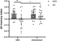
Effects of zatolmilast and sleep conditions on OLT performance in mice. The pooled data of the vehicle groups are the same as presented in Figure 2. (SD) Sleep deprivation, (NSD) non-sleep deprivation, (OLT) object location task, (VEH) vehicle. Data represent mean (+SEM). (*) P < 0.05, (**) P < 0.01.
This study examined the individual contribution of the PDE4 subfamilies PDE4B and PDE4D to the mitigation of the SD-induced memory consolidation deficit in object location memory as observed after nonselective PDE4 inhibition. To this end, we tested whether selective PDE4B inhibition using A-33 and selective PDE4D inhibition using zatolmilast could reverse the SD-induced memory deficit in the OLT in mice. The data showed that both A-33 and zatolmilast prevented the object location memory impairment after SD during the first 6 h of the light phase.
We explored the contribution of PDE4B by examining the potential of the PDE4B inhibitor A-33 to prevent sleep loss–induced deficits in object location memory. The findings of the current study provide support that A-33 has procognitive action and can prevent hippocampus-dependent memory deficits. In a previous study, we investigated the effects of A-33 in an SD animal model, showing that A-33 improved spatial pattern separation performance (Zhao et al. 2024). Of note, the spatial memory and pattern separation task has been found to be differently affected in humans (Blokland et al. 2023), indicating that they rely on different hippocampal structures. However, that study administered sleep restriction and drug treatment directly prior to the retrieval test. A-33 has also exhibited procognitive action in other deficit models. For example, similar to our study, a single dose of A-33 resulted in improved retrieval of conditioned fear memories as well as spatial memories in an animal model of TBI (Titus et al. 2016). Furthermore, in a cuprizone-induced multiple sclerosis mouse model, long-term administration of A-33 (twice daily for 10 days) preserved spatial memory (Schepers et al. 2023). Similarly, in an APPswe/PS1dE9 AD mouse model, long-term administration (20 days to 4 months) of A-33 improved both working and spatial memory performance (Rombaut et al. 2024). These studies support the procognitive potential of PDE4B inhibition, although the latter two studies administering chronic treatment may induce their effects through neuroprotection, whereas the acute studies target neuroplasticity mechanisms (Argyrousi et al. 2020). Taken together, these findings support our results and underscore that PDE4B inhibition protects cognitive performance, highlighting the PDE4B gene as a target for interventions aimed at preserving memory function against the negative effects of sleep loss.
Interestingly, in our study, non-sleep-deprived mice treated with A-33 exhibited a marginal reduction in discrimination performance, yet they are still able to discriminate between the object locations indicating that the mice still remembered the object location. This finding is comparable to another study where A-33 resulted in a nonsignificant lower performance in sham-treated animals, whereas the same dose prevented the memory deficits in TBI animals (Titus et al. 2016). This effect of A-33 in mice may be attributed to an inverted U-shaped dose-response effect that has been reported for PDE inhibitors previously (Prickaerts et al. 2002; Rutten et al. 2005, 2006). Thus, the dose applied for A-33 in this study appears to prevent the negative effects on object location memory after sleep loss, but could result in over- or understimulation of cAMP signaling in non-sleep-deprived animals. Since our study administered an even higher dose (3.0 mg/kg) compared to Titus et al. (2016) (0.3 mg/kg), and their study already observed an overstimulation in the animals in the control condition, these results are in line with previous observations. It should be noted, however, that the current study used only a single dose per compound, which limits the ability to determine dose-dependent effects and complicates the interpretation of subtype-specific contributions. Without a broader dose–response assessment, it is difficult to fully disentangle the relative potency and specificity of PDE4B versus PDE4D inhibition.
Regarding PDE4D, our findings demonstrate that the PDE4D inhibitor zatolmilast effectively prevented spatial memory impairments in OLT caused by SD. This is in line with the general assumption that PDE4D is the PDE4 subtype most relevant for cognition enhancement. We previously administered zatolmilast in an animal model of sleep loss showing protection against SD-induced deficits in spatial pattern separation (Zhao et al. 2024). In addition, memory improvements were observed for object recognition in the ORT after acute treatment in non-sleep-deprived animals (Zhang et al. 2017a), and other studies in which zatolmilast improved memory performance. While our study focused on acute intervention, zatolmilast has been more frequently studied using chronic dosing regimens. For example, daily administration of zatolmilast for 14 days improved learning and memory in the Morris water maze and Y-maze tests in an AD model induced by Aβ1–42 (Cui et al. 2019; Xu et al. 2019), indicating that the drug's efficacy is sustained over longer periods. Similarly, a 5 day daily treatment of zatolmilast reversed scopolamine-induced memory and cognitive deficits in humanized PDE4D mice (Wang et al. 2020). These findings align with our hypothesis and study outcome, highlighting the potential of PDE4D inhibition to protect against memory deficits under conditions of sleep loss.
While the expression patterns of PDE4 subfamilies show considerable similarity across species, there are notable differences between the expressions of the subfamilies in individual brain regions. PDE4A is expressed in several brain regions, with high levels in the cerebral cortex, hippocampus, and cerebellum (McPhee et al. 1995; Cherry and Davis 1999; Pérez-Torres et al. 2000; Johansson et al. 2012), suggesting a role in cognition and memory (Richter et al. 2013; Havekes et al. 2016a). PDE4B is widely distributed throughout the brain, but particularly expressed in the amygdala, thalamus, striatum, and hypothalamus (Richter et al. 2013; Tibbo and Baillie 2020). Its expression in the hippocampus is likely related to its procognitive action in the current study, given the OLT's dependence on the hippocampus and its sensitivity the sleep loss (Cherry and Davis 1999; Pérez-Torres et al. 2000; Shepherd et al. 2003; Heckman et al. 2020). In addition, PDE4B's significant presence in the amygdala, hypothalamus, and striatum suggests it also regulates anxiety and stress responses, critical for emotion regulation (Charney and Deutch 1996). PDE4D is like PDE4A highly expressed in the hippocampus, cortex, and cerebellum suggesting a role in cognition as well (Pérez-Torres et al. 2000; Gurney et al. 2015; Lusardi et al. 2024).
The distinct expression patterns of PDE4 subfamilies across brain regions are interesting as this may suggests that PDE4B and PDE4D inhibition could affect different brain networks but both are able to prevent SD-induced memory deficits. Alternatively, they could affect the same brain network or cells but have a distinct distribution within (different compartments) of cells, as we showed for PDE4D (Paes et al. 2023). An analogous situation could be hypothesized for PDE4B isoforms, i.e., in the same hippocampal cell, all PDE4A, PDE4B, and PDE4D isoforms work in concert for optimal signal transduction. Whether PDE4B and PDE4D inhibition indeed lead to a differential effect on brain networks would need to be investigated in further studies. If it is assumed that PDE4B and PDE4D affect different brain networks, this would suggest that a nonselective PDE4 inhibitors that target multiple subtypes, including PDE4A, PDE4B, and PDE4D, may offer a more comprehensive approach to rescue SD-induced memory deficits. A previous study, using the nonselective PDE4 inhibitor roflumilast, has indeed shown that roflumilast effectively restores memory after SD (Vecsey et al. 2009; Vanmierlo et al. 2016; Bolsius et al. 2023).
Finally, while previous studies using subtype- or nonselective PDE4 inhibitors have shown protection against a range of deficit models including sleep loss, it is important to emphasize that this can be established by targeting different memory processes (Heckman et al. 2020). Administering these inhibitors after learning trials helps preserve memory consolidation by stabilizing new information, while administering them before test trials enhances memory retrieval by maintaining synaptic integrity. While PDE4 inhibition during consolidation or prior to retrieval results in increased levels of cAMP, it is hypothesized that they result in the activation of different effectors, i.e., PKA or exchange protein directly activated by cAMP (EPAC), respectively. During consolidation, the cAMP/PKA pathway activates molecules like CREB and BDNF to strengthen memory (Amidfar et al. 2020; Ben Zablah et al. 2021), whereas during retrieval, the cAMP/EPAC pathway activates Rap proteins to maintain synaptic connections for recall (Ostroveanu et al. 2010). Our study focused on memory consolidation, administering both SD and the subtype-selective PDE4 inhibitors immediately after training to safeguard this process. In contrast, our previous work examining spatial pattern separation in mice, induced SD and administered PDE4 inhibitors immediately prior to testing, which protected against sleep loss–induced deficits in discrimination between similar spatial patterns (Heckman et al. 2020; Zhao et al. 2024). In another study, mice received sleep restriction during memory consolidation, while roflumilast was administered before retrieval, resulting in the successful restoration of SD-induced memory impairments in OLT (Bolsius et al. 2023). These findings highlight that there are multiple effective strategies for restoring memory after sleep loss, depending on which memory process is targeted.
The results from this study show that both PDE4B and PDE4D inhibition can prevent SD-induced memory impairments. This is not in line with our initial hypothesis assumed that targeting PDE4D would be more effective. Apparently, both subtypes are relevant, but the effects may be mediated via different brain and cellular networks. The broader effect of nonselective inhibition suggests that a more comprehensive modulation of cAMP signaling pathways, involving multiple PDE4 subtypes in different brain structures, is beneficial for mitigating the adverse effects of SD on memory. This insight highlights the potential of nonselective PDE4 inhibitors as therapeutic agents for cognitive impairments related to SD and other conditions affecting memory.
Data access
The data that support the findings of this study are available from the corresponding author upon reasonable request.
Acknowledgments
We thank Igor Magaraggia, Denise Hermes, Ellis Nelissen, and Richard Frijnts for their advice and guidance during the experimental operation and design phases. This work was supported by the Dobberke foundation of the Royal Netherlands Academy of Arts and Sciences (KNAW) (grant number Dobberke/2349/202105) and the Chinese Scholarship Council (CSC) (grant number 202108440210).
Footnotes
-
Article is online at http://www.learnmem.org/cgi/doi/10.1101/lm.054101.125.
- Received February 18, 2025.
- Accepted May 15, 2025.
This article is distributed exclusively by Cold Spring Harbor Laboratory Press for the first 12 months after the full-issue publication date (see http://learnmem.cshlp.org/site/misc/terms.xhtml). After 12 months, it is available under a Creative Commons License (Attribution-NonCommercial 4.0 International), as described at http://creativecommons.org/licenses/by-nc/4.0/.



