


Medial septal theta stimulation enhances spatial working memory performance in rats
- Corresponding author: amygriff{at}udel.edu
Abstract
Spatial working memory (SWM) relies on the integrity of the medial septum area (MSA) and its ability to drive theta (4–12 Hz) oscillations in the hippocampus. This study tested the hypothesis that optogenetic theta stimulation of the MSA would enhance choice accuracy on a hippocampus-dependent task in rats. We delivered either excitatory or control theta stimulation during the delay period (10 or 30 sec) of a delayed alternation (DA) task. We show that MSA theta stimulation improved choice accuracy on the 30 sec delay trials, providing strong support for the notion that MSA theta stimulation boosts SWM.
Spatial working memory (SWM) refers to the ability to temporarily store and manipulate spatial information to guide behavior. The hippocampus plays a crucial role in SWM, integrating spatial cues and maintaining location-based information over short delays (Olton 1976, 1979; Packard and McGaugh 1996; Lee and Kesner 2003). Rodent models of SWM offer ways of directly manipulating specific neural circuits with excellent temporal precision. One neural circuit that has consistently been shown to support SWM in rodents is the projection from the medial septal area (MSA) to the hippocampus (Freund and Antal 1988; Toth et al. 1993). The MSA contains three main populations of neurons, all of which send long-range projections to the hippocampus: GABAergic, parvalbumin (PV)-positive neurons, cholinergic, and glutamatergic neurons (Frotscher and Leranth 1985; Toth et al. 1993, 1997; Manseau et al. 2005; Simon et al. 2006). MSA PV-positive neurons project onto PV-positive hippocampal interneurons, providing rhythmic disinhibition of hippocampal pyramidal cells, which is thought to be responsible for pacing the 4–12 Hz hippocampal theta rhythm (Toth et al. 1997), a neural oscillation that is implicated in learning and memory processes (Lisman and Idiart 1995). This idea is supported by recent studies that have controlled theta rhythm frequency by delivering theta stimulation restricted to PV-positive MSA neurons (Varga et al. 2008; Zutshi et al. 2018). SWM deficits likely arise after MSA disruption because the MSA is critical for the generation of the hippocampal theta rhythm (Green and Arduini 1954; Kesner et al. 1989; Stewart and Fox 1990). There is a long history of work demonstrating that MSA lesions or inactivation result in profound SWM impairments (Sutherland and Rodriguez 1989; Mizumori et al. 1990; Poucet and Buhot 1994; Whishaw and Jarrard 1995). In a recent paper from our laboratory, we found that optogenetic silencing of the MSA during the delay period of a delayed alternation (DA) task significantly impaired choice accuracy. Moreover, we found that MSA silencing impaired choice accuracy on a SWM-dependent conditional discrimination task but did not affect the performance of an SWM-independent variant of the task (Gemzik et al. 2021).
Can the ability to drive hippocampal theta be leveraged to improve cognitive function? Recent studies have demonstrated that artificial stimulation of the MSA in human patients and animal models of neuropsychiatric disorders restored MSA-driven hippocampal oscillations and improved cognition (Suthana et al. 2012; Lee et al. 2013; Izadi et al. 2019, 2021; Zepeda et al. 2020; Ríos et al. 2022). These findings suggest that MSA theta stimulation is a potential means of enhancing SWM in nonclinical populations in addition to rescuing working memory impairments in clinical populations. Building upon evidence that the MSA is essential SWM, that specific types of pharmacological modulation of the MSA can either enhance or impair memory (Chrobak et al. 1989; Chrobak and Napier 1992; Givens et al. 1992; Wan et al. 1995; Herzog et al. 1996; Walsh et al. 1996; Sabolek et al. 2004), and that hippocampal theta rhythm restoration can recover learning (McNaughton et al. 2006), our study investigates the less-explored potential for optogenetic stimulation of the MSA to enhance SWM. We hypothesized that MSA theta stimulation would enhance SWM in rats. To test this hypothesis, we optogenetically delivered theta or control stimulation to the MSA during the delay period of a SWM-dependent DA task and varied the working memory demand by interleaving 10 and 30 sec delay trials within a testing session. Using a pan-neuronal promoter, we expressed an excitatory opsin in the entire MSA neuronal population, including theta-regulating GABAergic and cholinergic neurons (Smythe et al. 1992; Yoder and Pang 2005). Our specific prediction was that MSA theta stimulation delivered during the delay period would improve choice accuracy on the 30 sec working memory-intensive trials.
Subjects were 10 adult (six male and four female) Long–Evans hooded rats. Rats were housed in a temperature (∼21°C) and humidity-controlled room under a 12 h light–dark cycle. During the start of the experiment, rats were single housed to ensure implants were not damaged by cage mates. Food access was slightly restricted, maintaining rats within 90% of their ad libitum body weight. Rats were allowed to have full access to water throughout the experiment. All procedures in this experiment were conducted in accordance with the University of Delaware Institutional Animal Care and Use Committee.
Rats underwent a pretraining procedure, followed by surgery before starting to train on the DA task. Pretraining consisted of a 2 week process which included handling the rats to habituate them to the experimenter, introducing rats to the T-maze reward zones, and shaping running behavior on the maze as described previously (Hallock et al. 2013; Maisson et al. 2018; Gemzik et al. 2021). Surgery was performed in the same manner as described previously (Gemzik et al. 2021). Briefly, 1 µL of a viral suspension containing the neural activator channelrhodopsin (ChR2) that is activated by blue (but not red) light (AAV5-hSyn-hChR2(H134R)-EYFP) (3.1 × 10¹² virus molecules/mL) was infused into the MSA at DV coordinates −6.5, −6.3, −6.0, and −5.8 mm (Paxinos and Watson 2006) measured from the surface of the brain via four successive injections (0.30, 0.25, 0.25, and 0.20 µL), respectively. Next, an optic fiber implant (Thorlabs; 2.5 mm ceramic ferrule; 230 µm fiber diameter; nA 0.33) was placed directly dorsal to the MSA (AP: 0.7 mm, ML: 0.0 mm, DV: −5.5 mm). To confirm that we were driving hippocampal theta via MSA optogenetic theta stimulation as intended, two additional rats received recording electrode arrays composed of two (MSA) or four (dorsal hippocampus) staggered stainless steel wires (0.2032 mm coated; A-M Systems) placed into the MSA and dorsal hippocampus to measure local field potential (LFP) activity from both regions during MSA stimulation (Figs. 1, 2). After a 5 day recovery period, rats were trained on the DA task (see Fig. 1A). Training sessions consisted of 40 trials, all with a 10 sec delay between trials. Rats were trained until they reached 80% choice accuracy for two consecutive sessions. Once the training criterion was reached, rats underwent a short retraining period to familiarize them with running the task with the patch cable attached. After two consecutive days of maintaining a choice accuracy of 80% while tethered, testing began (see Fig. 1B). The Neuroscience Studio (Doric Lenses Inc.) application was used to operate the laser drivers and pulsing parameters during the testing sessions. The lasers were set to an output of ∼7 mW at the fiber tip, measured by a power meter (Thor Labs). After the power output for both the control (638 nm, red) and excitatory (460 nm, blue) lasers was set for the testing session, the stimulation frequency was set to 6 Hz using a square wave pulse pattern (50–50 duty cycle), allowing us to drive MSA theta (Fig. 2). Our choice to use a square waveform was based on the robust hippocampal theta entrainment demonstrated previously using this stimulation pattern (Blumberg et al. 2016; Mouchati et al. 2020; Quirk et al. 2021). We chose to use a stimulation frequency of 6 Hz for a couple of reasons. First, a recent study demonstrated enhanced spatial goal finding with 6 Hz MSA stimulation (Mouchati et al. 2020). Second, the delay period is associated with restricted movement and movement planning, both of which have been shown to be accompanied by low-frequency theta (Vanderwolf 1969; Whishaw and Vanderwolf 1973; Mouchati et al. 2020). The experiment had four conditions: two control stimulation (red laser) conditions, one for 10 sec delay (as in training) and one for 30 sec delay trials, and two excitatory stimulation (blue laser) conditions, one for 10 sec delay trials and one for 30 sec delay trials. A custom MATLAB script was used to pseudo-randomly generate and interleave the four types of trials and initiate laser stimulation as the rats entered the delay zone. Rats completed 40 daily trials (10 per trial type) across 3 days, with trial types counterbalanced within each session. Choice accuracy was averaged over the three testing sessions for each rat. At the end of testing, rats were perfused and brains sectioned for histological verification of fiber placements and viral expression.
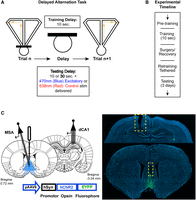
DA task schematic and experimental time line. (A) Schematic of the DA task. Rats alternated visits to the left and right goal arms of a T-maze to receive a food reward at the end of each maze arm. Between trials, the rats were confined to a box at the base of the maze stem for a delay period. The schematic also shows the four trial types that were interleaved within each testing session: 10 versus 30 sec delay and red (control) versus blue (excitatory) laser. On training sessions (before rats reached criterion), rats were given 10 sec delay trials only with no laser stimulation. (B) Brief overview of experimental time line. (C, left) Schematic depicting viral injection into MSA (light blue) along with an optic fiber (emitting blue light over MSA) and electrode array implant. (Bottom) Viral vector composition containing excitatory opsin used for stimulation. (Right) Example histology sections of DAPi-stained coronal slices of MSA and hippocampus. The yellow box in the top image indicates the location of the hippocampal recording array, and the yellow box in the bottom image indicates the location of the optic fiber implant over the MSA. Yellow–green fluorescence represents a localized restriction of viral spread throughout MSA and MSA projecting cells terminating in the dorsal hippocampus.
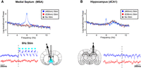
Optogenetically induced theta oscillations in the MSA and dorsal CA1. Top panels show the log-transformed power of MSA (A) and dorsal CA1 (B) LFP during bouts of 460 nm excitatory 6 Hz MSA theta stimulation (blue), 638 nm control stimulation (red), and no stimulation (gray). Stimulation was applied in two rats, each undergoing a single 31 min session with alternating 30 sec on/off cycles. Solid lines show the mean, with shading representing the standard error of the mean (SEM). Notice the large peak at the pulsed frequency of 6 Hz in both brain regions. Bottom panels show example traces of LFP activity from the same rat in response to excitatory versus control 6 Hz MSA theta stimulation for each brain region; cyan bars denote the 6 Hz pulsing paradigm with a 50–50 duty cycle.
To validate that the MSA stimulation was effective at driving hippocampal activity, we compared power spectra between bouts of excitatory and control stimulation in two rats. As expected, excitatory (but not control or no stimulation) was accompanied by a robust increase in 6 Hz power in both the MSA and dorsal CA1 (Fig. 2). It is unlikely that this 6 Hz oscillation observed in MSA in response to the optogenetic stimulation was volume conducted from the hippocampus. If the observed oscillations in MSA were solely volume conducted from the hippocampus, we would expect the amplitude to be larger in the hippocampus than in the MSA. However, we consistently observed the opposite: the amplitude of the theta rhythm was always larger in the MSA than in the dorsal hippocampus in response to the optogenetic stimulation. This suggests a local generation of 6 Hz oscillations within the MSA in response to optogenetic stimulation.
To examine the effects of 6 Hz MSA stimulation on DA choice accuracy, we performed a within-subjects repeated measures ANOVA, which revealed a significant laser (excitatory vs. control) × delay duration (10 vs. 30 sec) interaction (F(1,9) = 11.484, P = 0.008) (Fig. 3). There were no significant differences in choice accuracy between control and excitatory 10 sec stimulation trials (F(1,9) = 0.755, P = 0.921). As expected, due to the higher working memory demand for 30 versus 10 sec delay trials, choice accuracy was significantly lower on the 30 sec delay trials with control stimulation compared to both the control 10 sec delay (F(1,9) = 4.093, P = 0.004) and excitatory 10 sec delay conditions (F(1,9) = 2.862, P = 0.045). Strikingly, choice accuracy for 30 sec excitation stimulation trials was significantly better than choice accuracy on 30 sec control stimulation trials (F(1,9) = −3.621, P = 0.010) (d = 1.461) and not significantly different from control and excitatory stimulation on 10 sec delay trials (excitatory 30 sec vs. control 10 sec: [F(1,9) = −0.230, P = 0.921]; excitatory 10 sec vs. excitatory 30 sec: [F(1,9) = −1.162, P = 0.783]). Thus, the decrease in choice accuracy that is normally seen with an increase in working memory load was ameliorated by the 6 Hz MSA stimulation.
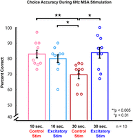
MSA theta stimulation facilitates choice accuracy on a spatial working memory task. Each data point represents the choice accuracy of each rat on the corresponding delay stimulation condition trial averaged over all 3 days of testing (rmANOVA: laser color × delay length, F(1,9) = 11.484, P = 0.008; post hoc Holm test, red/control 10 vs. 30 sec, P = 0.004; 10 sec red/control vs. blue/excitatory, P = 0.921; 30 sec red/control vs. blue/excitatory, P = 0.01).
Our findings are in line with others that have shown the efficacy of optogenetic MSA theta stimulation in driving hippocampal LFP activity (Oddie et al. 1994; Scarlett et al. 2004; Blumberg et al. 2016; Zutshi et al. 2018; Dannenberg et al. 2019; Mouchati et al. 2020; Quirk et al. 2021). Manipulation of the MSA in the theta range reliably drives hippocampal theta at the induced frequency for the duration of MSA stimulation (Fig. 2). These findings strongly suggest that the theta stimulation was effectively delivered during each stimulation condition, allowing us to be confident that we were reliably driving hippocampal theta activity in rats that were implanted with an optic fiber only (Fig. 3).
The stimulation frequency, while low for active movement, corresponds to theta frequencies observed during movement planning and anticipatory behavior (<7 Hz) and with low-speed locomotion (<2 cm/sec) in contrast to theta frequencies above 7 Hz observed during high-speed locomotion (>5 cm/sec) (Whishaw and Vanderwolf 1973; Oddie et al. 1997; Bender et al. 2015; Kennedy et al. 2022). It is also possible that the consistency of theta pacing, rather than the specific frequency used, is most important for SWM. Future studies could employ systematic manipulation of stimulation frequency, including those more typically associated with high-speed locomotion to provide a more comprehensive understanding of the role of theta stimulation of MSA in SWM.
We chose pan-neuronal stimulation to ensure greater stability in maintaining the stimulated frequency in the hippocampus as rats transition between behavioral states, compared to stimulating cholinergic or glutamatergic MSA projections (Mamad et al. 2015; Robinson et al. 2016). Pan-neuronal stimulation activates all or most neurons within a region, including excitatory and inhibitory neurons, as well as different subtypes of each. This makes it difficult to isolate the specific contributions of individual cell types to the observed effects. Hippocampal theta oscillations have been shown to be enhanced by selective activation of cholinergic (Vandecasteele et al. 2014), glutamatergic (Manseau et al. 2005; Huh et al. 2010; Robinson et al. 2016), and GABAergic (Zutshi et al. 2018; Quirk et al. 2021) neurons. However, a study using ChAT::Cre rats found that nonselective MSA theta burst stimulation, but not the selective stimulation of cholinergic MSA neurons, entrained hippocampal neurons during active movement (Mamad et al. 2015). Moreover, MSA theta stimulation is reported to be similarly effective in entraining hippocampal theta under either pan-neuronal or GABAergic PV+ MSA cell activation (Blumberg et al. 2016; Mouchati et al. 2020; Quirk et al. 2021). Interestingly, in vitro work demonstrates that driving specific cell subpopulations results in the absence of hippocampal cell activity patterns that occur endogenously. For instance, when comparing pan-neuronal theta stimulation to either GABAergic PV+ or cholinergic MSA stimulation, only pan-neuronal stimulation resulted in complex spike bursting activity in hippocampal CA1, an activity pattern linked to LTP (Kandel and Spencer 1961; Fox and Ranck 1975; Gao et al. 2021). Taken together, we believe our implementation of MSA 6 Hz pan-neuronal theta stimulation to be an effective means to investigate the effects of MSA theta stimulation on SWM performance. Future studies could explore the respective contributions of cholinergic, glutamatergic, and GABAergic MSA neurons in enhancing SWM in this paradigm.
Hippocampal theta synchronization with MSA theta is behaviorally dependent, modulated by both speed and cognitive demand (Mamad et al. 2015; Mouchati et al. 2020). During SWM tasks, where rats incorporate past trial information into upcoming choices (Zilli and Hasselmo 2008), distinct hippocampal neuronal populations exhibit sequential activity signaling future trajectories (Wood et al. 2000; Pastalkova et al. 2008). As rats undergo a delay period and their SWM is taxed, task-relevant information about the previous trial needs to be incorporated into the planning for the upcoming traversal. This process, where information from past trials influences current decisions, is consistent with a computational model showing that working memory and episodic memory can be implemented within a reinforcement learning framework to simulate performance on tasks that rely on remembering prior events, such as spatial alternation (Zilli and Hasselmo 2008). We hypothesize that 6 Hz MSA stimulation facilitated these sequential patterns, effectively promoting task-relevant hippocampal activity during the 30 sec delay compared to control trials. This enhancement might be attributed to theta rhythmic pacing, which separates encoding and retrieval dynamics (Hasselmo et al. 2002), enabling efficient episodic retrieval and supporting trial-to-trial integration (Siegle and Wilson 2014).
While artificial hippocampal theta could override endogenous signals, potentially altering neuronal firing, studies show that during MSA GABAergic theta stimulation, hippocampal cells maintain diverse frequencies (Zutshi et al. 2018). Consistent with this, we observed no observable decrease in other theta frequencies during MSA stimulation (Fig. 2). Furthermore, hippocampal spatial coding remains intact during MSA manipulations (Brandon et al. 2014; Zutshi et al. 2018; Mouchati et al. 2020), whereas temporal coding in hippocampal “time cells” is impaired (Wang et al. 2015). Time cells, crucial for SWM maintenance, form sequential patterns during delays (Manns et al. 2007; Pastalkova et al. 2008; Gill et al. 2011; Mankin et al. 2012; MacDonald et al. 2013; Rangel et al. 2014), though their necessity in some tasks is debated (Yong et al. 2022). How MSA theta stimulation affects time cell sequences, especially with varying SWM demands, remains unclear. However, it is possible that MSA-generated theta provides temporal scaffolding, stabilizing task-relevant hippocampal sequential activity during extended delays.
In contrast to our findings, some recent studies report null or impaired SWM with MSA theta stimulation (Quirk et al. 2021; Zhang et al. 2021). This could be due to several factors, such as variations in animal training procedures, the specific neuronal populations targeted, and the stimulation frequency used. Our large effect size may stem from our novel introduction of interleaved 30 sec delay trials on the first test day, which significantly heightened working memory demands. Moreover, our interleaved design required continuous SWM updating, unlike a blocked trial design in which all trials of a single condition are presented consecutively (Quirk et al. 2021; Zhang et al. 2021). Our use of pan-neuronal activation could also explain the discrepancy between our results and those of some of the prior studies.
Future studies could explore interactions with other SWM-related regions, like the medial prefrontal cortex, given its synchronization with the hippocampus during high SWM demand (Myroshnychenko et al. 2017; Stout et al. 2024). Future work could also investigate the effects of delivering MSA stimulation during different phases of the task other than just the delay period using similar methods to those used in a previous thalamic silencing study from our laboratory (Maisson et al. 2018).
In summary, 6 Hz MSA stimulation during the delay period significantly improved choice accuracy in an SWM task, supporting our hypothesis. This memory enhancement provides a foundation for developing cognition-enhancing neural circuit manipulations.
Data access
Data will be made available upon request to the corresponding author.
Acknowledgments
This research was supported by the National Institute of Mental Health of the National Institutes of Health under award number R21 MH117687 to A.L.G.
Footnotes
-
Article is online at http://www.learnmem.org/cgi/doi/10.1101/lm.054075.124.
- Received October 18, 2024.
- Accepted March 6, 2025.
This article is distributed exclusively by Cold Spring Harbor Laboratory Press for the first 12 months after the full-issue publication date (see http://learnmem.cshlp.org/site/misc/terms.xhtml). After 12 months, it is available under a Creative Commons License (Attribution-NonCommercial 4.0 International), as described at http://creativecommons.org/licenses/by-nc/4.0/.



