


Domain-selective and sex-dependent regulation of learning and memory in mice by GIRK channel activity in CA1 pyramidal neurons of the dorsal hippocampus
- Haichang Luo1,
- McKinzie Frederick1,
- Ezequiel Marron Fernandez de Velasco,
- Jenna Osterlund Oltmanns,
- Courtney Wright and
- Kevin Wickman
- Corresponding author: wickm002{at}umn.edu
-
↵1 These authors contributed equally to this work.
Abstract
G protein-gated inwardly rectifying K+ (GIRK) channels mediate the postsynaptic inhibitory effect of many neurotransmitters in the hippocampus and are implicated in neurological disorders characterized by cognitive deficits. Here, we show that enhancement or suppression of GIRK channel activity in dorsal CA1 pyramidal neurons disrupted novel object recognition in mice, without impacting open field activity or avoidance behavior. Contextual fear learning was also unaffected, but extinction of contextual fear was disrupted by suppression of GIRK channel activity in male mice. Thus, the strength of GIRK channel activity in dorsal CA1 pyramidal neurons regulates select cognitive task performance in mice.
A growing body of evidence has implicated G protein-gated inwardly rectifying K+ (GIRK) channels in learning and memory (Luo et al. 2022). GIRK channels are highly expressed in brain regions that support cognitive function, including the hippocampus (HPC) (Luján and Aguado 2015), where they mediate the G protein-dependent postsynaptic inhibitory effect of neurotransmitters such as GABA, serotonin, and adenosine (Lüscher et al. 1997). GIRK channels are found in the somatodendritic compartment of glutamatergic pyramidal neurons (Koyrakh et al. 2005) and GABAergic interneurons (Booker et al. 2013) in the HPC. In CA1 pyramidal neurons, GIRK channels colocalize with the GABAB receptor (GABABR) in dendritic spines (Kulik et al. 2006) and serve as a break on glutamatergic neurotransmission (Otmakhova and Lisman 2004).
Neuronal GIRK channels are tetramers formed by variable assembly among three homologous subunits–GIRK1, GIRK2, and GIRK3 (Luo et al. 2022). While the loss of GIRK1 or GIRK2 eliminates GIRK channel activity in HPC pyramidal neurons, GIRK3 ablation has no evident impact (Lüscher et al. 1997; Koyrakh et al. 2005). Ectopic expression of GIRK3, however, suppresses GIRK channel activity in expression systems (Kofuji et al. 1995) and neurons (McCall et al. 2019). The dominant-negative influence of GIRK3 may reflect its contribution to GIRK channel trafficking. GIRK3 harbors a lysosomal targeting sequence absent in other subunits (Ma et al. 2002), and a PDZ interaction that promotes an interaction with sorting nexin 27, which can shepherd GIRK channels from the cell surface to early endosomes (Lunn et al. 2007).
Gain-of-function and loss-of-function mutations involving GIRK channels have been linked to cognitive impairment (Kleschevnikov 2022; Luo et al. 2022). For example, KCNJ6/GIRK2 is located in the critical segment of human Chromosome 21 that is trisomic in Down syndrome (DS), a congenital disorder characterized by intellectual disability (Kleschevnikov 2022). Ts65Dn mice, a well-studied model of DS (Reeves et al. 1995), exhibit deficits in HPC-dependent cognitive function and elevated GIRK channel activity in CA1 pyramidal neurons (Best et al. 2012), and genetic suppression of GIRK2 rescues cognitive deficits in male Ts65Dn mice (Kleschevnikov et al. 2017).
Decreased GIRK channel activity is also associated with cognitive impairment. For example, exomes from three unrelated human patients with Keppen-Lubinsky syndrome, a rare disease characterized by intellectual disability (Basel-Vanagaite et al. 2009), exhibit de novo mutations in KCNJ6/GIRK2 that impair GIRK channel function (Masotti et al. 2015). Downregulation of KCNJ6/GIRK2 is evident in human brains with Alzheimer's disease (AD) (Zhao et al. 2021), and KCNJ6/GIRK2 is one of three genes in the only AD-associated gene module common to White, Hispanic, and African American populations (Dai et al. 2022). Surface levels of GIRK channels are decreased in the CA1 of 12-month-old male APPswe/PS1dE9 mice (Martín-Belmonte et al. 2022), a well-studied AD model (Jankowsky et al. 2004). Furthermore, the GIRK channel activator ML297 can rescue HPC-dependent cognitive performance in male rodent models of AD (Sánchez-Rodríguez et al. 2017, 2020).
Collectively, evidence suggests that enhanced or diminished GIRK channel activity contributes to cognitive dysfunction. Studies to date, however, have lacked anatomic and cellular precision and consideration of sex as a biological variable. Here, we used Trpc4-Cre(+) mice (Okuyama et al. 2016) and Cre-dependent AAV vectors to overexpress GIRK2 or GIRK3 in CA1 pyramidal neurons in male and female adult mice, with the goal of assessing the impact of bidirectional manipulation of GIRK channel activity on HPC-dependent cognitive function (Fig. 1).
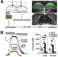
Bidirectional manipulation of GIRK channel activity in dorsal CA1 pyramidal neurons. (A) Depiction of the experimental timeline. Trpc4-Cre(+) mice received intra-dCA1 infusions of high titer (1 × 10¹² genocopies/mL) AAV8-hSyn-DIO-GFP (control), AAV8-hSyn-DIO-GIRK2-IRES-GFP, or AAV8-hSyn-DIO-GIRK3-IRES-GFP. The scope and accuracy of viral targeting were evaluated for all animals after behavioral testing (validation). Images on the right show bilateral viral-driven GFP expression in the dorsal (–1.90 mm A/P) but not ventral (−3.10 mm A/P) CA1 of a male Trpc4-Cre+ mouse injected with the GFP control vector; scale, 500 µm. Only mice showing bilateral GFP expression in the dCA1 were included in the final analysis. In total, 26 male (11 GFP/control, 8 GIRK2, and 7 GIRK3) and 19 female (7 GFP/control, 6 GIRK2, and 6 GIRK3) mice were included in the final data sets. (B) Whole-cell currents (Vhold = −60 mV) evoked by the GABABR agonist baclofen (200 µM), and their reversal by the GABABR-selective antagonist CGP55845 (2 µM), in GFP-expressing pyramidal neurons from Trpc4-Cre+ mice injected with GFP control, GIRK2, or GIRK3 vectors; scale, 100 pA/50 sec. The bar graph on the right summarizes baclofen-evoked currents in dorsal CA1 pyramidal neurons from male GFP/control (n = 7), GIRK2 (n = 4), GIRK3 (n = 4), and female GFP/control (n = 3), GIRK2 (n = 4), and GIRK3 (n = 5) viral treatment groups. Data are presented as mean ± SEM. Symbols (Dunnett's multiple comparisons): (*) P < 0.05, (**) P < 0.01, (****) P < 0.0001 (within sex).
All experiments were approved by the University of Minnesota Institutional Animal Care and Use Committee. Trpc4-Cre(+) mice (70–80 days) received bilateral infusions of Cre-dependent AAV vectors harboring GFP (control), GIRK2, or GIRK3 in the dorsal CA1 (dCA1) subregion of the HPC (from bregma: –1.90 mm A/P, ± 1.45 mm M/L, –1.45 mm D/V) (Fig. 1A). Following a 2–3 week recovery period, mice were used to validate viral efficacy via slice electrophysiology or processed in a behavioral test battery. In cognitive disorders like DS and AD, declarative or episodic-like memory is impaired (Pause et al. 2013). dCA1 pyramidal neurons are essential to this type of memory (Fanselow and Dong 2010) and are susceptible to dysregulation in AD (Llorens-Martin et al. 2014). Our test battery began with a 2-day habituation period and an open field (OF) test to assess locomotor activity and avoidance behavior (Seibenhener and Wooten 2015), followed by two tasks that are dCA1-dependent and evaluate declarative memory: object recognition (OR) (Ásgeirsdottir et al. 2020) and contextual fear conditioning (CFC) (Ji and Maren 2008; Kim and Cho 2020; Ratigan et al. 2023). All apparati and objects, as well as shock chambers, were cleaned with 70% ethanol and wiped dry between sessions. Data were analyzed in GraphPad Prism (v10.2.3) using two-way ANOVA with sex and viral treatment as factors or using two-way ANOVA with repeated measures, as appropriate. As sex differences were observed in some measures, we present data from male and female subjects separately throughout for consistency.
To evaluate the functional impact of the viral manipulations, we measured whole-cell currents evoked by baclofen in GFP-positive dCA1 pyramidal neurons (Fig. 1B), as previously described (Vo et al. 2021). GIRK channels mediate most (∼80%) of the baclofen-induced current in CA1 pyramidal neurons (Koyrakh et al. 2005). As expected, GIRK2 and GIRK3 overexpression enhanced and suppressed baclofen-induced currents, respectively, in male and female mice (Fig. 1B) (sex: F(1,21) = 1.526, P = 0.2303; treatment: F(2,21) = 66.48, P < 0.0001; interaction: F(2,21) = 1.416, P = 0.2650).
In the OF test (Fig. 2A), there was no effect of sex or viral treatment, or interaction between factors, on time spent in the field center (Fig. 2B) (sex: F(1,39) = 0.3194, P = 0.5752; treatment: F(2,39) = 0.1197, P = 0.8875; interaction: F(2,39) = 0.7370, P = 0.4851) or total distance traveled (Fig. 2C) (sex: F(1,39) = 0.6712, P = 0.4176; treatment: F(2,39) = 0.9133, P = 0.4096; interaction: F(2,39) = 0.8853, P = 0.4207). Thus, the strength of GIRK channel activity in dCA1 pyramidal neurons does not impact general locomotion or avoidance behavior in the OF.
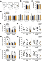
Enhancement or suppression of GIRK channel activity disrupts novel OR. (A) Depiction of OF and OR tests. Mice were placed in an OF (40 × 40 cm white plastic box) illuminated by cool white LED lights (70–90 LUX, 4000 K). Total distance traveled and time spent in the field center (20 × 20 cm) were tracked by ANY-maze software during the 10 min session. Mice were returned to their home cage for 3 h after the OF session. For OR, mice were returned to the OF which now contained two identical objects: 50 mL conical tubes filled with a pink solution and positioned diagonally with cap down at standardized positions A and B. Subjects were allowed to explore the environment during the 10 min training session. Orientation of the mouse head toward an object and within 2 cm of the object was considered as object interaction; total interaction time was recorded for each object during training, along with distance traveled. Mice were then returned to their home cage for 3 h, after which they were placed back in the OF which now contained one familiar object (point A) and one novel object, a 25 cm culture flask containing a yellow solution with cap up (point B). Total distance traveled, along with object interaction times, were measured during the 10 min testing session. The percentage of time spent interacting with the object at position B was compared across groups. Only subjects exhibiting >10 sec total object interaction times during training and testing sessions were included in E–J: 4/11 males treated with the control vector were excluded based on this criterion. (B) Time spent in the center of the OF environment during OF. (C) Total distance traveled during OF. (D) Percentage of time spent in quadrant/zone 1 and zone 3 during the OF test (left), as well as during the OR training (middle) and testing (right) sessions. Symbols (Tukey's multiple comparisons): (*) P < 0.05. (E) Total distance traveled during the OR training ([††] P < 0.01; main effect of sex) and testing sessions. Symbols (Šídák's multiple comparisons): (*) P < 0.05 (within treatment); (#) P < 0.05 (within sex). (F) Total distance traveled across OR training and testing sessions in male and female mice. Symbols (Šídák's multiple comparisons): (*) P < 0.05 (within treatment). (G) Total object interaction time during OR training (Šídák's multiple comparisons: [*] P < 0.05, [**] P < 0.01) and testing session. (H) Total object interaction time across OR sessions in male (Tukey's multiple comparisons: [*] P < 0.05) and female (Šídák's multiple comparisons: [*] P < 0.05, within session) mice. (I) Percentage of time spent interacting with the object at position B relative to total object interaction time during OR training and testing sessions. Symbols (Tukey's multiple comparisons): (*) P < 0.05, (**) P < 0.01 (within sex). (J) Percentage of time spent interacting with the object at position B across OR sessions in male (Tukey's multiple comparisons: [*] P < 0.05, [**] P < 0.01) and female (Tukey's multiple comparisons: [**] P < 0.01) mice.
Subjects were next evaluated for OR using the same apparati (Fig. 2A). Importantly, there was no main effect of sex (F(1,78) = 0.1335, P = 0.7159), viral treatment (F(2,78) = 0.1392, P = 0.8703), or zone (F(1,78) = 0.0288, P = 0.8658) on total time spent in zones 1 or 3 during the OF test (Fig. 2D, left), indicating that none of the groups exhibited a bias for either zone where objects would be positioned. Similarly, there was no main effect of sex (F(1,78) = 2.372, P = 0.1276), viral treatment (F(2,78) = 0.3222, P = 0.7255), or zone (F(1,78) = 0.0461, P = 0.8306) on total time spent in zones 1 or 3 during the OR training session (Fig. 2D, middle), indicating that none of the groups exhibited a bias toward one of the identical objects or an object/zone combination. While there were no main effects of sex, viral treatment, or zone during the OR testing session, there was interaction between viral treatment and zone (F(2,78) = 3.978, P = 0.0266) during the OR testing session (Fig. 2D, right). Males in the control viral treatment group spent more time in zone 3, which contained the novel object (Fig. 2G).
We also evaluated the total distance traveled, total object interaction time, and the percentage of time spent interacting with the object at position B; these parameters were compared across sex and viral treatment within the training and testing sessions (Fig. 2E,G,I), as well as across sessions and treatment within each sex (Fig. 2F,H,J). Total distance traveled during training (sex: F(1,35) = 10.14, P = 0.0030; treatment: F(2,35) = 0.6392, P = 0.5338; interaction: F(2,35) = 0.01654, P = 0.9836) and testing (sex: F(1,35) = 20.60, P < 0.0001; treatment: F(2,35) = 6.959, P = 0.0029; interaction: F(2,35) = 0.1379, P = 0.8717) sessions was higher for females, and there was a main effect of viral treatment on this parameter during the testing session (Fig. 2E). There was also an impact of session for both male (session: F(1,19) = 12.41, P = 0.0023; treatment: F(2,19) = 1.883, P = 0.1795; interaction: F(2,19) = 1.067, P = 0.3638) and female (session: F(1,16) = 10.41, P = 0.0053; treatment: F(2,16) = 1.337, P = 0.2904; interaction: F(2,16) = 1.091, P = 0.3597) mice (Fig. 2F); diminished GIRK channel activity (GIRK3 overexpression) was associated with reduced distance traveled.
During training, there was an interaction between sex and viral treatment (F(2,35) = 3.555, P = 0.0393); female controls spent more time in total object interaction than females treated with GIRK2 or GIRK3 vectors, or their male counterparts (Fig. 2G). In contrast, there was no main effect of sex (F(1,35) = 2.824, P = 0.1018), viral treatment (F(2,35) = 2.718, P = 0.0800) or interaction between factors (F(2,35) = 0.01063, P = 0.9894) in the testing session (Fig. 2G). There was an interaction between session and viral treatment (F(2,19) = 4.767, P = 0.0210) in male mice, with the control animals showing increased total object interaction during testing (Fig. 2H). A main effect of treatment was found in females across sessions (session: F(1,16) = 0.02092, P = 0.8868; treatment: F(2,16) = 3.667, P = 0.0489; interaction: F(2,16) = 2.539, P = 0.1102); however, with diminished GIRK channel activity correlating with decreased object interaction during training.
There was no group difference in the percentage of time spent interacting with the object at position B during training (sex: F(1,35) = 0.1964, P = 0.6603; treatment: F(2,35) = 0.3482, P = 0.7084; interaction: F(2,35) = 0.7372, P = 0.4857) (Fig. 2I). During the testing session, however, there was a main effect of viral treatment on this measure (sex: F(1,35) = 1.415, P = 0.2422; treatment: F(2,35) = 14.21, P < 0.0001; interaction: F(2,35) = 0.03719, P = 0.9635) (Fig. 2I). For both males and females, GIRK2 and GIRK3 overexpression was associated with reduced time spent with the novel object at position B. An interaction between session and viral treatment was detected in males (F(2,19) = 4.147, P = 0.0320) and females (F(2,16) = 7.575, P = 0.0048) (Fig. 2J); controls interacted more with the object in position B during the test as compared to the training session, and relative to GIRK2- and GIRK3-treatment groups during the testing session. Thus, enhancing or suppressing GIRK channel activity in dCA1 pyramidal neurons disrupts novel OR in male and female mice.
Collectively, the OR data suggest that the deficits in novel OR observed in mice with bidirectional GIRK channel manipulation have different underlying causes in male and female mice. Notably, the total distance traveled by females was greater than that of male counterparts during OR training and testing sessions. Females also interacted more with the objects during these sessions. Interestingly, enhancing or suppressing GIRK channel activity in dCA1 pyramidal neurons correlated with reduced total object interaction by females, but only during the training session. The reduced object interaction seen in female mice following GIRK2 or GIRK3 overexpression may be due to decreased detection, attention, and/or motivation when the two identical (novel) objects were introduced to the apparatus for the first time (Antunes and Biala 2012). These animals may also be engaged in competing behaviors. Regardless of the underlying cause, reduced object interaction during training may impede the development of object familiarity and result in decreased recognition of the novel object in females during testing. In male mice, object interaction during training was unaffected by viral treatment. As such, the impairment in novel OR seen during the test session is more likely attributable to a failure to remember the familiar object.
Previous work has shown that a single intracerebroventricular injection of either the GIRK channel activator ML297 or the GIRK channel blocker tertiapin between training and testing sessions led to deficits in novel OR in male C57BL/6J mice (Djebari et al. 2021). Our results support these findings while contributing novel cellular and sex-specific insights related to the GIRK channel contribution to novel OR. Here, we show that bidirectional manipulation of GIRK channel activity in dCA1 pyramidal neurons disrupts novel OR in male and female mice, further supporting the contention that an optimal range of GIRK channel activity in the dorsal HPC is required for intact OR in mice.
Following OF and OR tests, mice were evaluated in a context-only fear conditioning test (Fig. 3A). As summarized in Figure 3B, no main effects of sex or viral treatment, or interaction between the factors was detected during pre-shock (sex: F(1,39) = 0.06913, P = 0.7940; treatment: F(2,39) = 1.511, P = 0.2333; interaction: F(2,39) = 1.347, P = 0.2718) or post-shock (sex: F(1,39) = 0.02693, P = 0.8705; treatment: F(2,39) = 0.1718, P = 0.8428; interaction: F(2,39) = 1.891, P = 0.1645) intervals on acquisition day, or during context test I (sex: F(1,39) = 0.001501, P = 0.9693; treatment: F(2,39) = 1.879, P = 0.1663; interaction: F(2,39) = 0.5697, P = 0.5703). Interestingly, males in the GIRK3 treatment group showed elevated freezing during context test II (Fig. 3B) (interaction: F(2,39) = 3.525, P = 0.0391). This is consistent with the analysis of freezing across the three sessions for males (interaction: F(4,46) = 5.717, P = 0.0008) and females (session: F(1.722,27.56) = 97.96, P < 0.0001; treatment: F(2,16) = 3.490, P = 0.0552; interaction: F(4,32) = 0.5635, P = 0.6909) (Fig. 3C). For both males and females, all viral treatment groups exhibited elevated freezing in context test I as compared to pre-shock, and all treatment groups but the male GIRK3 overexpression group showed decreased freezing in context test II compared to test I.
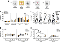
Suppression of GIRK channel activity impairs extinction of CFC in male mice. (A) Depiction of CFC. Mice were introduced to a footshock chamber (Med Associates) for 5 min (pre-shock) before delivery of a footshock (0.5 mA/2 sec). Mice remained in the chamber for 1.5 min after the shock (post-shock), at which point they were returned to their home cages and housing room. Mice were reintroduced to the chamber 24 h (context test I) and 96 h (context test II) later for 5 min test sessions. The percentage of time spent freezing (freezing %) during acquisition (pre-shock and post-shock), as and during context tests I and II, were determined and compared across groups. (B) Freezing % during preshock, post-shock, test I, and test II. Symbols (Tukey's multiple comparisons): (***) P < 0.001. (C) Freezing % across the 5 min pre-shock, test I, and test II sessions in male (Tukey's multiple comparisons: [*] P < 0.05, [**] P < 0.01, [***] P < 0.001, [****] P < 0.0001) and female (Tukey's multiple comparisons: [*] P < 0.05, [**] P < 0.01, [***] P < 0.001) mice. (D) Freezing % of male and female mice by minute during test I. (E) Freezing % of male and female mice by minute during test II. Symbols (Dunnett's multiple comparisons: [*] P < 0.05 vs. GFP).
We also analyzed freezing behavior during each minute of context test I and test II. Freezing during each minute of test I (Fig. 3D) in males (time: F(3.258,74.94) = 1.449, P = 0.2333; treatment: F(2,23) = 1.063, P = 0.3617; interaction: F(8,92) = 0.2693, P = 0.9743) and females (time: F(3.273,52.38) = 7.075, P = 0.0003; treatment: F(2,16) = 1.102, P = 0.3561; interaction: F(8,64) = 0.5757, P = 0.7940), as well as freezing during each minute of test II (Fig. 3E) in females (time: F(2.493,39.90) = 4.903, P = 0.0081; treatment: F(2,16) = 1.797, P = 0.1976; interaction: F(8,64) = 0.7512, P = 0.6464), revealed no treatment differences. In contrast, elevated freezing in GIRK3-treated males was evident throughout test II (Fig. 3E) (time: F(3.186,73.28) = 4.320, P = 0.0063; treatment: F(2,23) = 5.456, P = 0.0011; interaction: F(8,92) = 1.785, P = 0.0899). Collectively, these data suggest that the formation and recall of a contextual fear memory is not impaired by enhancing or suppressing GIRK channel activity in dCA1 pyramidal neurons, but diminished GIRK channel activity impairs the extinction of fear learning in a sex-dependent manner.
Interestingly, our earlier studies using delay (Victoria et al. 2016) or trace (Marron Fernandez de Velasco et al. 2017) fear conditioning protocols in a forebrain-specific GIRK2 knockout mouse line showed that cue-associated CFC was impaired, while cued fear was intact. Here, we used a CFC protocol without cue and found that enhancing or suppressing GIRK channel activity in dCA1 pyramidal neurons does not impact baseline freezing (pre-shock), fear response to the shock (post-shock), or fear association with the context (test I). These results are reminiscent of studies involving mice with loss-of-function mutations in the α5 subunit of the GABAA receptor (GABAAR), which showed that only certain aspects of fear learning were impacted by the genetic manipulations of inhibitory tone in the HPC, and that outcomes were dependent on the genetic models and protocols used (Crestani et al. 2002; Martin et al. 2010; Engin et al. 2015, 2020). For example, constitutive ablation of α5-GABAAR did not impact CFC, but it did enhance trace fear conditioning (Martin et al. 2010). Though a subsequent study demonstrated enhanced contextual fear in constitutive/global α5-GABAAR knockout mice, this phenotype was not observed in mice lacking α5-GABAAR in CA1 pyramidal neurons (Engin et al. 2020). Ablation of α5-GABAAR in CA1 pyramidal neurons in male mice did correlate, however, with impaired context discrimination during extinction (Engin et al. 2020). Since fear generalizes widely while extinction is stimulus-specific, the impaired context discrimination during extinction implicates better recognition of fear-conditioned stimuli at the expense of mnemonic precision (Laing and Dunsmoor 2023); namely, generalized fear to the fear-associated context and other context(s) similar to it, as well as diminished precision in extinction memory. Notably, input from the anterior cingulate cortex to the dorsal HPC has been implicated in contextual fear generalization in mice (Nagayoshi et al. 2022).
Although prior work has revealed no sex differences in dorsal HPC and amygdala c-Fos activity during CFC consolidation, males exhibit stronger c-Fos activity in dorsal HPC during contextual fear memory retrieval and generalization, whereas females exhibit preferential recruitment of basal amygdala (Keiser et al. 2017). Sex differences in contextual fear generalization have also been reported in rats and are linked with HPC computation (Trott et al. 2022). In human studies (Bartholomew et al. 2022; Wen et al. 2022), sex-divergent extinction of fear conditioning has also been noted. For example, functional MRI pointed to distinct functional activation and connectivity between the sexes during extinction learning, suggesting sex-dependent cognitive processing (Wen et al. 2022). Sex hormones have been implicated in the sex differences related to the extinction of conditioned fear, as well as the development and persistence of post-traumatic stress disorder (Milad et al. 2006; Glover et al. 2013; Bartholomew et al. 2022). Our data suggest that the inhibitory tone in the dorsal HPC is part of the sex-divergent neuronal mechanisms in the regulation of contextual fear extinction.
In sum, we found that persistent enhancement or suppression of GIRK channel activity in dCA1 pyramidal neurons leads to the disruption of novel OR in males and females, albeit via distinct mechanisms. Moreover, suppression of GIRK channel activity in dCA1 pyramidal neurons impaired the extinction of CFC, but only in males. Our study adds to the growing body of evidence indicating that an optimal level of GIRK channel activity is required for cognitive processing and suggests that targeting GIRK channel activity may be a viable therapeutic development for cognitive impairment in multiple neurological disorders.
Acknowledgments
The authors would like to thank Dr. Susumu Tonegawa for providing the Tg(Trpc4-Cre)383Stl mouse line, and John C. Brent IV and Anna Souders for care of the mouse colony. This project was supported by National Institutes of Health (NIH) grants to K.W. (R01 NS128039). Support for AAV vector design and production came from the University of Minnesota Viral Vector and Cloning Core and the University of Minnesota Center for Neural Circuits in Addiction (P30 DA048742).
Footnotes
-
Article is online at http://www.learnmem.org/cgi/doi/10.1101/lm.054022.124.
- Received May 10, 2024.
- Accepted September 15, 2024.
This article is distributed exclusively by Cold Spring Harbor Laboratory Press for the first 12 months after the full-issue publication date (see http://learnmem.cshlp.org/site/misc/terms.xhtml). After 12 months, it is available under a Creative Commons License (Attribution-NonCommercial 4.0 International), as described at http://creativecommons.org/licenses/by-nc/4.0/.



