


Stress enhances memory for previously encoded events depending on stressor recall
- 1Department of Cognitive Psychology, Institute of Psychology, University of Hamburg, 20146 Hamburg, Germany
- 2Donders Institute for Brain, Cognition and Behaviour, Radboud University Medical Center, 6525 EN Nijmegen, The Netherlands
- Corresponding author: lars.schwabe{at}uni-hamburg.de
Abstract
Stressful events are typically well remembered, but their effects on memory for surrounding neutral events and the underlying mechanisms remain less clear. We hypothesized that stress would enhance memory for events surrounding the stressor, contingent on the memory of the stressor itself. Additionally, we predicted that memory for neutral events would be modulated by pairing them with stressor-related cues. To test these hypotheses, 122 healthy participants encoded a series of images before and after experiencing a stressful or control episode. During encoding, images were preceded by cues from stressor or control contexts. Memory for the stress or control episode and the encoded images was tested 24 h later. Our results showed enhanced memory prioritization, reflected in better memory for central versus peripheral features, for the stressful compared to the control episode. Exposure to the stressful episode further enhanced memory for neutral images preceding the stressor. However, this memory boost occurred only in participants with enhanced memory prioritization for the stressor. Memory for stimuli encoded after the stressor remained unaffected, and there was no evidence for the proposed cueing mechanism. These findings indicate that stressful events enhance memory consolidation only when these events themselves are distinctly represented in memory.
When we are exposed to stressful events, a myriad of physiological changes take place in our body, including the release of catecholamines and glucocorticoids (mainly cortisol in humans; Joëls and Baram 2009). Through the action of these stress mediators on prefrontal and medial-temporal areas, stressful events can exert a profound impact on learning and memory processes (Diamond et al. 2007; Roozendaal et al. 2009; Schwabe et al. 2022). In general, stress effects on memory are thought to be time-dependent, closely tied to the temporal profile of action of catecholamines and glucocorticoids, with stress enhancing memory for events that are encoded in the temporal and spatial context of the stressor but impairing memory for information encoded outside the context of the stressor (Joëls et al. 2006, 2011; Smeets et al. 2007; Schwabe et al. 2022). Accordingly, it has been repeatedly shown that memory is typically enhanced for a stressful episode itself, in particular for its central features (Vogel and Schwabe 2016; Kalbe et al. 2020; Bierbrauer et al. 2021; Lin et al. 2022; Stanek et al. 2024), or for events encoded shortly before the stressor (Cahill et al. 2003; Smeets et al. 2009; Shields et al. 2017). For events encoded after a stressor, findings are more heterogenous, with some studies reporting enhancing (Domes et al. 2002; Schwabe et al. 2008; Vogel and Schwabe 2016; Goldfarb et al. 2019) and others impairing effects (Kirschbaum et al. 1996; Elzinga et al. 2005; Quaedflieg et al. 2013) of preencoding stress on subsequent memory (for a meta-analysis, see Shields et al. 2017). Given the relevance of stress effects on memory, for instance, in educational or clinical settings (Vogel and Schwabe 2016; De Quervain et al. 2017), understanding the mechanisms underlying stress effects on subsequent memory of surrounding events is important.
Previous research suggested that the influence of stress on subsequent memory for events surrounding the stressor may depend on the emotionality of the encoded information (Cahill et al. 2003; Payne et al. 2007, 2006; Schwabe et al. 2008) or on the temporal distance between stressor exposure and encoding (Joëls et al. 2006; Zoladz et al. 2011; Cadle and Zoladz 2015; Shields et al. 2022). Both of these factors are generally in line with the synaptic and behavioral tagging hypotheses, which assume that initially weakly encoded information becomes effectively consolidated if a significant encounter, such as a stressor, occurs in close temporal proximity (Frey and Morris 1997; Ballarini et al. 2009; Moncada et al. 2015). Another potential mechanism that might underlie stress effects on the memory of events preceding or following a stressor is the linking of emotionally neutral information to a stressful experience via stressor-related cueing. More specifically, a recently proposed cueing mechanism suggests that briefly reactivating a memory engram of an experience allows connecting this experience to newly encoded information (Josselyn and Frankland 2018). Whether cueing a stressful event during the encoding of neutral information modulates the memory for this information, and whether a potential cueing mechanism operates only prospectively (affecting the memory for events encoded after the stressor) or also retrospectively (affecting the memory for events encoded before the stressor) is completely unknown.
Both the proposed tagging mechanism and the potential cueing mechanism through which stressful events might impact the memory of surrounding neutral events should rely on the memory enhancement for the stressful event itself. If the memory of the stressful episode itself is not enhanced, then it would seem unlikely that this stressor could affect memory formation for surrounding events, whether through tagging or cueing mechanisms. Since memory formation for the stressful event is enhanced by the glucocorticoid response elicited by this event, presumably in interaction with noradrenergic arousal (Roozendaal et al. 2006), the memory enhancement for the stressful episode, as well as its impact on surrounding events, should be linked to the glucocorticoid release in response to the stressor. While a potential cueing (as well as a tagging) mechanism would assume that strong memory for the stressful events should also strengthen memory for surrounding events if cued by stressor-relevant information, it has also been argued that the consolidation of the stressful episode may compete with the consolidation of stressor-unrelated information encoded before or after the stressful event (Diamond et al. 2005; Joëls et al. 2006). According to this alternative view, a strong memory for the stressful event itself would undermine memory formation for subsequently encoded information. Although highly relevant for understanding the mechanisms underlying stress effects on memory formation, the relationship between memory for a stressful event itself and its effects on memory formation for surrounding events remains poorly understood.
In the present study, we aimed to elucidate the mechanisms underlying the impact of stressful events on the memory for surrounding neutral events. Specifically, we tested whether cueing a stressful event during the encoding of neutral information before or after the stressor modulates the memory for the neutral information. We reasoned that cueing the stressful context would result in reactivation of the stressful experience, thus extending the memory enhancement for the stressful experience to unrelated, neutral information. Therefore, images that were cued by a stressor context should be better remembered than images cued by a neutral context. We further hypothesized that a stressful episode would influence the memory for surrounding events only if this episode itself is distinctly represented in memory. We predicted that this effect would depend on the individual glucocorticoid response to the stressor. For information encoded before the stressor, we expected a stress-related memory enhancement based on the known beneficial effects of stress and glucocorticoids on consolidation (Cahill et al. 2003; Roozendaal et al. 2006; Shields et al. 2017). For information encoded after the stressor, however, we predicted a stress-related memory impairment, due to the potential competition with the consolidation of the stressful event itself.
Results
To assess how memory formation for the stressor itself and for surrounding neutral information relate to one another and whether cueing the stressor context enhances the memory for surrounding information, 122 healthy participants completed two encoding sessions before and after a stressful (or control) episode (Trier Social Stress Test [TSST]) (Fig. 1; Kirschbaum et al. 1993). This treatment episode was enriched by several central and peripheral objects (Kalbe and Schwabe 2020; Lin et al. 2022). Objects were considered to be central (e.g., a coffee mug that a panel member drank from) or peripheral (e.g., a poster), depending on whether the panel interacted with the objects or not. Each of the encoding sessions consisted of 160 trial unique images of neutral objects. At the start of each trial, we presented one of three context types to probe the effects of stressor context cueing on the encoding of stressor-unrelated (neutral) information: (1) stressor context, a photograph of the room where the stress/control treatment took place, (2) lure context, a photograph of a very similar room, or (3) control context, an unrelated room, for example, a supermarket aisle. We measured subjective and physiological stress responses throughout the experiment through blood pressure, pulse, salivary cortisol, and mood questionnaires. Approximately 24 h after stress manipulation and encoding, participants completed free recall and recognition tests assessing their memory for the encoded images as well as for the stressful (or control) episode.
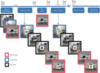
Experimental procedure. On day 1, participants first saw images of several rooms that would later be cued as contexts in the image encoding task. Participants then completed an image encoding task before and after a stressful encounter, or a control treatment. In the image encoding tasks, we first cued a context that was either the same room where the stressful encounter took place (stressor context), a lure context that looked similar to the stressor context, or an unrelated control room (e.g., a kitchen); indicated with colored frames for illustrative purposes. The participants were presented with 320 images in total (160 before the stressor and 160 thereafter). We also collected questionnaires and salivary cortisol throughout day 1 to track subjective and physiological stress responses. On day 2, 24 h later, participants completed free recall and recognition memory tasks for the encoded images as well as the stressful/control encounter.
Successful stress induction
Subjective, autonomic, and endocrine measures confirmed the successful stress induction by the TSST (Fig. 2).
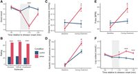
Successful stress induction. After the stress versus control treatment, participants in the stress condition rated their mood as significantly worse (A), found the treatment to be more difficult, unpleasant, and stressful (B), and had elevated blood pressure (C,D) and pulse (E). Stressed participants also showed elevated salivary cortisol immediately after, 25 min, and 40 min after treatment onset (F). Error bars represent standard error of the mean (SEM). (*) P < 0.05 and (***) P < 0.001, for stress compared to control group.
Mixed-design ANOVAs revealed a significant time × condition (stress vs. control) interaction for all mood ratings (all F > 6.64, all P < 0.001, all η2 > 0.019). Follow-up tests using pairwise comparisons showed that immediately after the treatment, participants of the stress group rated their mood as less awake (Supplemental Fig. S1A), calm (Supplemental Fig. S1B), and good (Fig. 2A) compared to control participants [all t(120) > 3.914, all P < 0.001]. Furthermore, stressed participants rated their mood as less calm at the end of day 1 [t(120) = 2.490, P = 0.014]. There was no difference in these parameters between the groups at the start of day 1 or before the treatment (all |t| < 0.965, all P > 0.336; Supplemental Table S1). Moreover, participants in the stress group rated the treatment as significantly more difficult, stressful, and unpleasant than those in the control group (all F > 91.12, all P < 0.001, all η2 > 0.435; Fig. 2B).
At the autonomic arousal level, mixed-design ANOVAs revealed significant time × condition interactions for pulse, systolic, and diastolic blood pressure (all F > 17.56, all P < 0.001, all η2 > 0.031; Fig. 2C–E). Follow-up pairwise comparisons showed that during the treatment, participants in the stress group had a significantly higher pulse, systolic, and diastolic blood pressure relative to control participants (all |t| > 2.842, all P < 0.006), while there was no difference between the stress and control groups at the start of day 1 (all |t| < 1.557, all P > 0.122).
Finally, the exposure to the TSST led also to a significant increase in salivary cortisol. A mixed-design ANOVA showed significant main effects of time point [F(1.83, 205.09) = 8.75, P < 0.001, η2= 0.023] and condition [F(1, 112) = 12.52, P < 0.001, η2= 0.072], as well as a significant interaction between these factors [F(1.83, 205.09) = 16.03, P < 0.001, η2= 0.042; Fig. 2F]. Follow-up analyses showed that cortisol was significantly higher in stressed participants, relative to controls, at the offset of the stressful episode, 25 min after the onset of the stressful episode, and at the end of the experiment on day 1, ∼ 40 min after the onset of the stressor (all |t| > 2.492, all P < 0.015). There were no differences between the conditions at baseline on day 1 or right before the stress/control treatment (both |t| < 0.695, both P > 0.489).
Enhanced memory for the stressful episode
We tested participants’ memory for the stressful (vs. control) episode 24 h later. At the time of memory testing, groups did not differ in subjective stress levels (all |t| < 1.591, all P > 0.113), autonomic arousal (all |t| < 1.190, all P > 0.239), or salivary cortisol [t(112) = 0.982, P = 0.328; Supplemental Table S1]. In a recognition test, we presented images of central and peripheral objects that were present during the treatment as well as of novel objects. For each image, participants selected whether the object was present in the treatment room (old) or not (new).
Using a mixed-design ANOVA with condition and object type (central vs. peripheral) as predictors, we found a significant main effect of object type [F(1, 118) = 401.12, P < 0.001, η2= 0.543], as well as a significant interaction of condition and object type [F(1, 118) = 8.04, P = 0.005, η2= 0.023, Fig. 3A]. Pairwise comparisons, however, revealed no significant differences between the stress and control group for central [t(118) = −1.715, P = .089] or peripheral items [t(118) = 1.643, P = 0.103]. We next computed a centrality bias index for each participant by subtracting the hit-rate for peripheral objects from the hit-rate for central objects and compared stress and control participants using a t-test. The centrality bias reflects the prioritization of memory and was significantly higher in the stress group than in the control group [t(118) = −2.838, P = 0.005, d = −0.518, Fig. 3C]. The false alarm rate did not differ between groups [stress: M = 0.106, SD = 0.083; control: M = 0.120, SD = 0.112; t(110.890) = 0.756, P = 0.451, d = 0.138].
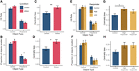
Memory for the stressful episode. Participants in the stress condition tended to remember more central and less peripheral objects in a recognition task (A,E) and free recall task (B,F) than those in the control group. There was a significantly higher centrality bias (difference between recognized or recalled central and peripheral objects) in the stress group than in the control group, both in the recognition test (C) and in the free recall test (D). Specifically, stressed participants with a high salivary cortisol response had a significantly higher centrality bias compared to participants in the control group (G). A similar, but nonsignificant, pattern was also shown in the free recall task (H). Error bars represent SEM. (*) P < 0.05, (**) P < 0.01.
In the next step, we tested for a potential role of the stress-induced cortisol response in stress effects on memory of the treatment episode. To this end, we divided stressed participants into two groups based on their cortisol response to the stressor and compared each to the control group. Participants with an increase in cortisol above 1.5 nmol/L were considered to be high responders (n = 29), the rest were considered low responders (n = 29) (Miller et al. 2013). A mixed-design ANOVA revealed a significant interaction of responder group and object type [F(2, 109) = 3.99, P = 0.021, η2= 0.026, Fig. 3E], but no follow-up tests were significant (all |t| < 1.899, all P > 0.143, Tukey-corrected). We then used a one-way ANOVA to test for differences in centrality bias depending on cortisol response (high vs. low vs. control). There was a significant difference in centrality bias depending on the group [F(2, 109) = 3.99, P = 0.021, η2= 0.068, Fig. 3G], and pairwise comparisons indicated that high cortisol responders had a significantly higher centrality bias than control participants [t(109) = −2.711, P = 0.021, Tukey-corrected], while the differences between high responders and low responders and between low responders and controls remained nonsignificant (both |t| < 1.654, both P > 0.228, Tukey-corrected). The lack of a difference between the low responders and controls may however be due to the effect being too small to detect in this comparison (Fig. 3G). To test whether autonomic nervous system activity during the stressor influenced the extent of this centrality bias, we computed separate linear models predicting the centrality bias with blood pressure, pulse, and group, as well as the interaction between them. No interactions reach significance (all |β| < 0.003, all P > 0.434), suggesting that the autonomic response was not directly associated with the prioritization of stressor memory.
In addition to the recognition test, participants completed also a free recall test for the stressful (vs. control) episode. In this free recall test, participants were asked to verbally list all objects they remembered seeing in the treatment room. A mixed ANOVA revealed a significant main effect of object type [F(1, 118) = 176.54, P < 0.001, η2= 0.371] and a significant interaction of condition and object type [F(1, 118) = 7.36, P = 0.008, η2= 0.024] on free recall performance (Fig. 3B). Pairwise comparisons revealed that stressed participants recalled significantly fewer peripheral objects than the control participants [t(118) = 2.121, P = 0.036]. There was no difference between stressed and control participants in free recall memory for central objects [t(118) = −1.441, P = 0.152]. Same as for the recognition test data, we further calculated a centrality bias by subtracting the proportion of recalled peripheral objects from the proportion of recalled central objects. This centrality bias was significantly higher in the stress compared to the control group [t(117.83) = −2.713, P = 0.008, d = −0.493, Fig. 3D].
We next tested whether free recall memory depended on the cortisol response (high vs. low vs. control). We found a significant effect of object type [F(1, 110) = 161.58, P < .001, η2= 0.361] as well as an interaction effect of responder group and object type [F(2, 110) = 3.62, P = 0.030, η2= 0.025, Fig. 3F]. Pairwise comparisons, however, showed no significant differences (all |t| < 1.881, all P > 0.149, Tukey-corrected). A one-way ANOVA comparing centrality bias depending on cortisol response (high vs. low vs. control) showed an effect of group on centrality bias [F(2, 110) = 3.62, P = 0.030, η2 = 0.062, Fig. 3H]. However, none of the follow-up tests approached significance (all |t| < 2.214, all P > 0.073, Tukey-corrected). To test the impact of ANS activity on centrality bias, we again ran separate linear models for pulse and blood pressure, which did, however, not yield any significant interactions between group and autonomic arousal parameters (all |β| < 0.003, all P > 0.153).
Notably, although groups did not differ in the overall false alarm rate, we re-ran the recognition memory mixed ANOVAs with d′ instead of the hit-rate, which left the pattern of results mostly unchanged, yet even showed a significant difference between stressed and control participants for central objects (see Supplemental Material).
Memory for events surrounding the stressful episode
We next assessed the probability of remembering each individual stimulus image from the pretreatment and posttreatment encoding phases with mixed-design ANOVAs and GLMMs. We first ran a mixed-design ANOVA to analyze the effect of condition and phase (pretreatment vs. posttreatment) on the hit-rate.
This model revealed a significant effect of phase [F(1, 120) = 87.51, P < 0.001, η2= 0.033] as well as a significant interaction of phase and condition [F(1, 120) = 15.32, P < 0.001, η2= 0.006]. The significant interaction between phase and condition was driven by a memory enhancement for stimuli encoded before the treatment in the stress group. As shown in Figure 4A, participants remembered overall more of the items from the pretreatment than from the posttreatment encoding session, but this difference was more pronounced in the stress group than in the control group [control: t(120) = 3.847, P < 0.001, stress: t(120) = 9.383, P < 0.001]. However, pairwise comparisons between the stress and control groups for the pretreatment [t(120) = −1.568, P = 0.120] and posttreatment encoding [t(120) = 0.093, P = 0.926] sessions separately remained nonsignificant.
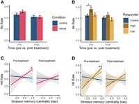
Memory for events occurring before or after the stressor or control treatment. Stressed participants tended to remember more objects encoded before the stressor (A), yet this pattern did not reach significance. This was driven by high cortisol responders recognizing more images that were encoded before the stressor (B). When stressed participants showed memory prioritization for the stressful episode, they recognized significantly more images than control participants (C). This was especially the case for stressed participants showing a pronounced cortisol response to the stressor (D). Error bars show SEM. (*) P < 0.05, (**) P < 0.01.
We next tested for a potential role of the stress-induced cortisol response in stress effects on the memory for surrounding neutral events. A mixed-design ANOVA revealed a significant effect of phase [F(1, 111) = 91.55, P < 0.001, η2= 0.036] and a significant interaction of phase and responder group [high vs. low vs. control; F(2, 111) = 7.99, P < 0.001, η2= 0.007, Fig. 4B]. Tukey-corrected pairwise comparisons showed that stressed participants with a high cortisol response recognized significantly more stimuli encoded prestress, than the control group [t(111) = −2.588, P = 0.029]. No other pairwise comparisons reached significance (all |t| < 1.890, all P > 0.146). To test whether autonomic nervous system activity (blood pressure and pulse) during the stressor predicted memory for events surrounding the stressor, we ran separate mixed linear models with condition (stress vs. control), phase (pretreatment vs. posttreatment), and autonomic nervous system activity, as well as an interaction term, as predictors. Only the interaction term of group × phase × systolic blood pressure approached significance (β = −0.027, P = 0.059; both other |β| < 0.023, both other P > 0.086). Follow-up simple slopes analyses assessing the effect of stress at high, moderate, and low systolic blood pressure in the pretreatment or posttreatment revealed no significant differences between the stress and control groups (all |β| < 0.031, all P > 0.397, Holm–Bonferroni-corrected). The false alarm rate did not differ between conditions [t(111.060) = −0.494, P = 0.622, d = –0.089] or depending on the cortisol response [F(2, 111) = 0.89, P = 0.411, η2= 0.016]. The pattern of results remained largely unchanged when using d′ instead of hit-rate as outcome measure in the recognition memory mixed ANOVAs (see Supplemental Material).
With respect to the potential cueing of memory by the stressor context, we ran a GLMM with phase, condition, and cue type (treatment vs. lure vs. control), as well as distance to treatment as predictors. We found no interaction of condition, cue type, and phase or distance (all |β| < 0.192, all P > 0.196). The reduced model with two-way interactions of the variables again showed no interaction of cue type and condition (both |β| < 0.022, both P > 0.771). Moreover, there was no effect of cue type on hit probability (both |β| < 0.100, both P > 0.118), indicating that images were equally likely to be recognized irrespective of whether they were cued by a stressful context or not. Thus, cueing a stressful context did not affect the memory for subsequent neutral information. We next added the cortisol response (high vs. low vs. control) to the model and repeated the analyses. There was no interaction of cue type, responder group, and phase or distance (all |β| < 0.286, all P > 0.132). The reduced model with two-way interactions of the variables again showed no interaction of cue type and responder group (all |β| < 0.078, all P > 0.401). Predicted probabilities are presented in the Supplemental Material (stress vs. control, Supplemental Table S2; high cortisol vs. low cortisol vs. control, Supplemental Table S3).
In addition to the recognition memory task, participants were asked in a free recall task to verbally list all objects they recalled seeing in the pretreatment and posttreatment encoding tasks. We used GLMMs to analyze the probability of an item being recalled, with the same predictors as for the recognition task. We found no interaction of condition, cue type, and phase or distance (all |β| < 0.110, all P > 0.665). The reduced model included two-way interactions and again showed no interaction of cue type and condition (both |β| < 0.197, both P > 0.148). We next repeated the analyses with the responder group (high vs. low vs. control) as a predictor. Similar to the analyses above, there was no interaction of cue type, responder group, and phase or distance (all |β| < 0.360, all P > 0.227). The reduced model with two-way interactions of the variables showed a significant interaction of low responders and treatment cue (β = 0.346, P = 0.049). However, follow-up simple slopes analyses showed no significant effects of the response group for treatment cues (all |β| < 0.374, all P > 0.118). Predicted probabilities are presented in the Supplemental Material (stress vs. control, Supplemental Table S4; high cortisol vs. low cortisol vs. control, Supplemental Table S5).
Memory for events surrounding a stressful episode depends on the memory prioritization for the stressful episode itself
We next analyzed whether memory for the stressful episode itself would moderate the influence of stress on the memory for surrounding events. We reasoned that stress would affect the memory for surrounding events only if there is also a memory prioritization (i.e., centrality bias) for the stressful episode itself. To this end, we ran a linear mixed model with condition (stress vs. control), phase (pretreatment vs. posttreatment), and centrality bias as predictors. We also included state anxiety score as a covariate in order to control for baseline differences in state anxiety on day 2 (see below). We found a significant phase × condition × centrality bias interaction (β = −0.035, P = 0.025). We followed up this significant interaction with simple slopes analyses at 1 SD below and above mean centrality bias. Simple slopes analyses revealed that stress significantly enhanced memory for items encoded before the treatment, if participants had a high centrality bias, that is, more pronounced memory for central aspects of the stressful episode (β = 0.136, P = 0.036, Fig. 4C). The effect of condition was nonsignificant in all other simple slopes analyses (at moderate and low centrality bias levels, in the posttreatment encoding phase, all |β| < 0.066, all P > 0.306; Holm–Bonferroni-corrected).
To zoom into the effect of stress further, we next divided stressed participants into high and low cortisol responders and reran the linear mixed model presented above. Simple slopes analyses revealed that high cortisol responders who also had a high centrality bias showed a significant memory enhancement for items encoded before the stressor (β = 0.196, P = 0.006). Low cortisol responders also showed this effect at a descriptive level, but did not differ from the control group after Holm–Bonferroni-correction (β = 0.130, P = 0.248, Fig. 4D). None of the other simple slopes analyses showed a significant effect of responder group (all |β| < 0.102, all P > 0.135). We next ran an exploratory analysis comparing low cortisol responders to high responders, and found no significant difference in their memory performance (all |β| < 0.047, all P > 0.340). These findings suggest a memory enhancement for information encoded before the stressor that is dependent on memory prioritization of the stressful episode, but largely independent of the stress-related cortisol response. The pattern of results remained mostly unchanged when using d′ instead of hit-rate as outcome measure (see Supplemental Material). In particular, the significant interaction effect between condition, phase, and centrality bias as well as the interaction between cortisol response and phase remained. With d′ as the outcome measure, however, the post hoc analysis contrasting stressed and control participants with a high centrality bias for material encoded before the stressor was not significant anymore. However, the difference between high cortisol responders and control participants with a high centrality bias remained.
Lastly, we assessed whether stressor cueing would enhance memory for participants with an elevated centrality bias. In this model, we found no significant interaction of cue type, condition, centrality bias, and encoding phase (both |β| < 0.164, both P > 0.295).
Control variables
To control for baseline differences between stressed and control participants, we measured chronic stress, anxiety or depressive symptoms, state anxiety, sleep duration, and quality. Groups did not differ in any of these variables (all |t| < 1.001, all P > 0.301, see Supplemental Table S1) except for state anxiety at the beginning of experimental day 2 [t(118.51) = −2.370, P = 0.019, d = −0.431]. Participants in the stress group had higher state anxiety at the beginning of day 2 than controls, which may be due to different expectations based on the stressful event experienced on day 1. In order to control for this difference between groups, we included the state anxiety score on day 2 as an additional predictor in our final linear mixed models.
Discussion
Acute stress is known to be a powerful modulator of memory (Joëls et al. 2006; Diamond et al. 2007; Schwabe et al. 2022). Although it is well established that stressful events are typically well-remembered (Sandi and Pinelo-Nava 2007; Vogel and Schwabe 2016; Kalbe et al. 2020; Bierbrauer et al. 2021) and that they can interfere with the retrieval of unrelated information (De Quervain et al. 1998; Shields et al. 2017), the impact of these events on the memory for surrounding neutral events and the cognitive mechanisms involved remain elusive. In this study, we probed a cueing mechanism whereby stress-induced memory alterations would selectively affect surrounding neutral information cued by the stressful encounter. In addition, we tested how the effect of stress on the memory for surrounding events relates to the memory of the stressor itself. While our data did not support the proposed cueing mechanism, they demonstrated a memory enhancement for events encoded before the stressor, which was directly linked to the memory enhancement for the stressful episode itself.
Significant increases in subjective stress responses, autonomic arousal, and salivary cortisol validated the successful stress induction through the enriched TSST. Consistent with previous studies, the stressful episode was more distinctly represented in memory, compared with the nonstressful control episode (Kalbe et al. 2020; Bierbrauer et al. 2021; Lin et al. 2022; Stanek et al. 2024). Specifically, we obtained a memory prioritization or centrality bias for the stressful episode, reflected in a more pronounced difference in the memory for central and peripheral features of the episode. Previous research has suggested a trade-off of memory during stress, whereby arousal enhances the memory for high-priority information but impairs the memory for low-priority information (Mather 2007; Sakaki et al. 2014). A potential mechanism behind this trade-off effect is the proposed arousal-driven reconfiguration of large-scale neural networks. In particular, arousal has been suggested to induce a shift from default- or executive control networks toward the salience network, resulting in the prioritized encoding of highly salient information at the expense of less relevant, peripheral information (Hermans et al. 2011, 2014; Schwabe et al. 2022). However, it is to be noted that this large-scale network reconfiguration was shown to be driven by noradrenergic arousal (Hermans et al. 2011) and that we did not find associations between the memory prioritization (i.e., centrality bias) for the stressful episode and parameters of autonomic arousal in the present study. Moreover, several studies that assessed this trade-off in the past did not show an improvement for central feature memory at the expense of peripheral features (Kalbe and Schwabe 2020; Bierbrauer et al. 2021; Lin et al. 2022), suggesting that the process might be more dynamic and depend on additional factors (Kensinger et al. 2007).
In addition to the memory prioritization for the stressful episode, stress affected the memory for neutral information encoded before the stressor. Exposure to the stressful episode enhanced the subsequent memory of information encoded before the stressor. Importantly, however, this memory boost occurred only if the stressful episode itself was distinctly represented in memory, as reflected in the centrality bias. Interestingly, the memory for information encoded before the stressor did not differ between stressed participants with a low versus high cortisol response when these participants showed a pronounced centrality bias for the stressful episode, suggesting that if the stressful event itself is distinctly represented in memory, it may impact the subsequent memory of previously encoded events, even in the absence of a pronounced cortisol response. The centrality bias in memory of a stressor has recently been linked to distinct representational changes in the basolateral amygdala (Bierbrauer et al. 2021), the same region shown to integrate glucocorticoid and noradrenergic actions to modulate memory processes in other brain areas (Roozendaal et al. 2009; McGaugh 2015). Based on these data, an interesting question for future research is whether the representational changes in the basolateral amygdala are necessary for potential stress (hormone) effects on memory, as has been suggested in the case of stress-induced effects on gist-memory (Adolphs et al. 2005), or whether these changes result from the interaction of glucocorticoids and noradrenergic arousal.
While memory for stimuli encoded before the stressor was enhanced in stressed participants, memory for stimuli encoded after stressor exposure remained unaffected. This result is generally in line with a meta-analysis suggesting that the effect of preencoding stress on memory is rather inconsistent (Shields et al. 2017). It has been proposed that stress effects on subsequent encoding depend on the temporal distance between stressor and encoding (Joëls et al. 2006; Zoladz et al. 2011). Thus, it could be argued that there may have been differential, potentially opposite, effects of stress on subsequent encoding of stimuli presented shortly after the stressor exposure or after a longer time delay. However, our results showed no modulation of the stress effect by the distance of a stimulus to the stressor. Our data did show, however, that memory performance was lower in the posttreatment phase for all participants, regardless whether they experienced a stressful or neutral encounter, which may have been due to fatigue or an already high memory load. This overall low memory performance for stimuli encoded after the treatment may have obscured any effects of stress on subsequent encoding.
The memory enhancement for stimuli encoded before the stressor when there is also a memory prioritization for the stressor aligns well with the behavioral tagging hypothesis, which predicts that emotionally arousing events can boost memory for previously weakly encoded events (Ballarini et al. 2009). In the present study, we probed an additional cueing mechanism, assuming that neutral stimuli would be linked to the stressor and therefore preferentially stored in memory if the stressor was cued at the time of stimulus encoding. Our results did not confirm this proposed cueing mechanism: cueing a stressful encounter did not modulate subsequent memory for stimuli that were encoded before or after the stressor. This finding suggests that the proposed cueing mechanism may not be relevant for stressor effects on surrounding neutral events. There is, however, an alternative explanation for the absence of a cueing effect in our experiment, related to the used contextual stressor cues. Accumulating evidence indicates that contextual information is less well processed under stress (Schwabe et al. 2009; Kaouane et al. 2012; Simon-Kutscher et al. 2019) and that stress induces a shift from a hippocampus-based system, which is relevant for contextual memory, toward a dorsal striatal system, which focuses on single cues (Kim et al. 2001; Vogel et al. 2016; Wirz et al. 2017). Thus, if spatial and contextual cues are less well processed under stress, these cues might not be well-suited to effectively cue the stressor. Future studies are required to test the proposed cueing mechanism using central aspects of the stressor, such as faces of the panel members in the TSST. Moreover, the effectiveness of the stressor context cueing may have been reduced in the present study by the relatively frequent presentation of the stressor context cue (16 times in total), which might have resulted in habituation to this cue. Finally, it is important to note that we used exclusively emotionally neutral images in the encoding sessions before and after the stressor. Stressor-cueing effects on the memory for surrounding information might only occur if the surrounding information involves some degree of emotional arousal, as suggested by studies showing that stress or glucocorticoid effects on memory require concurrent noradrenergic arousal (Roozendaal et al. 2006; Joëls et al. 2011).
In sum, we demonstrate that the impact of stress on memory for stimuli preceding the stressor is contingent on the recall of the stressor itself. Our results show a centrality bias in the memory of the stressful episode, indicated by a pronounced differentiation between the storage of central and peripheral features of the event. While cueing the stressful encounter did not affect the later recall of surrounding information, memory for information encountered before the stressor was enhanced. This enhancement, however, was observed only in stressed individuals who exhibited a pronounced centrality bias in their memory of the stressful encounter. Collectively, our findings provide novel insights into the relationship between memory for the stressor itself and its impact on memory for surrounding (neutral) events. Specifically, these findings suggest that stress-related consolidation enhancements require a distinct memory representation of the stressful event itself.
Materials and Methods
Participants and experimental design
One-hundred twenty-seven healthy volunteers participated in the experiment (68 female, mean age = 24.09 years, SD = 3.62 years). Participants were screened in a standardized interview for the following exclusion criteria: lifetime history or current neurological or psychological disorders, hormonal contraceptive use, or medication intake. Only nonsmokers were included, and women were not tested during their periods, because these factors are known to influence cortisol concentrations. The sample size was based on a power analysis using G-Power (version 3.1.9.7) assuming a small effect size of partial η2 = 0.02, and a power of 0.85. We accounted for a dropout rate of ∼15%. Five participants were excluded due to being extreme outliers in the recognition memory task about material encoded before or after the treatment (> 3 SD from the mean, or false alarm-rate > hit-rate, n = 4) or due to experimenter error (n = 1), thus resulting in a final sample size of 122 participants (66 female). In the free recall task, one further outlier was identified and excluded. In the analysis on memory for the stressful episode, two further participants were identified as outliers in recognition memory performance (> 3 SD from mean) and were excluded from the subsequent analysis (stress: n = 59, control: n = 61). Based on the same cutoff, one participant was excluded from the free recall data analysis (stress: n = 60, control: n = 61). All participants provided written informed consent before the beginning of the experiment, and the study was approved by the local ethics committee of the University of Hamburg. Participants received a moderate monetary compensation for their participation.
In a between-subjects design, participants were pseudorandomly assigned to a stress (n = 61; 32 female, age: M = 24.00 years, SD = 4.21 years; 29 male, age: M = 23.90 years, SD = 3.27 years) or control group (n = 61; 34 female, age: M = 23.30 years, SD = 2.72 years; 27 male, age: M = 24.40 years, SD = 3.20 years) to ensure a comparable distribution of men and women in both conditions.
Experimental procedure and tasks
The experiment took place over 2 days, separated by approximately 24 h. Testing took place between 13.00 and 19.00 in order to control for the diurnal rhythm of cortisol. The experiment consisted of four parts: the context familiarization task, pretreatment encoding task, stress (or control) episode, and posttreatment encoding task. All tasks were programmed in Matlab (version R2019b, 9.7.0.1319299) with Psychtoolbox (version 3.0.16).
Day 1: context familiarization task
At arrival on day 1, we measured salivary cortisol using a Salivette (Sarstedt), blood pressure (OMRON M500, OMRON Healthcare Europe), mood (Mehrdimensionale Befindlicheitsfragebogen [MDBF], Steyer et al. 1997) and state anxiety (State-Trait Anxiety Inventory-State [STAI-S], Laux 1981) using standardized questionnaires. We also measured the number of hours that participants slept the night before the experiment.
The participants then completed the context familiarization task, which served to familiarize participants with all context images that were used as cues in the subsequent encoding phases in order to prevent that the memory cueing was confounded by context familiarity. On each trial of the context familiarization task, participants saw an image of a context (i.e., room) for 3 sec on a computer screen. There were ten different context images. Each was presented six times during the familiarization task, with the order of the context images being fully randomized. The 10 context images belonged to one of three types of context cues: treatment context (n = 1), lure context (n = 1), and control contexts (n = 8). The treatment context cue was an image of the room where the stress (or control) manipulation would take place later. The lure context was included in order to control for the possibility that the treatment room was remembered better simply due to its distinctiveness. The lure room looked similar to the treatment room: the furniture set-up and objects in the room were similar, as well as the angle from which the picture was taken. Thus, this lure room would reference a context that was visually similar to the treatment room, but was not associated with the treatment itself. Importantly, it was counterbalanced across participants and groups whether the stress (or control) manipulation would take place in one or the other context; that is, one room served as treatment context for some participants but as lure context for others (and vice versa). This allowed us to test memory cueing effects that were independent of the specific room cue but solely related to the fact that the experimental treatment took place in that room or not. The control rooms were eight photographs of rooms or places that were independent of the treatment or lure rooms. These were photographs of, for example, a supermarket aisle, a meeting room, or a gym. We presented control images in 80% of the trials and the stress context image in only 10% of the trials in order to prevent habituation to the stressor-context cue.
Day 1: Pretreatment encoding session
Immediately after the context familiarization task, participants completed the pretreatment encoding task. The stimuli in this task consisted of 160 trial-unique images of neutral items. The images were sourced from a variety of online databases (Brodeur et al. 2010; Hovhannisyan et al. 2020). At the start of each trial, one of the context cues was presented for 3 sec such that it covered the whole screen. Then, the stimulus image was presented on top of the context cue for 2 sec. In order to control for participants’ attention and promote deeper encoding, participants were instructed to press a button if the item fits into a shoebox. The size of the stimulus image was 30% of the screen height. Thus, the context cue was still visible in the background during the image presentation. The trials were separated by a fixation cross presented for 2.5–3 sec. This trial structure allowed us to cue a specific context—which was linked to the subsequent treatment or not—at each trial, and then present unrelated information while continuing to cue the context.
The room cue presentation was pseudorandomized: the encoding session was broken into ten blocks of 16 trials, within which the order of the context cues was randomized. Each room cue was presented once per block. The same context cue was never shown twice in a row.
Participants were instructed to memorize the stimulus images, as their memory for these images would be tested on the next day, alongside their memory for the room cue that was presented during that stimulus image. The pretreatment encoding task lasted ∼ 21 min. During encoding, we measured pupil dilation (SMI RED250, SensoMotoric Instruments), skin conductance, and ECG (BIOPAC MP150, BIOPAC Systems Inc.). These physiological measures are not analyzed in this paper and thus will not be reported further.
Day 1: Stress and control manipulation
Immediately after the pretreatment encoding task, participants gave a second saliva sample, we measured blood pressure and pulse (OMRON M500) and the participants completed the mood questionnaire (MDBF) again. Participants were then escorted to a different room where they underwent either the stress or control treatment.
To induce stress in half of the participants, we used the TSST (Kirschbaum et al. 1993). In the TSST, the participant is brought into a room with a nonreinforcing panel of two experimenters wearing lab coats. The participant is told to imagine they are interviewing for their dream job, and that the panel will evaluate their performance. The participant is videotaped and can see themselves on a large screen. The TSST consists of three parts: (1) a 3 min preparation phase, during which the participant can make some notes about the upcoming speaking task, (2) a 5 min free speech, during which the participant has to argue why they are the ideal candidate for their dream job, and (3) a 5 min mental arithmetic task, during which the participant has to count as fast as possible backward from 2043 in steps of 17. We measured blood pressure and pulse twice during the mental arithmetic task (Dinamap, Critikon).
The remaining half of the participants took part in the control treatment. The control treatment is structured similarly to the TSST, but the panel acts in a friendly manner and does not wear lab coats. The participant is not being videotaped and the speaking task is replaced with an informal discussion with the panel, during which the participant can freely choose a topic (e.g., a book they like). The mental arithmetic task is replaced with a counting game played together with the panel. Blood pressure and pulse were measured twice during the counting game.
As described above, the TSST or control treatment took place in one of two rooms (room A/room B), in order to rule out a confounding of the fact that the treatment took place in that room and a potentially different memorability of that room in the mnemonic context cueing.
The TSST and control manipulation were enriched by placing several objects in the room where the treatment took place. The panel members interacted with some of the objects (central objects), while others remained in the periphery (peripheral objects). Central objects included, for example, a cup that one of the panel members drank from, or a stapler that the panel member used to staple some notes together. The peripheral objects included, for example, a poster on the wall, a plant, and an umbrella. In total, there were twelve peripheral and ten central objects. The object interactions were exactly the same in the TSST and control manipulation.
Day 1: Posttreatment encoding session
After the TSST or control manipulation, participants were escorted back to the room where the preencoding task had taken place. Here, they gave a third saliva sample and completed a mood questionnaire (MDBF) as well as a questionnaire assessing the difficulty and stressfulness of the TSST or control manipulation. We also measured blood pressure and pulse (OMRON M500).
About 5 min after the end of the TSST (or control manipulation), participants completed the posttreatment encoding task. This task was structurally identical to the encoding task that took place before the treatment, but all stimulus images were new. During the posttreatment encoding session, we obtained a fourth saliva sample, because salivary cortisol was expected to peak ∼ 25 min after stressor onset.
After completing the posttreatment encoding task, about 45 min after treatment onset, participants gave a fifth saliva sample and completed a mood questionnaire, and we measured blood pressure and pulse (OMRON M500).
Day 2: Free recall test for images
Approximately 24 h later, participants returned for memory testing. Participants first completed mood (MDBF), sleep, and anxiety questionnaires (STAI-S), gave a final saliva sample, and we measured blood pressure and pulse (OMRON M500) to rule out that groups differed in their stress level before memory testing.
First, we assessed participants’ memory for the encoded pictures in a free recall test. In this task, participants were given ten minutes to name all item images they remembered seeing during the encoding sessions on day 1. The participants were instructed to verbally list all images they remembered, while the responses were written down by the experimenter and audio-recorded. After completing data collection, the free recall data was analyzed by identifying all mentioned images in the pretreatment and posttreatment encoding tasks. If the mentioned image had not been presented, it was recorded as a false alarm. If the image was not clearly identifiable, it was recorded as a miss and was not further analyzed.
Day 2: Recognition test for images
Next, we assessed participants’ recognition memory of the encoded images. Here, participants saw all 320 images that were presented on day 1 (“old”) as well as 160 new ones in randomized order. Participants were asked to indicate with the computer mouse for each image whether the image was old or new. They could select between two levels of certainty (rather old/definitely old/rather new/definitely new). If the participant selected either “old” answer, they were shown three context cues and asked to select which one had been presented while the image was shown. For old images (i.e., hits), two additional context cues were randomly shown. For new images (i.e., false alarms), three room cues were randomly shown. This task was self-paced.
Day 2: Free recall and recognition test for the stressful or control episode
We next assessed participants’ memory for the TSST or control manipulation via free recall and recognition memory tasks. In the free recall task, participants were asked to verbally recall details about the TSST from the previous day. These details included questions asked by the panel, the rules of the mental arithmetic task and their own performance, and listing the objects that were present in the room. This task was self-paced.
In the recognition memory task, participants were shown images of all central and peripheral objects from the room where they completed the TSST or control treatment, pictures of the panel members’ faces, as well as 22 lure objects and two lure panel members’ faces. For each image, participants were asked whether it was old or new. They could select between three levels of certainty (maybe, sure, very sure). This task was self-paced. Finally, participants were debriefed about the purposes of the study and paid for their participation.
Salivary cortisol sampling and analysis
As outlined above, salivary cortisol was measured in total six times for each participant, using Salivette collection devices (Sarstedt), and were stored at −18°C until analysis at the end of data collection. The thawed and centrifuged samples were analyzed using chemiluminescence immunoassay (IBL, Tecan). Cortisol was assessed as the fraction of free cortisol (nmol/L). In total, cortisol samples from eight participants (five control) were not analyzed due to insufficient volume of saliva. Seven (six control) participants were missing only one sample. For these participants, we used regression imputation to estimate the salivary cortisol concentration in the missing sample (Tabachnick and Fidell 2019). In the remaining 114 complete data sets (58 stress), salivary cortisol was log-transformed and analyzed with a mixed-design ANOVA.
Control variables
We assessed potential group differences in chronic stress and depressive symptoms using standardized questionnaires (TICS, Schulz and Schlotz 1999; IDAS-II, Wester et al. 2021) at the beginning of the experiment.
Statistical analysis of behavioral data
To verify successful stress induction, mixed-design ANOVAs and Welch's t-tests were used to assess mood, blood pressure, pulse, and cortisol (nmol/L) throughout testing, with condition (stress vs. control), and time point of measurement as predictors. We used Holm–Bonferroni to correct for multiple testing in regressions and Tukey's HSD for pairwise comparisons where more than two groups were compared. When sphericity was violated, the Greenhouse–Geisser correction was applied. We compared stress and control participants in autonomic nervous system activity (blood pressure and pulse) at baseline on both days as well as during the treatment. The blood pressure and pulse measurements from before the stressor, after the stressor, as well as at the end of day 1 were not analyzed, as we were mainly interested in comparing stress vs. control participants’ pulse and blood pressure during the treatment, as well as controlling for baseline differences between groups. Due to technical failure, three blood pressure measurements were missing during the stress treatment (n = 2) and at the start of day 2 (n = 1).
Memory performance was analyzed using ANOVAs and generalized linear mixed regression models in RStudio (version 4.2.2), using the afex (Singmann et al. 2024), emmeans (Lenth et al. 2024), rstatix (Kassambara 2023), lme4 (Bates et al. 2014), and lmerTest (Kuznetsova et al. 2017) packages. To assess memory for the stressful episode, the participant indicated for each image whether it was old or new and how certain they were (maybe, sure, very sure). We analyzed the data with mixed-design ANOVAs with group (stress vs. control) and object type (central vs. peripheral) as predictors. If memory differed depending on the room (room A/room B), it was included as a covariate. We considered only high-confidence hits (sure old/very sure old). We opted to analyze hit-rate rather than d′ as we included two kinds of object types (central vs. peripheral) and two encoding phases (pre- vs. posttreatment) in the experiment, and lures could not be separated into one of the two categories. We thus ran a control analysis checking for a difference between stress and control participants’ false alarm rate. As response bias still may obscure the memory effects, we further ran control analyses with d′ in accordance with the signal detection framework, here subtracting the z-scored false alarm rate for the participant from the z-scored hit-rates for central and peripheral objects (see Supplemental Material). We next computed a hit-rate separately for pretreatment and posttreatment encoding for stress and control participants, and ran a mixed-design ANOVA to analyze the effect of stress on the memory for information encoded before or after the stressor. We again acknowledge the possibility of response bias confounding a possible memory effect here, and thus computed d′ with the z-scored false alarm rate for the participant, subsequently subtracting it from the z-scored hit-rate for material encoded before or after the stressor (see Supplemental Material). We also ran linear mixed models to assess the effect of stressor memory and cortisol response on the hit-rate for information encoded before or after the stressor. Finally, we predicted the probability of a hit with cue type (treatment vs. lure vs. control), condition (stress vs. control), phase (pre- vs. posttreatment) and distance to treatment (number of trials from treatment onset and offset, respectively). Room (room A/room B) was included as a covariate to control for potential differences between the treatment rooms. A random intercept was included for each participant. Effect sizes are reported as generalized eta squared (η2, ANOVAs) and as Cohen's d (d, t-tests).
Acknowledgments
We gratefully acknowledge the support of Carlo Hiller with the programming of the experimental tasks and the assistance of Maria Parda, Katja Senger, Katharina Ringeisen, Celine Haas, Malte Kühn, and Nadja Steckelberg during data collection. This work was funded by the Deutsche Forschungsgemeinschaft (DFG), RTG “Emotional Learning and Memory,” grant 449640848.
Footnotes
-
Article is online at http://www.learnmem.org/cgi/doi/10.1101/lm.053987.124.
- Received July 17, 2024.
- Accepted October 7, 2024.
This article is distributed exclusively by Cold Spring Harbor Laboratory Press for the first 12 months after the full-issue publication date (see http://learnmem.cshlp.org/site/misc/terms.xhtml). After 12 months, it is available under a Creative Commons License (Attribution-NonCommercial 4.0 International), as described at http://creativecommons.org/licenses/by-nc/4.0/.



