


Sleep disruption by memory cues selectively weakens reactivated memories
- 1Department of Psychology, Cognitive Neuroscience Program, Northwestern University, Evanston, Illinois 02139, USA
- 2Fluid Interfaces Group, Massachusetts Institute of Technology, Cambridge, Massachusetts 60208, USA
- Corresponding author: nathanww{at}media.mit.edu
Abstract
A widely accepted view in memory research is that recently stored information can be reactivated during sleep, leading to memory strengthening. Two recent studies have shown that this effect can be reversed in participants with highly disrupted sleep. To test whether weakening of reactivated memories can result directly from sleep disruption, in this experiment we varied the intensity of memory reactivation cues such that some produced sleep arousals. Prior to sleep, participants (local community members) learned the locations of 75 objects, each accompanied by a sound naturally associated with that object. Location recall was tested before and after sleep, and a subset of the sounds was presented during sleep to provoke reactivation of the corresponding locations. Reactivation with sleep arousal weakened memories, unlike the improvement typically found after reactivation without sleep arousal. We conclude that reactivated memories can be selectively weakened during sleep, and that memory reactivation may strengthen or weaken memories depending on additional factors such as concurrent sleep disruption.
A prevalent view in the neuroscience of memory is that reactivation during sleep functions to stabilize memories and reduce forgetting (Pavlides and Winson 1989; Sirota et al. 2003; Born and Wilhelm 2012; Paller et al. 2020). This understanding reflects a combination of human research and rodent research, but memory reactivation in human and rodent sleep has been inferred using different methods. In rodent sleep, hippocampal place cells fire in sequences representing the routes traversed by the animal during previous wake (Skaggs and McNaughton 1996). This phenomenon has been termed hippocampal replay and has been repeatedly observed (Foster 2017). In human sleep, metabolic activation (Peigneux et al. 2004) and EEG activity (Schönauer et al. 2017; Schreiner and Staudigl 2020) were shown to reflect the type of information learned before sleep. Subsequent studies have applied additional methods to yield EEG evidence of memory reactivation during sleep (Belal et al. 2018; Cairney et al. 2018; Wang et al. 2019).
Other experimental approaches, such as administering disruptive stimulation, have also been used to support the view that memory reactivation during sleep facilitates memory storage. For example, disrupting reactivation in the rodent hippocampus via electrical stimulation impairs learning (Girardeau et al. 2009; Ego-Stengel and Wilson 2010). In these studies, periods of reactivation were identified by the presence of sharp wave ripples, a stereotyped discharge in the hippocampus that occurs coincident with place cell replay (Buzsáki 2015; Laventure and Benchenane 2020). Because the same stimulation did not produce memory impairments when delivered outside of sharp wave ripple periods, these results suggest that memory was impaired due to disruption of the ripple-associated reactivation process.
In human studies, a procedure known as targeted memory reactivation (TMR) has been used extensively to probe the effects of memory reactivation during sleep (Oudiette & Paller 2013). In a typical TMR study, to-be-learned information is associated with a sensory stimulus (e.g., a specific sound), which is subsequently presented during sleep without waking the participant. A recent meta-analysis has substantiated these sorts of results (Hu et al. 2020). In within-subject designs, memory is measured both before and after a sleep period, and some to-be-remembered items (but not others) are reactivated during sleep. Reactivated items are typically remembered better on the postsleep test than items not reactivated. In between-subject studies, participants who receive reactivation during sleep perform better on postsleep tests than subjects who receive irrelevant sounds during sleep.
The TMR procedure relies on the premise that sensory stimulation can be delivered during sleep without producing awakening or arousal from sleep. However, sleep may indeed be disrupted under some circumstances. Göldi and Rasch (2019) described a TMR procedure delivered in participants’ homes, unsupervised by laboratory personnel. They found that when participants reported that reactivation cues disturbed their sleep, reactivated items were remembered less well than items not reactivated. However, there were no measures of sleep physiology to assess the possible disruption of sleep. Subsequently, in a laboratory study, Whitmore et al. (2022a) found that participants with shallow sleep and large numbers of arousals evident in EEG recordings did not show the normal benefits of TMR and in some cases showed weakening of reactivated memories.
What might explain these effects? We hypothesized that reactivation combined with sleep disruption introduces errors into memory traces. This model rests on two claims. The first claim is that memory engrams can be modified when memories are reactivated during sleep, thereby allowing for strengthening and consolidation. The second claim is that if sleep does not continue normally during reactivation but is instead disrupted, consolidation can go awry, inducing errors in the reactivated memory trace.
Our primary prediction was that in a TMR protocol where sleep is frequently disrupted by loud memory cues, reactivated items should be forgotten more than nonreactivated items. Such an effect could manifest in two ways: The effect of sleep disruption might be item-specific, with forgetting occurring only for items that are reactivated in immediate temporal proximity to a disruption. Alternatively, forgetting might occur for all reactivated items compared with nonreactivated items.
To test the effects of sleep disruption and its item specificity, we conducted a TMR experiment with 24 participants in which some of the TMR cues were played at a high intensity calibrated to be just loud enough to disrupt sleep (Fig. 1), and others were played at a typical quiet intensity. We then tested whether items reactivated during sleep were forgotten more than nonreactivated items and whether the effect was item-specific or occurred for all items.
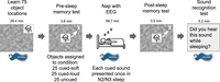
Illustration of experimental procedure, with mean duration for each phase. Participants learned locations of 75 objects paired with sounds on a Perlin noise background. Bioelectric recording setup was next (not shown; mean time 29.1 min, SEM = 1.4 min). Participants then took a memory test where they moved objects (illustrated here by white arrow and X) from the center to the correct location. Following memory testing, participants slept while object sounds were presented in N2 and N3 sleep. Following sleep, participants performed a second memory test, identical to the first. Finally, we played each of the 75 sounds, and participants indicated whether they had heard each sound during sleep. Teapot image is from Brodeur et al. (2010) under a Creative Commons license.
Results
Spatial recall accuracy was matched across conditions before sleep and declined after sleep, especially for cued objects
At the presleep test, participants recalled locations with a mean error of 6.8 cm (SEM = 0.5). As shown in Figure 2, recall accuracy did not differ between cued-loud, cued-soft, and uncued objects (F(2,46) = 1.22, P = 0.30). This lack of a difference was expected due to the procedure for assigning objects to conditions and the fact that cueing during sleep had yet to occur.
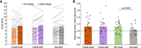
(A) Mean spatial recall error on presleep and postsleep tests (lighter and darker shades, respectively). Error bars represent the standard error of the mean (SEM) for the postsleep error minus presleep error within subjects. (B) Forgetting ratio (fold change in spatial error) for all conditions. Error bars represent the SEM across participants. Statistical significance was determined using a two-tailed t-test.
Recall error increased at the postsleep test to a mean of 7.5 cm (SEM = 0.5), which was significantly worse than presleep (t(23) = 2.89, P = 0.008). We first tested our prediction of greater forgetting for cued objects. We found that forgetting was indeed higher for cued objects compared with uncued objects (t(23) = 2.39, P = 0.025). Next, we tested our second prediction that forgetting would be higher for objects cued with loud sounds. A one-way ANOVA found no differences in forgetting between uncued, cued-loud, and cued-soft items (F(2,46) = 1.64, P = 0.21), and therefore no evidence that cued-loud objects were forgotten more than cued-soft objects (Fig. 2).
Forgetting was increased when cues generated an arousal
To understand the effects of arousal during sleep on reactivation, we categorized cued objects according to whether arousal was apparent during the 10 sec after a stimulus was presented. Arousals were common for both types of sound. Arousals occurred following 43.0% of the soft sounds and 57.9% of the loud sounds.
As shown in Figure 3, forgetting was greater for objects that were cued with arousal than for uncued objects. On the presleep test, recall did not differ by condition (F(2,44) = 2.05, P = 0.14). An ANOVA comparing forgetting ratio for uncued objects, objects cued with arousal, and objects cued without arousal revealed a significant effect (F(2,44) = 3.51, P = 0.039). Post-hoc testing using two-tailed t-tests of all pairs with Bonferroni correction (alpha per test = 0.017) showed a significant difference between objects cued with arousal and uncued objects (t(22) = 3.28, P = 0.003). No significant differences were found in the other pairs (objects cued without arousal vs. uncued, or objects cued with arousal vs. objects cued without arousal).
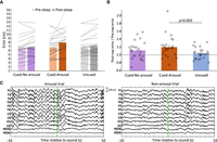
(A) Mean error for uncued objects and objects cued with/without arousal. Error bars represent the SEM for postsleep error minus presleep error within subjects. (B) Forgetting ratio for these three conditions. Error bars represent the standard error of the mean across participants. Statistical significance was determined using a two-tailed t-test. On average, 50.6% of cues caused arousal (SEM = 2.5%, range = 21.5%–76.0% across participants). (C) An example of an arousal trial (left) and a nonarousal trial (right). The green line indicates the onset of sound presentation. While both trials contain sleep spindles and K complexes, the arousal trial shows a long-lasting spectral perturbation indicative of arousal.
Precue alpha power may predict the effect of cueing on memory
Previous research has shown that effects of TMR on memory depend on EEG factors like spindle activity prior to the cue (Antony et al. 2018b) and broadband EEG activity following the cue (Schreiner and Rasch 2015; Whitmore et al. 2022a). Therefore, we tested whether EEG spectra at Cz in the 10 sec prior to the cue or 10 sec after the cue predicted the memory effects of cues.
We divided the spectrum into 21 2.5-Hz-wide bins, spanning 0–52.5 Hz (Fig. 4). Increased power in the high alpha bin (10–12.5 Hz) before the cue was associated with more forgetting of the cued object (t(1,405.8) = 2.25, P = 0.025). However, following FDR multiple-comparisons correction, this correlation was nonsignificant (P = 0.52).
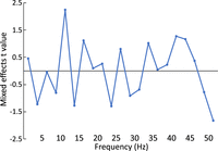
Correlations between the spectrum in the 10 sec prior to the cue and the memory fate of the cued item. Positive values indicate that greater power in a band was associated with more forgetting.
Subjective recall of sleep cues did not influence memory performance
Because participants sometimes report hearing cue sounds during sleep, we examined possible relationships between subjective reports of hearing a sound during sleep and changes in the corresponding object location memories. At the end of the experiment, participants were presented with each cue sound and asked whether they remembered hearing it while sleeping. On average, participants reported definitely hearing 11.3% of the sounds played during sleep (SEM = 2.7%) and possibly hearing 27.9% of the sounds (SEM = 3.8%). We combined these two categories for this analysis. There was no significant difference in forgetting ratio (t(23) = 0.88, P = 0.39) between cued objects with sounds heard during sleep (mean forgetting ratio = 1.2, SEM = 0.2) and cued objects with sounds not heard during sleep (mean forgetting ratio = 1.1, SEM = 0.0).
Cues were presented mostly in N2 and N3 sleep
To verify the sleep stages in which cues were presented, we performed offline sleep staging following standard procedures. Offline staging showed that 93.5% of cues were presented in stage 2 or 3. Full sleep details are shown in Table 1.
Sleep statistics from offline sleep staging
Sleep was highly disrupted compared with a typical memory reactivation experiment
To verify that our experiment produced a high level of sleep disruption, we compared the sleep fragmentation index (Haba-Rubio et al. 2004) between participants in our study and participants in Antony et al. (2018a), which used a similar nap period but presented only quiet sounds. Mean sleep fragmentation was significantly higher in this study compared with the previous study (unpaired t-test; t(81) = 4.5, P < 0.001), with mean SFI of 21.6 ± 1.9 in the current study and mean SFI of 12.6 ± 1.0 in the study by Antony et al. (2018a), indicating that the protocol functioned as intended in disrupting sleep.
Discussion
Results from this experiment confirmed our primary hypothesis that TMR in the context of sleep disruption yields forgetting for reactivated items. We did not observe memory differences between items reactivated with loud and soft sounds. Rather, forgetting was similar for all memories reactivated during the nap.
Cued-loud and cued-soft items may have been subject to similar forgetting because arousals were triggered so often by both types of cues. Furthermore, repeated sleep interruptions during the nap may have lowered the arousal threshold. Our EEG analysis suggests that forgetting occurs when a cue triggers arousal, as indexed by EEG changes, and that the degree of arousal is more important than the objective intensity of the cue.
We found no memory-enhancing effects of TMR despite the strong prevalence of memory improvement in prior TMR studies (Hu et al. 2020). An important implication is thus that TMR studies may fail to find memory benefits if the procedures do not adequately avoid sleep arousal. Here, spatial recall accuracy for objects reactivated without arousal within 10 sec after the cue was comparable with that for uncued objects, with a slight, nonsignificant increase in error. There was also no significant difference in forgetting between objects reactivated with and without arousal, although forgetting was highest for objects reactivated with arousal, moderate for objects reactivated without arousal, and lowest for nonreactivated objects. We propose that this pattern might arise from two sources. First, very small arousals may not be reliably detectable by EEG measures but still cause forgetting. Second, memory disruption from an arousal may spread to other recently reactivated or related memories, as demonstrated by Schechtman et al. (2023), who found that reactivating one memory in sleep causes reactivation of other memories encoded in the same context.
Our results also indicate that sleep state prior to a cue may influence the fate of a memory when it is reactivated. When cues were presented during periods of sleep with relatively high alpha power, the corresponding object locations tended to be recalled less accurately—although this effect did not survive correction for multiple comparisons, so further evidence on this point is needed. Given that increased alpha power during sleep is commonly thought to reflect decreased sleep depth and potential arousability (McKinney et al. 2011), a reasonable interpretation is that cues presented in periods of light sleep are more likely to trigger arousal and weakening.
We also found that participant reports of hearing cues during sleep did not significantly predict memory fate. In contrast, Göldi and Rasch (2019) found that only participants who reported sleep disturbed by cues showed worsening of memory induced by TMR. This difference between experiments may reflect differences in the questions used to assess sleep. Göldi and Rasch (2019) asked participants whether any sounds woke them, whereas we asked whether participants remembered hearing each sound and correlated responses with memory for specific objects. Asking about waking (as opposed to memory for hearing the sound) may therefore provide a better proxy of TMR-induced arousal.
Our results can also be considered in relation to various circumstances of selective memory disruption, which defy the principle that retrieval strengthens memory (Roediger and Butler 2011). Misanin et al. (1968) showed that a retrieval cue followed by electroconvulsive stimulation in rats produced forgetting of the reactivated information. Retrieval in the context of protein synthesis inhibition also produces forgetting (Nader et al. 2000). In our research in patients with epilepsy, we observed memory weakening due to TMR when prominent epileptiform activity occurred in the hippocampus during sleep (Creery et al. 2019). All of these cases of memory weakening may stem from retrieval followed by dysfunctional consolidation. Interrupting sleep while cueing reactivation may prevent memory stabilization and, in the extreme, produce a destabilizing effect, leading to less accurate memories.
In two TMR studies, a second sound presented shortly after a TMR cue abolished the typical memory benefits of TMR (Schreiner et al. 2015; Farthouat et al. 2017). Notably, these studies did not report a weakening effect. However, it is possible that the mechanism is similar. That is, weak or unreliable disruption of consolidation in some studies may abolish the TMR effect, whereas strong disruption of consolidation in other studies, like the current study, may lead to forgetting.
We acknowledge the limitation that many factors likely influenced memory performance after sleep in this study. Some passive decay of memory could certainly have occurred over the delay period. Sleep inertia could also contribute to lower performance at test 2 compared with test 1. However, these factors would be expected to impact reactivated and nonreactivated items equally, as performance in these conditions was matched prior to sleep. Therefore, the selective memory change from test 1 to test 2 for reactivated items cannot easily be attributed to such other factors.
An outstanding question is how changes in the timing of sleep, stimulation, and memory processing would influence the effects of sleep disruption with TMR. In this study, we used a short nap followed by an immediate memory test because this paradigm reliably detects memory benefits from TMR (Rudoy et al. 2009; Creery et al. 2015; Vargas et al. 2019). However, the effects of sleep disruption may be different if memory is measured after a longer delay, as in some TMR studies (Cairney et al. 2018; Whitmore et al. 2022b). If the period of sleep had been longer, as in typical nocturnal sleep, the additional sleep might have counteracted the effect of sleep disruption on memories. It is also unclear whether effects of sleep disruption depend on the age and type of memory; future studies could examine whether sleep disruption produces similar effects for remote memories or emotional memories.
In sum, the present results add to the literature on memory by showing that it is possible for memory reactivation during sleep to lead to a relative weakening, not only strengthening. Weakening may be particularly perpetuated when sleep is disrupted. The ability to systematically produce both strengthening and weakening may prove useful for understanding the discrete memory reactivation processes that operate during sleep. Potential practical applications could also be explored in future research, such as using TMR to weaken memories of traumatic events. A further possibility that should be examined in detail is that individuals with low-quality sleep may experience memory difficulties when memory reactivation occurs in proximity to sleep disruption, regardless of the cause of sleep disruption.
Materials and Methods
Participants
Participants were recruited from Northwestern University and the surrounding community and consisted of 11 males and 13 females 18–30 yr old (mean = 21.6, SD = 3.8). We recruited individuals in this age range without a sleep disorder who felt able to sleep in the afternoon in the laboratory. Our sample size (N = 24) was selected to be comparable with that used in other sleep reactivation experiments. To increase the likelihood of falling asleep, we asked participants to get 1 h less sleep than normal the night before and to avoid nicotine and caffeine on the day of the study. After participants arrived in the laboratory and gave written informed consent, the following six phases transpired: initial learning, bioelectric recording setup, presleep memory test, sleep, postsleep memory test, and a test to assess which sounds (if any) were heard during the nap. Participants received monetary compensation at the conclusion of the experiment. Figure 1 shows an overview of the procedure, which was approved by the Northwestern University Institutional Review Board.
Procedure
Participants learned the locations of 75 pictures of common objects (Brodeur et al. 2010) presented individually on a Perlin noise background (Fig. 1). The learning consisted of two parts. In the first part, participants were shown each object in its correct location and then immediately asked to move the object from the center of the screen to the correct location. Participants were shown the correct location after placement and received visual feedback (either “correct” or “incorrect” with the correct location of the object shown). Correct responses were defined as those placed <3 cm from the correct location. The sound associated with each object was played when it first appeared on the screen and when participants made a correct response.
After performing this procedure for all 75 objects, participants began the second part, which required learning to criterion. Objects were presented in the center of the screen, and participants were asked to move them to the correct location. As in the first part, sounds were presented when the object first appeared and when the participant made a correct response. Objects were presented in a random order, constrained so that the same object could not be shown twice in a row unless it was the only object remaining. After placing the object, participants received feedback in the same manner as during the first part. If the participant placed the object center within 3 cm of the correct location, the criterion was considered achieved and the object was not shown again; otherwise, the object was included in the rotation. Learning ended when the participant correctly placed all the objects.
Bioelectric recording
Following learning, we attached bioelectric recording electrodes for EEG (electroencephalography), EMG (electromyography), and EOG (electro-oculography). Data were recorded using a Neuroscan Synamps2 system referenced to the right mastoid with 26 scalp channels plus horizontal and vertical EOG, and chin EMG. Data were recorded at 1000 Hz with a high-pass filter at 0.1 Hz and a low-pass filter at 100 Hz.
Presleep test
After bioelectric recording setup, participants completed a presleep memory test. In this test, objects were presented in random order, and the participant attempted to place each object at its correct location. No feedback or sounds were presented during the presleep test, and each object was tested once.
After the test was complete, objects were divided into three sets comprising 25 to be cued with loud sounds during sleep, 25 to be cued with soft sounds, and 25 not cued. Objects were assigned to sets so as to match presleep memory performance across sets. In this procedure, the objects were first ranked by accuracy and sequentially assigned to sets so that each set received an equal mix of high-, medium-, and low-accuracy objects. We verified that recall performance did not differ between sets of objects before sleep using a one-way ANOVA comparing mean error for objects in the uncued, cued-soft, and cued-loud conditions.
Sleep period
Participants slept on a futon in the same chamber where they completed the behavioral tasks. When participants reached stage N2 sleep (determined by the experimenter's real-time sleep staging), their initial arousal threshold was determined by presenting a probe sound (bike bell) not related to the memory task. If the sound did not elicit an arousal, the intensity was raised and the sound was presented again, repeating this procedure until an arousal occurred. The intensity that prompted an arousal was used as the initial intensity for the sounds in the loud set.
After finding the arousal threshold, we waited for the participant to return to stable N2 sleep and then began presenting cue sounds. Sounds were presented in random order, with loud and soft sounds intermixed. Loud sounds were presented at a mean intensity of 43 dBa, with intensity continually adjusted to reliably produce brief arousals but avoid prolonged awakenings, defined as more than 1 min of wake or N1 following the cue. Quiet sounds were presented at a low intensity (mean 28 dBa) and adjusted to avoid arousal. The mean intensity of quiet sounds was 31.7% of the initial arousal threshold (SEM = 4.3%) and 6.9% of the mean intensity of loud sounds (SEM = 0.6%). Decibel values were determined by testing using Decibel X on a Redmi Note 9 placed at the location of the participant's head.
Sounds were presented with at least a 10-sec interstimulus interval. If a sound triggered an arousal, cueing was paused until the participant returned to stable N2 or N3 sleep. Each sound was presented only once, allowing us to correlate each object's spatial memory fate with the sleep physiology surrounding sound presentation. After all sounds were presented, the participant was allowed to sleep for 5 min before they were awakened.
Immediately after the participant awoke, we informed them retrospectively that we had played sounds during the nap and asked them whether they remembered hearing any sounds. This was the first time that participants were explicitly told anything about sounds during sleep.
Postsleep test and sound recognition test
Participants performed the postsleep test for object locations, which was identical to the presleep test, ∼5 min after awakening. Following the postsleep test, we presented the 75 object-associated sounds one at a time. Participants were asked to indicate whether they heard each sound during their sleep, with three possible responses: definitely yes, possibly, and definitely no.
Memory performance measurement
We defined the forgetting ratio across the sleep period as postsleep error (in centimeters)/presleep error (in centimeters). We computed a three-level repeated-measures ANOVA to compare forgetting ratio for cued-loud objects, cued-soft objects, and uncued objects. We also computed a two-tailed repeated-measures t-test to compare forgetting ratios for all cued objects with uncued objects.
EEG data processing
EEG data were analyzed in EEGLAB 2020.0 (Delorme and Makeig 2004). Prior to analysis, we visually inspected the data and replaced EEG channels with poor signal quality using interpolation. No other data preprocessing or cleaning was performed. Data from one participant were excluded from arousal and EEG spectrum analyses due to a technical failure that prevented stimulus times from being recorded.
Sleep staging
We first scored the sleep automatically using YASA, a validated automated sleep staging algorithm (Vallat and Walker 2021). An experienced sleep scorer (N.W. Whitmore) then verified and corrected the automated sleep staging using AASM rules (Iber et al. 2007). YASA requires an EEG, EOG, and EMG channel for staging; we performed staging using three channels: left HEOG to right mastoid, Cz to right mastoid, and left mastoid to right mastoid. We visually inspected these channels prior to automatic staging to verify good signal quality; if the signal on Cz was inadequate, we interpolated it using EEGLAB spherical interpolation. Signal on the HEOG and left mastoid channels was adequate for all participants, so no correction was required.
Arousal analysis
To measure the effects of EEG arousal on memory, we performed offline manual scoring to classify each sound cue as arousal-provoking or non-arousal-provoking. During this classification, the rater was blinded to the type of cue (loud vs. soft). The rater examined a segment of time from 10 sec before to 10 sec after each cue and scored the cue as arousal-provoking if an arousal meeting AASM criteria (Iber et al. 2007) occurred in the 10 sec after the cue. We computed the forgetting ratio for three conditions: cued objects that produced an arousal, cued objects that did not produce an arousal, and uncued objects. We then tested whether the forgetting ratio differed for objects cued with arousal, objects cued with no arousal, and uncued objects in a three-level repeated-measures ANOVA.
Between-study comparison of arousal rate
To test whether our participants had overall high levels of sleep disruption, we compared the mean sleep fragmentation index (Haba-Rubio et al. 2004) between our participants and a cohort of participants in a TMR experiment not intended to produce arousal (Antony et al. 2018a) using a two-tailed t-test. Sleep fragmentation index is defined as the number of awakenings or sleep stage shifts per hour of sleep.
Effects of cue perception on memory fate
To assess the impact of remembering perceiving sounds during sleep on memory fate, we compared forgetting ratio for objects where participants reported hearing the associated sound to forgetting ratio for objects where participants did not report hearing the sound. In this analysis, sounds were considered heard if the participant reported definitely or possibly hearing them during the sleep period. We tested whether forgetting ratio differed for cued heard and cued unheard objects using a two-tailed repeated-measures t-test.
EEG predictors of memory fate
To identify EEG features associated with enhancement or weakening of memory, we correlated the forgetting ratio for each cued object with the power spectrum from the 10 sec before the cue. Spectra were computed from electrode Cz using the spectopo function in EEGLAB v2020.0. Each spectrum consisted of 21 2.5-Hz-wide frequency bins, spanning 0–52.5 Hz. Correlations were performed using a mixed model in JMP 15, with participant as a random factor, and spectral power in each bin as a fixed factor.
Acknowledgments
We are grateful for funding from the National Institutes of Health (R01-NS112942, T32-NS047987, and T32-MH06756) and a McKnight Memory and Cognitive Disorders Award from the McKnight Endowment Fund for Neuroscience.
Footnotes
-
Article is online at http://www.learnmem.org/cgi/doi/10.1101/lm.053615.122.
- Received May 27, 2022.
- Accepted February 14, 2023.
This article is distributed exclusively by Cold Spring Harbor Laboratory Press for the first 12 months after the full-issue publication date (see http://learnmem.cshlp.org/site/misc/terms.xhtml). After 12 months, it is available under a Creative Commons License (Attribution-NonCommercial 4.0 International), as described at http://creativecommons.org/licenses/by-nc/4.0/.



