


Lateral entorhinal cortex neurons that project to nucleus accumbens mediate contextual associative memory
- Hayley M. Kuhn1,
- Luis Colon Serrano2,
- Grace A. Stys1,
- Brianna L. Smith1,
- Julia Speckmaier3,
- Brenique D. Dawson4,
- Brooklynn R. Murray1,
- Jin He5,
- Alfred J. Robison1 and
- Andrew L. Eagle6
- 1Department of Physiology, Michigan State University, East Lansing, Michigan 48824, USA
- 2University of Puerto Rico-Cayey, Puerto Rico 00736, USA
- 3Heinrich Heine University Düsseldorf, 40225 Düsseldorf, Germany
- 4University of the Virgin Islands, St. Thomas, Virgin Islands 00802
- 5Department of Biochemistry, Michigan State University, East Lansing, Michigan 48824, USA
- 6Department of Neuroscience, The University of Texas at Dallas, Richardson, Texas 75080, USA
- Corresponding author: andrew.eagle{at}utdallas.edu
Abstract
The lateral entorhinal cortex (LEC) contains glutamatergic projections that innervate the nucleus accumbens (NAc) and may be involved in the encoding of contextual associations with both positive and negative valences, such as those encountered in drug cues or fear conditioning. To determine whether LEC-NAc neurons are activated by the encoding and recall of contexts associated with cocaine or footshock, we measured c-fos expression in these neurons and found that LEC-NAc neurons are activated in both contexts. Specifically, activation patterns of the LEC-NAc were observed in a novel context and reexposure to the same context, highlighting the specific role for LEC-NAc neurons in encoding rather than the valence of a specific event-related memory. Using a combination of circuit-specific chemogenetic tools and behavioral assays, we selectively inactivated LEC-NAc neurons in mice during the encoding and retrieval of memories of contexts associated with cocaine or footshock. Chemogenetic inactivation of LEC-NAc neurons impaired the formation of both positive and negative context-associated memories without affecting the retrieval of an established memory. This finding suggests a critical role for this circuit in the initial encoding of contextual associations. In summary, LEC-NAc neurons facilitate the encoding of contextual information, guiding motivational behaviors without directly mediating the hedonic or aversive properties of these associations.
The nucleus accumbens (NAc), a central hub of reward processing, intricately links with brain circuits governing motivational states and motivated behaviors (Mogenson et al. 1980; Meredith et al. 2008; Sesack and Grace 2010; Klawonn and Malenka 2018). Motivated behaviors are intentional actions by organisms to achieve goals or fulfill needs; examples include feeding, exploration, social interaction, reproduction, avoidance, and drug-seeking behaviors. Various factors, including an organism's physiological state, the surrounding environment, and past experiences, drive motivated behaviors by generating cues that undergo multiple stages of evaluation and encoding. Unless the experience is novel, assessment and encoding are influenced by learning and retrieval mechanisms based on previous associations of an experience (i.e., associative memory) (Simpson and Balsam 2016).
Memory intricately intertwines with valence, salience, encoding, and retrieval; collectively shaping motivated behaviors. The emotional significance of experiences (valence) significantly affects memory formation and retrieval, promoting the behaviors of the positively valenced memory (Berridge 2019). Salience, the prominence of stimuli, impacts memory due to its connection with emotional experiences and influences which behaviors will be performed (Cowan et al. 2021). Encoding, the transformation of input into a retrievable format, is driven by motivation, favoring emotionally charged events (Tye 2018). Past experiences stored in memory further shape motivated behaviors, reinforced by the NAc (Rescorla R 1972), thus implicating the NAc in memory driving motivated behaviors. The NAc also receives innervation from many brain regions that process memory encoding and retrieval. These primarily include glutamatergic inputs from the amygdala, prefrontal cortex, ventral tegmental area, and ventral hippocampus (Britt et al. 2012; Li et al. 2018). These circuits/pathways are critical in mediating various aspects of motivated behavior, including drive, salience, properties of reward, behavioral inhibition, and memory. Reward and punishment enhance the encoding of memory, and this correlates with increased activation in reward (ventral tegmental area and NAc) and memory regions (e.g., hippocampus and cortex) (Shigemune et al. 2013). Ventral tegmental area and NAc also respond to different levels of valence in a monetary incentive delay task (Carter et al. 2009). Understanding how these memory regions mediate NAc function is critical to knowing how memories underlie motivated behavior.
The entorhinal cortex (EC), comprising medial (MEC) and lateral (LEC) divisions, functions as a conduit (Insausti and Amaral 2008) connecting the cortex to the hippocampus and is pivotal for memory establishment and novelty discernment. The MEC has been established in spatial memory (Fyhn et al. 2004); however, the LEC is less well understood, although we do know the LEC assumes a crucial role in associative memory (Wilson et al. 2013b). Unlike the hippocampus and MEC, LEC lacks place or grid cells (Hargreaves et al. 2005) and only appears to indirectly affect spatial representation (Van Cauter et al. 2012). Rather, the LEC encodes temporal and contextual information, synchronizes experiences, nonspatial memory, object-context memory, and novel-object context associations (Deshmukh and Knierim 2011; Wilson et al. 2013a; Tsao et al. 2018; Kuruvilla et al. 2020). There is clear evidence that the EC plays a role in contextual memory (Baldi and Bucherelli 2014; Kang and Han 2021; Marks et al. 2021); however, the contribution of LEC in contextual memory underlying motivated behavior is less well understood.
Newer evidence indicates that LEC may be encoding reward information (Lee et al. 2021; Soma et al. 2023; Issa et al. 2024). These findings indicate that the LEC may be a region poised to mediate motivated behavior. Notably, the LEC innervates the NAc (Krayniak et al. 1981; Phillipson and Griffiths 1985; Sørensen 1985; Aylward and Totterdell 1993; Finch et al. 1995; Totterdell and Meredith 1997; Friedman et al. 2002; Barrientos et al. 2018; Ma et al. 2020). We hypothesized that LEC, via NAc projections, encodes memories that underlie motivated behavior. In the current study, we characterize LEC-NAc neurons using both behavioral assays and chemogenetics to evaluate this projection's role in different valenced contexts, and its potential role in memory formation underlying motivated behavior.
Results
LEC-NAc neurons are activated by novel contexts, regardless of what occurs in that context
LEC neurons, including layer 5, are activated by exposure to a novel context (VanElzakker et al. 2008). However, whether LEC-NAc neurons are similarly activated by contexts is unknown. There are also questions regarding the specificity of activation in the context of various experiences (drug reward, fear dependent, novel context, etc.). To address this question, we utilized a circuit tagging approach (Fig. 1A) to label LEC-NAc neurons with GFP. To assess LEC-NAc activation during encoding of a contextual memory (Fig. 1B), mice underwent cocaine-context conditioning (Coc CPP), saline-context conditioning (Sal CPP), footshock-context conditioning (context + FS), or context exposure (Context). We also included naive mice that were left in their homecage (Home) for the duration of the experiment. One hour after the last exposure to these contexts, we collected formalin-fixed mouse brain tissue that was later processed for c-fos immunohistochemistry to measure neuronal activity. No differences were observed in GFP + LEC-NAc neurons between groups (Fig. 1D,E, left). Exposure to all contexts increased c-fos in LEC, including LEC-NAc neurons (Fig. 1C–E), compared to Home mice (Fig. 1D, middle: one-way ANOVA F2,41 = 8.121, P = 0.0011; Fig. 1D, right: one-way ANOVA F2,41 = 3.303, P = 0.0468; Fig. 1E, middle: one-way ANOVA F2,49 = 8.346, P = 0.0008; one-way ANOVA Fig. 1E, right: F2,49 = 7.400, P = 0.0016; [*] P < 0.05, [**] P < 0.01, [***] P < 0.001 by Holm–Sidak post hoc tests). Interestingly, c-fos expression was increased by novel context but it did not seem to differ whether conditioning was paired with cocaine or saline (Fig. 1D) or whether a context was associated with footshock or no footshock (Fig. 1E). These results suggest that a novel context increases c-fos expression in LEC-NAc neurons regardless of the experience associated with that context.
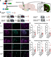
LEC-NAc neurons are activated by novel conditioning contexts. (A) Circuit-tagging method to label LEC-NAc neurons with GFP. Cre-dependent Rosa26eGFP-L10a male and female mice received intracranial infusion of a retrograde Cre virus (HSVrg-hEf1α-Cre) to drive expression of GFP in LEC-NAc neurons. (B) Experimental approach schematic and timeline of brain extraction for c-fos immunohistochemistry. (C) Representative 20 fluorescent images from LEC showing GFP + cells (left, magenta), c-fos + cells (middle, cyan), and merged image (right). White arrows indicate overlap and denote c-fos + GFP + LEC-NAc neurons. (D) Quantification of cell counts in each ROI (per mm2) expressing GFP (left), c-fos (middle), and colabeled (right) in Home (n = 3 mice), Sal CPP (n = 5 mice), and Coc CPP groups (n = 5 mice). (E) Quantification of cell counts in each ROI (per mm2) expressing GFP (left), c-fos (middle), and colabeled (right) in Home (n = 3 mice; same control used in D), Context (n = 4 mice), and Context + FS groups (n = 5 mice). One-way ANOVA followed by Holm–Sidak post hoc comparisons; (*) P < 0.05, (**) P < 0.01, (***) P < 0.001.
LEC-NAc neurons are activated by reexposure to contexts
We next sought to determine whether memory recall would activate LEC-NAc neurons. We hypothesized that reexposure to a context associated with cocaine or footshock would not activate LEC-NAc neurons. To test this hypothesis, we used a circuit-tagging approach (Fig. 2A) and tested recall of contextual memory (Fig. 2B). Circuit-tagged mice were tested for cocaine CPP (Coc CPP Test), saline CPP (Sal CPP Test), or underwent cocaine CPP conditioning, but did not undergo posttest retrieval (No CPP Test). A separate cohort of controls was tested for contextual fear memory (CFC Test), no shock context reexposure (context test), or underwent contextual fear conditioning (CFC) but did not undergo a test for recall (No CFC Test). We also included Home mice as a control group. One hour after the recall test (or 24 h after conditioning, e.g., No CPP Test and No CFC Test groups), we collected formalin-fixed mouse brain tissue that was later processed for c-fos immunohistochemistry to measure neuronal activation to context exposure (Fig. 2C–E). No differences were observed in GFP + LEC-NAc neurons between groups (Fig. 2D,E, left). No CPP Test did not differ from Home mice in c-fos expression in LEC or specifically in LEC-NAc neurons. However, both Sal CPP Test and Coc CPP Test mice had increased c-fos in LEC, including LEC-NAc neurons, compared to Home and No CPP Test groups (Fig. 2D, middle: one-way ANOVA F3,116 = 46.28, P < 0.0001; Fig. 2D, right: one-way ANOVA F3,116 = 12.71, P < 0.0001; [**] P < 0.01, [****] P < 0.0001 by Holm–Sidak post hoc tests), and, interestingly, there were no differences in c-fos between Coc CPP Test and Sal CPP Test. Contrary to our hypothesis that recall would not activate LEC-NAc neurons, these findings suggest that reexposure to conditioned contexts, regardless of the type of drug conditioning, activates LEC-NAc neurons.
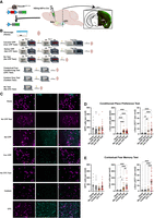
LEC-NAc neurons are activated by retrieval of context. (A) Circuit-tagging method to label LEC-NAc neurons with GFP. Cre-dependent Rosa26eGFP-L10a male and female mice received intracranial infusion of a retrograde Cre virus (HSVrg-hEf1α-Cre) to drive expression of GFP in LEC-NAc neurons. (B) Experimental approach schematic and timeline of brain extraction for c-fos immunohistochemistry. (C) Representative 20 fluorescent images from LEC showing GFP + cells (left, magenta), c-fos + cells (middle, cyan), and merged image (right). White arrows indicate overlap and denote c-fos + GFP + LEC-NAc neurons. (D) Quantification of cell counts in each ROI (per mm2) expressing GFP (left), c-fos (middle), and colabeled (right) in Home (n = 6 mice), No CPP Test (n = 5 mice), Sal CPP Test (n = 5 mice), and Coc CPP Test groups (n = 5 mice). (E) Quantification of cell counts in each ROI (per mm2) expressing GFP (left), c-fos (middle), and colabeled (right) in Home (n = 6 mice; same control used in D), No CFC Test (n = 6 mice), Context Test (n = 5 mice), and CFC Test groups (n = 6 mice). One-way ANOVA followed by Holm–Sidak post hoc comparisons; (*) P < 0.05, (**) P < 0.01, (***) P < 0.001, (****) P < 0.0001.
Contextual fear memory recall also increased activation in LEC (Fig. 2E, middle: one-way ANOVA F3,102 = 23.10, P < 0.0001; Fig. 2E, right: one-way ANOVA F3,102 = 28.43, P < 0.0001; [*] P < 0.05, [***] P < 0.001, [****] P < 0.0001 by Holm–Sidak post hoc tests). No CFC Test did not differ from Home; however, we observed significantly more c-fos expression in total LEC neurons in both our Context Test and CFC Test groups (Fig. 2E, middle), with significantly greater c-fos expression in the CFC Test group compared to Context Test group. Furthermore, we observed significantly more c-fos expression in LEC-NAc neurons in the CFC Test group compared to all other groups (Fig. 2E, right). These findings suggest that LEC neurons, including LEC-NAc neurons, are activated by the reexposure to a context, with further increased activation when that context has been previously associated with footshock. These results provide compelling evidence that LEC-NAc neurons display c-fos-based neuronal activation in response to the recall of novel environments, which may be (excluding contextual fear recall) irrespective of the valence of the experience.
LEC-NAc neuron activity is necessary for the encoding of a drug-context associative memory
To validate our c-fos data, we tested whether LEC-NAc neurons are necessary for contextual associative memory. Contextual associations between positive (appetitive) and negative (aversive) stimuli may be important for predicting information about the occasioned stimuli that occurs in specific contexts. The LEC is important for contextual associative memory (Wilson et al. 2013b; Morrissey and Takehara-Nishiuchi 2014; Basu and Siegelbaum 2015). LEC neurons encode information about cues, and potentially some aspects of rewarding stimuli (Igarashi et al. 2014). Supporting this, cocaine cues activate the LEC in cocaine self-administering rats (Kufahl et al. 2009) and cocaine-dependent human subjects (Bonson et al. 2002). However, the role of LEC, and specifically LEC-NAc neurons, in contextual associative memory is unknown.
We used a chemogenetic approach to test the hypothesis that LEC-NAc neurons play a role in contextual associative memory, specifically in the encoding and retrieval of contextual memory to drive a motivated behavior. We used a circuit-specific viral vector strategy to express inhibitory Gi DREADDs in LEC-NAc neurons (Fig. 3A). The cocaine-conditioned place preference (CPP) paradigm involves both valuation of the drug (valence) and memory for the specific context (e.g., compartment) paired with a drug (Sanchis-Segura and Spanagel 2006). For this reason, we used this task to determine whether LEC-NAc neurons are necessary for making cocaine-context associations. Mice expressing Gi DREADDs or mCherry (control) in LEC-NAc neurons were tested for cocaine CPP. Chemogenetic inactivation of LEC-NAc neurons throughout cocaine conditioning and the cocaine-free posttest retrieval impaired a place preference for cocaine (Supplemental Fig. S1a–e). mCherry mice spent significantly more time within the cocaine-paired chamber compared to the saline-paired chamber. However, Gi mice with chemogenetic inactivation of LEC-NAc neurons throughout conditioning and posttest retrieval spent an equivalent amount of time in both chambers, indicating no preference for the drug.
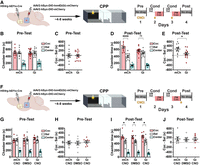
LEC-NAc neurons are necessary for the encoding, but not the retrieval of a cocaine-context association. (A) Circuit-specific DREADD approach to inhibit LEC-NAc neurons during cocaine-context conditioning. (B) Pretest time spent within the prospective cocaine-paired chamber (Coc), prospective saline-paired chamber (Sal), and center chamber in mCherry-expressing controls (mCh) and Gi-DREADD-expressing mice (Gi). There were no differences between Coc and Sal chambers in each group. Two-way RM ANOVA, P > 0.05. (C) Pretest preference (Coc time minus Sal time) within each group. There were no pretest differences between groups. t-test, P > 0.05. (D) Posttest chamber time in each group. mCh mice spent more time in Coc compared to Sal; however, Gi mice spent an equivalent amount of time in both chambers. Two-way RM ANOVA followed by Holm–Sidak post hoc comparisons, (****) P < 0.0001. (E) Posttest preference within each group. Gi mice had impaired preference for the Coc chamber (relative to Sal) compared to mCh controls. t-test, P < 0.05. (F) Circuit-specific DREADD approach to inhibit LEC-NAc neurons during cocaine-context posttest. (G) Pretest time spent within the prospective Coc, Sal, and center chambers in mCh controls (mCh-CNO), Gi mice that receive vehicle during posttest (Gi-DMSO), and Gi mice that receive CNO during posttest (Gi-CNO). There were no differences between Coc and Sal chambers in each group. Two-way RM ANOVA, P > 0.05. (H) Pretest preference within each group. There were no pretest differences between groups. t-test, P > 0.05. (I) Posttest chamber time in each group. All groups spent more time in Coc compared to Sal. Two-way RM ANOVA, (*) P < 0.05. (J) Posttest preference within each group. There were no differences between groups. One-way ANOVA, P > 0.05.
We also tested whether chemogenetic activation of LEC-NAc neurons in the absence of cocaine is inherently rewarding (Supplemental Fig. S2a-e) and found that it is not. Furthermore, chemogenetic activation of LEC-NAc neurons does not enhance cocaine CPP performance (Supplemental Fig. S3a-i). Our finding that chemogenetic inactivation of LEC-NAc neurons impairs cocaine place preference does not appear to be confounded by locomotor effects, as both activation and inactivation of LEC-NAc neurons did not change cocaine-induced locomotor activity or the sensitization to cocaine-induced locomotor activity (Supplemental Fig. S4a-b). Together, these findings suggest that LEC-NAc neuronal activity is necessary for cocaine-context memory, yet it remained unclear whether inactivation of LEC-NAc neurons during the conditioning (i.e., encoding) or during the posttest retrieval (i.e., recall) contributed to the impairment.
To determine whether the LEC-NAc neurons are contributing to the encoding of cocaine-context memory, we again expressed Gi DREADDs (or mCherry in controls) in LEC-NAc neurons (Fig. 3A). However, for this experiment, we aimed to specifically inhibit this circuit during the cocaine conditioning (i.e., during encoding). CNO was administered 30 min prior to each cocaine conditioning session (Fig. 3A, right). No side preferences at pretest were observed between groups, as expected (Fig. 3B: GroupXChamber F1,24 = 0.016, P > 0.05; Fig. 3C: t24 = 0.126, P > 0.05). At the posttest, mCherry control mice spent significantly more time within the cocaine-paired chamber compared to the saline-paired chamber (Fig. 3D: GroupXChamber F1,23 = 6.461, P = 0.018; [****] P < 0.0001). Conversely, Gi mice spent an equivalent amount of time in both the cocaine- and saline-paired chamber, indicating no preference for the drug, and preference was disrupted in Gi mice compared to mCherry mice (Fig. 3E: t23 = 2.542, P = 0.0182). This suggests that LEC-NAc neurons are necessary for the encoding of a cocaine-context associative memory.
We next tested whether LEC-NAc neurons are necessary for the retrieval of a cocaine-context memory. We used a similar approach to the previous experiment but administered CNO 30 min prior to the posttest (Fig. 3F). No preexisting side preferences were observed in the pretest (Fig. 3G: GroupXChamber F2,20 = 0.146, P = 0.8653; Fig. 3H: one-way ANOVA F2,20 = 0.146, P = 0.8653;). However, at the posttest, mCherry controls (mCh-CNO), Gi mice receiving vehicle (Gi-DMSO), and Gi mice receiving CNO (Gi-CNO) all developed a preference for the cocaine-paired chamber (Fig. 3I: ME of Chamber F1,20 = 11.220, P = 0.0032, GroupXChamber F2,20 = 0.726, P = 0.4960). There were no differences in preference between groups (Fig. 3J, P > 0.05). This finding indicates that LEC-NAc neurons are not necessary for the retrieval of a cocaine-context association. Along with the encoding results, these findings suggest that LEC-NAc neuronal activity is important during encoding of cocaine conditioning, but is not necessary for the recall of a cocaine-context associative memory.
LEC-NAc neuron activity is necessary for the encoding of contextual fear
Following validation of the LEC-NAc neurons during encoding of cocaine conditioning, we were interested in whether the LEC-NAc neurons are important in the association of context with a negatively valenced stimulus, that is, footshock. Previous work has identified a role for LEC in both cued and contextual memory. Specifically, lesions and tetrodotoxin-inactivation of general EC (i.e., not specific to LEC) during conditioning impairs contextual fear memory (Majchrzak et al. 2006; Baldi et al. 2013). Furthermore, LEC lesions 1 day after conditioning specifically impaired contextual fear memory (East et al. 2022). However, the specific role of LEC-NAc neurons in contextual fear memory is unknown, and we addressed this question using our circuit-specific inhibitory DREADD approach (Fig. 4A). mCherry and Gi mice were injected with CNO 20 min prior to CFC. The following day, mice were placed back into shock context and evaluated for time spent freezing. Gi mice spent significantly less time freezing compared to mCherry mice across minutes (Fig 4B: ME of group F1,32 = 4.804, P = 0.0032, GroupXMinute F7,224 = 1.799, P = 0.0885) and for the entire duration (Fig. 4C: t32 = 2.192, P = 0.0358). This finding suggests that LEC-NAc neurons are involved in the encoding of CFC.
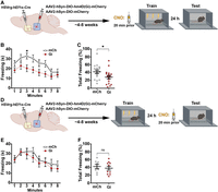
LEC-NAc neurons are necessary for the encoding, but not the retrieval of contextual fear memory. (A) Circuit-specific DREADD approach to inhibit LEC-NAc neurons during footshock-context conditioning. (B) Contextual fear memory test freezing time across 1 min bins in mCherry controls (mCh) and Gi-DREADD mice (Gi). Gi mice had decreased freezing across the test bins compared to mCh controls. Two-way RM ANOVA followed by Holm–Sidak post hoc comparisons, (*) P < 0.05. (C) Percent of freezing time for the entire test within each group. Gi mice spent less % time freezing compared to mCh mice. t-test, (*) P < 0.05. (D) Circuit-specific DREADD approach to inhibit LEC-NAc neurons during contextual fear memory test. (E) Contextual fear memory test freezing time across 1 min bins in mCh controls and Gi mice. There were no differences between groups. Two-way RM ANOVA, P > 0.05. (F) Percent of freezing time for the entire test within each group. There were no differences between groups. t-test, P > 0.05.
To determine whether LEC-NAc neurons are involved in the recall of contextual fear memory, we used mCherry and Gi mice that underwent CFC, and the next day CNO was administered 20 min prior to the test for contextual fear memory recall (Fig. 4D). mCherry and Gi mice had no significant differences in the amount of time spent freezing across minutes (Fig. 4E: P > 0.05) and total duration (Fig. 4F: P > 0.05), suggesting that LEC-NAc inactivation did not impair contextual fear memory recall. This is in line with our findings from cocaine-context associative memory and suggests that LEC-NAc neuronal activity is essential for contextual associative memory encoding, but not contextual memory recall. Importantly, LEC-NAc inactivation itself does not appear to alter anxiety-like behavior, social interaction, or object recognition memory (Supplemental Fig. S5a-j). LEC-NAc neuron activity is therefore critical for the consolidation of contextual associations to valenced stimuli.
Discussion
We show here for the first time that LEC-NAc neurons are important in the encoding of contextual associations with both positive and negative valenced stimuli, such as those encountered with drug cues or fear conditioning. Specifically, our findings reveal that the activity of LEC-NAc neurons is necessary for the encoding of cocaine CPP and contextual fear but may be superfluous during recall. This is based on our finding that the inactivation of LEC-NAc neurons during the retrieval of cocaine CPP and contextual fear memory had no effect. This suggests a selective role for LEC-NAc neurons in the initial memory formation of contextual associations. Interestingly, novel contexts increase c-fos expression in the LEC-NAc pathway, regardless of the positive or negative association with the experience, suggesting that these neurons play a role in the encoding of the context, but potentially not specific event-related memories. This is in line with findings that EC is activated by novelty, whereas output regions, for example, hippocampus, may be regulating the consolidation of memory (Maass et al. 2014; Caban Rivera et al. 2023). Therefore, LEC activity may be passing signals to output regions, such as the hippocampus and NAc, about the importance of a novel context, and that contextual information may be integrated with valence information in other brain regions (Ge et al. 2017; Woods et al. 2018; Hainmueller and Bartos 2020; Marks et al. 2021; Bilash et al. 2023). However, a limitation of this conclusion is that other immediate early genes show differential expression in response to differences in valence. For example, NPAS4 expression is preferentially increased in medial prefrontal cortex by cocaine and natural rewards, compared to shock and restraint (Ye et al. 2016). This suggests that some immediate early genes respond to different valenced events, and therefore c-fos may not be sensitive to valence. The differential expression of immediate early genes, as well as other measures of activation, is also observed in NAc MSNs (Bobadilla et al. 2020) and amygdala (O'Neill et al. 2018; Zhang et al. 2021). Therefore, other measures of activation, for example, in vivo imaging or other immediate early gene analyses, are required to adequately support this conclusion.
The activation of LEC-NAc neurons by contexts raises intriguing questions about the downstream effects within the NAc. One potential point of downstream activation could involve plasticity at NAc medium spiny neurons (MSN) synapses. The LEC projection neurons to the accumbens are glutamatergic (Finch et al. 1995) and glutamatergic inputs to the NAc from other regions throughout the brain have been implicated in plasticity within NAc MSNs after both rewarding stimuli and aversive stimuli (Lee et al. 2013; Tukey et al. 2013; Ma et al. 2014; Pascoli et al. 2014). Information encoded by the LEC-NAc neurons regarding the context in which a stimulus occurs could lead to synaptic modifications within the NAc (Turner et al. 2018). It is also important to note that the LEC preferentially sends projections to the NAc core and lateral shell, compared to the medial shell (Totterdell and Meredith 1997), and LEC projections synapse onto both D1 and D2 NAc MSNs (Barrientos et al. 2018; Li et al. 2018; Ma et al. 2020). Differences in synaptic plasticity across D1 and D2 NAc MSNs can differentially modulate behavioral outcomes and potential susceptibility to psychiatric disease risk (Kupchik and Kalivas 2017; Bariselli et al. 2019; Fox and Lobo 2019; Allichon et al. 2021); however, it is unclear whether LEC inputs onto NAc MSNs mediate plasticity, and whether they do so differently across these different regions and cell types. Nevertheless, our findings show that LEC-NAc neurons modulate contextual associative memory and lay the foundation for exploring the mechanism(s) of LEC effects on NAc MSNs.
LEC sends projections to the NAc (Krayniak et al. 1981; Phillipson and Griffiths 1985; Sørensen 1985; Aylward and Totterdell 1993; Finch et al. 1995; Totterdell and Meredith 1997; Friedman et al. 2002; Barrientos et al. 2018; Ma et al. 2020), however little is known beyond that. LEC-NAc neurons are likely pyramidal (Finch et al. 1995) and are primarily observed in layers 2–3 (Totterdell and Meredith 1997). We have also observed that LEC-NAc neurons are glutamatergic (Supplemental Fig. S6). However, it is unclear whether this specific subpopulation of LEC neurons also sends collaterals to other regions that may be involved in contextual memory formation (e.g., hippocampus) that may have contributed to the effects observed in our DREADD inhibition experiments. EC pyramidal neurons do send collaterals between layers of the EC, or between MEC and LEC (Tamamaki and Nojyo 1993; Canto et al. 2008; Nilssen et al. 2019), as well as hippocampus; however, we know very little about NAc-projecting LEC neurons. Our own findings show that while LEC-NAc neurons do send collaterals to dorsal HPC, these dual projections are rare (Supplemental Fig. S7). This does not preclude LEC-NAc projections also sending collaterals elsewhere. Alternatively, these neurons could also convey information back to layer 2/3 fan cells and other pyramidal neurons (Canto et al. 2008; Nilssen et al. 2019). Finally, older studies have identified medial EC neurons that also send projections to the NAc (Krayniak et al. 1981), yet very little investigation of the function of medial EC-NAc neurons has been done. Based on the known role of medial EC in context and spatial memory (Fyhn et al. 2004; Morrissey and Takehara-Nishiuchi 2014), we may see similar effects on contextual fear memory and cocaine CPP as we observed in our LEC-NAc experiments. Because so little is known about the EC-NAc pathways, it will be important for future studies to investigate the function of these separate pathways and full circuitry of these projections, both within the local EC circuit, and between other brain regions.
We found that LEC-NAc neurons are necessary for the encoding of memory and that they do not appear to directly mediate the hedonic or aversive properties of memory associations. Additionally, since activation of these neurons is not inherently rewarding, we can hypothesize that these neurons are conveying information that is necessary for a multiplex memory association, such as integrating information about potentially relevant contexts. This would align with literature indicating that the NAc itself is integrating motivational salience and reward value from various inputs (e.g., LEC) to guide motivated behaviors (Cooper and Knutson 2008; Schmidt et al. 2019; Richter et al. 2020; Vázquez et al. 2022).
We demonstrate here the significance of LEC-NAc circuitry in mediating motivated behaviors driven by associated memories. The LEC has been implicated in associative memory formation (Deshmukh and Knierim 2011; Wilson et al. 2013a; Tsao et al. 2018; Kuruvilla et al. 2020). Our findings expand on this body of evidence to suggest that the subpopulation of LEC-NAc neurons may serve as a conduit for conveying contextual associative information crucial for guiding motivated behaviors. By associating stimuli with specific contexts, LEC-NAc neurons may influence the motivation to pursue rewards or avoid unpleasant stimuli, thereby shaping behavioral responses in diverse environmental contexts.
Materials and Methods
Animals
All experiments were approved by the Institutional Animal Care and Use Committee at Michigan State University in accordance with AAALAC. Male and female C57Bl/6J mice (3–5/cage, 7–8 week-old upon arrival from Jackson Labs) were allowed at least 5 days to acclimate to the facility prior to any experimental procedures. Rosa26eGFP-L10a mice were a generous gift from the laboratory of Dr. Gina Leinninger at Michigan State University. Unless otherwise stated, all mice were group housed in a 12:12 h light–dark cycle with ad libitum food and water. Temperature (22°C) and humidity (50%–55%) were held constant in animal housing and behavioral testing rooms.
Stereotaxic surgery and viral vectors
Stereotaxic surgery was conducted as previously described (Eagle et al. 2020). For circuit-tagging experiments, retrograde Cre vector (HSVrg-hEf1α-Cre; 0.5 μL; Gene Delivery Core, Massachusetts General Hospital) was infused into NAc (+1.6 AP, ±1.5 ML, −4.4 DV relative to bregma, 10° angle). For DREADD experiments, the same retrograde Cre vector was infused into NAc and control vector (AAV2-hSyn-DIO-mCherry; 0.5 μL; UNC Vector Core) or DREADD Gi vector [AAV2-hSyn-DIO-hM4D(Gi)-mCherry; 0.5 μL; Addgene] was infused into the LEC (−3.3 AP, ±4.1 ML, −4.6 DV relative to bregma, 0° angle). All experimental procedures commenced at least 4–8 weeks following surgeries.
Behavioral testing
Behavior was collected using an IR-CCD camera (Panasonic) and analyzed using automated video tracking software (CleverSys or AnyMaze). Animals were transported to the behavioral testing rooms in their homecages and allowed 30 min to habituate to the room before testing began.
Cocaine CPP
CPP was conducted as previously described (Gajewski et al. 2019). Briefly, mice were tested for cocaine CPP in a three-chamber CPP box (San Diego Instruments). On day 1, mice received a pretest (no cocaine or saline), where they were allowed to explore the entire box for 15–20 min. On days 2–3, mice received injections of saline paired with one chamber in the morning for 30 min and injections of cocaine (7.5 mg/kg; IP) paired with the opposite chamber for 30 min in the afternoon (chamber counterbalanced by group). On day 4, mice were again tested in a posttest (no cocaine or saline) and allowed to freely explore the entire box.
Contextual fear conditioning
CFC was conducted in a conditioning chamber as previously described (Eagle et al. 2015). For conditioning sessions, mice were placed in the chamber for 30 sec, followed by delivery of three mild electric footshocks (0.8 mA, 1 sec duration), each separated by a 60 sec intershock interval in which no shock was delivered. Mice remained another 30 sec in the chamber following the last shock delivery. The percentage of time spent freezing was video scored by two blind, independent observers, and the average score between observers was calculated as freezing. Freezing was defined as the lack of skeletal movement for a period >1 sec.
Chemogenetic experiments
Gi (and control) mice were treated with clozapine-N-oxide (CNO; 0.3 mg/kg; IP) dissolved in 5% DMSO. CNO was injected 20 min prior to testing and conditioning, depending on the experiment. Mice were left in their homecages during the 20 min period until the experiment began.
Immunohistochemistry
Immunofluorescent analysis was conducted as previously described (Eagle et al. 2020). Mice were transcardially perfused with cold PBS, followed by 10% formalin. Brains from all immunostaining experiments were postfixed 24 h in 10% formalin, cryopreserved in 30% sucrose in PBS, and sliced frozen on an SM2010R microtome (Leica) into 35 μm sections. Immunohistochemistry was performed using primary antibodies against c-fos (2250S; 1:1000; Cell Signaling Technology) and GFP (ab5450; 1:4000; Abcam); and secondary antibodies (1:200; Jackson Immunoresearch) conjugated to fluorescent markers (AlexaFluor 488; Cy3; Cy5). Fluorescent images were visualized on an Olympus FluoView 1000 filter-based laser scanning confocal microscope or a Nikon Eclipse Ni-U Upright Fluorescent Microscope. Coexpression of GFP and c-fos were quantified using ImageJ (NIH) software by an experimenter blinded to conditions. Raw cell counts from each ROI (0.16 mm2) were normalized to the number of cells per 1 mm2.
Statistics and reproducibility
For statistical analyses, we used one-way and two-way ANOVAs (with some including repeated measures or mixed effects) followed by Holm–Sidak corrected post hoc comparisons in the case of significant omnibus effects, and independent samples t-tests. Alpha criterion was set to 0.05. To ensure reproducibility, separate cohorts were included in behavioral testing. All statistical analyses were performed using Prism software (GraphPad Software).
Acknowledgments
We would like to thank Dr. Gina Leinninger and Dr. Irving Vega, co-directors of the Bridge to PhD in Neuroscience Program (BPNP-ENDURE) at MSU, Dr. Susanne Hoffman-Benning of the MSU Molecular Biology German Student Exchange Program, Dr. Alice Stanford, Director of the University of the Virgin Islands Undergraduate Research Training Initiative for Student Enhancement (U-RISE), and Dr. Ryan Sweeder of the Lyman Briggs College at MSU. We would also like to thank Ken Moon for his incredible support in maintaining our colony of transgenic mice. Images created with BioRender.com.
Footnotes
-
[Supplemental material is available for this article.]
-
Article is online at http://www.learnmem.org/cgi/doi/10.1101/lm.054026.124.
- Received June 4, 2024.
- Accepted October 21, 2024.
This article is distributed exclusively by Cold Spring Harbor Laboratory Press for the first 12 months after the full-issue publication date (see http://learnmem.cshlp.org/site/misc/terms.xhtml). After 12 months, it is available under a Creative Commons License (Attribution-NonCommercial 4.0 International), as described at http://creativecommons.org/licenses/by-nc/4.0/.



