


Chemogenetic activation of the ventral subiculum–BNST pathway reduces context fear expression
- 1Department of Biology, Barnard College of Columbia University, New York, New York 10027, USA
- 2Department of Neuroscience and Behavior, Barnard College of Columbia University, New York, New York 10027, USA
- Corresponding author: ebauer{at}barnard.edu
-
↵3 These authors contributed equally to this work.
Abstract
An inability to reduce fear in nonthreatening environments characterizes many anxiety disorders. The pathway from the ventral subiculum (vSUB) to the bed nucleus of the stria terminalis (BNST) is more active in safe contexts than in aversive ones, as indexed by FOS expression. Here, we used chemogenetic techniques to specifically activate the vSUB–BNST pathway during both context and cued fear expression by expressing a Cre-dependent hM3D(Gq) receptor in BNST-projecting vSUB neurons. Activation of the vSUB–BNST pathway reduced context but not cued fear expression. These data suggest that the vSUB–BNST pathway contributes to behavioral responses to nonaversive contexts.
Anxiety disorders are often characterized by exaggerated fear responses to both threatening and neutral stimuli (Jovanovic and Ressler 2010; Lopresto et al. 2016). Numerous studies of the neural correlates of defensive behaviors in response to threat have identified contributions from areas such as the basolateral and central nuclei of the amygdala, the bed nucleus of the stria terminalis (BNST), and the prelimbic cortex (Sotres-Bayon and Quirk 2010; Herry and Johansen 2014; Bauer 2023). It is equally important to identify neural pathways that are active when animals are in a safe environment. Indeed, one hallmark of posttraumatic stress disorder (PTSD) is that intrusive thoughts and memories recur in “safe” contexts. Understanding how animals shift from exploratory to defensive behaviors can shed light on how the networks underlying these behaviors become disordered in psychiatric illnesses.
The ventral subiculum (vSUB), the main output of the ventral hippocampus, has been implicated in exploration as well as spatial learning (Burns et al. 1996; Floresco et al. 1997; Shankaranarayana Rao et al. 2001; Torromino et al. 2019). Within the vSUB, populations of neurons project to forebrain targets, the amygdala, and the anterior BNST (Canteras and Swanson 1992; Radley and Sawchenko 2011; Wee and MacAskill 2020; Urien et al. 2022). The BNST in turn processes adaptive and pathological anxiety (Walker et al. 2003), as well as unpredictable stressful events (Goode et al. 2019). Lesions or reversible inactivation of the BNST with muscimol impair the expression but not the acquisition of context fear conditioning (Duvarci et al. 2009; Pelrine et al. 2016). Additionally, expression of both FOS and ARC proteins increases in the anterolateral BNST in male rodents following context fear expression (Lemos et al. 2010; Urien et al. 2021; Urien and Bauer 2022). Lesions or inactivation of the vSUB impair context fear memory (Maren 1999; Biedenkapp and Rudy 2009).
High-frequency stimulation of vSUB inputs to the anteromedial BNST reduces anxiety in both basal and anxiogenic situations (Glangetas et al. 2017). Moreover, we have recently demonstrated that fewer BNST-projecting vSUB neurons express FOS when animals are exposed to an aversive context than when they are in a nonaversive context (Urien et al. 2022). These data suggest that the vSUB–BNST pathway contributes to the encoding of responses to anxiolytic contexts. Thus, to determine whether direct manipulation of the vSUB–BNST pathway modulates the expression of context fear conditioning, we expressed a CRE-dependent designer receptor activated by designer drug (DREADD) hM3D(Gq) in vSUB neurons. A retrograde AAV containing CRE (rgAAV-CRE-GFP) infused into the BNST allowed for expression of CRE recombinase in all neurons projecting to the BNST. AAV-hSyn-DIO-hM3D(Gq)-mCherry infused into the vSUB contained the floxed inverted sequence of hM3D(Gq), which was reoriented in the presence of CRE, allowing for expression of hM3D(Gq) specifically in BNST-projecting vSUB neurons. Our results suggest that activation of hM3D(Gq) in vSUB–BNST neurons prior to context fear expression reduced behavioral freezing in the aversive context. In contrast, activation of hM3D(Gq) prior to cued fear expression did not reduce fear expression.
Adult male Sprague Dawley rats (350–550 g; Charles River Laboratories) were housed in pairs with ad libitum food and water and maintained on a 12-h light–dark cycle. A total of 55 animals was used, of which 40 were analyzed and 15 were excluded due to misplaced injections during surgery. All procedures were approved by Columbia University's Animal Care and Use Committee in accordance with the National Institutes of Health Guide for the Care and Use of Laboratory Animals.
General surgery procedures were identical to those described previously (Urien et al. 2022). Using a 1-µL Hamilton syringe, 0.5 µL of AAV-CRE-GFP (Addgene 105540-AAVrg) or saline was infused into the BNST of both hemispheres (AP = −0.12, ML = ±1.5, and DV = −6.2), lowered at a 20° angle to avoid the ventricles, and left in place for 5 min to prevent diffusion of the adenovirus along the needle track. During the same surgery, a different 1-µL Hamilton syringe was lowered into the vSUB of both hemispheres at three locations (AP = −5.5, ML = ±3.6, and DV = −7.6; AP = −5.5, ML = ±4.2, and DV = −7.75; and AP = −5.5, ML = ±5.0, and DV = −7.9), and 0.25 µL of AAV hm3dGq-mCherry (Addgene 44361-AAV9) or saline as a control was infused at each location (Fig. 1A). Animals with misplaced injections were not included in the analysis. Animals recovered for 4 wk before behavioral testing or perfusion.
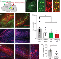
Cre-dependent hM3D(Gq) infusion into the vSUB specifically activates the vSUB–BNST pathway. (A) Schematic of dual-viral technique. (B) Example of CRE injection into the BNST. Scale bar, 100 µm. (C) Example of CRE (left), hM3D(Gq) (middle), and merged image (right) in the vSUB. Scale bar, 25 µm. (D) Extent of CRE and hM3D(Gq) expression in the vSUB. Dotted lines delineate the border of the vSUB. Scale bars, 100 µm. (E) FOS+ cells as a percentage of NeuN+ cells in the vSUB across four groups of animals. The asterisk represents a significant difference between animals receiving both viruses and the hM3D(Gq) agonist J60, animals receiving both viruses and saline injection, and animals receiving either the CRE virus alone or the hM3D(Gq) virus alone and J60 injections (P < 0.05). (F) Expression of FOS (blue) and NeuN (pink) throughout the vSUB in an example animal. Scale bars, 100 µm. (G) Expression of hM3D(Gq) and FOS (blue) in the vSUB. Scale bars, 100 µm. (H) The number of double-labeled FOS+/hM3D(Gq) cells as a percentage of hM3D(Gq)+ cells. Asterisks indicate a significant difference in FOS expression in animals receiving both viral infusions and injection of the hM3D(Gq) agonist J60 compared with animals receiving both viral infusions and injection of saline (P < 0.001).
Before proceeding with behavioral testing, we confirmed the feasibility of our approach in 21 animals, three of which were excluded due to misplaced injections during surgery. Four weeks after surgery, animals received injections of either the hM3D(Gq) agonist JHU37160-dihydrochloride (J60; HelloBio HB6261) at 0.1 mg/kg i.p. or 0.5 mL of 0.9% sterile saline. The J60 agonist has been used in several behavioral paradigms at concentrations ranging from 0.1 to 1 mg/kg (Zhang et al. 2020; Heinsbroek et al. 2021; Huang et al. 2021; Li and Hollis 2021). Higher concentrations of J60 (3 mg/kg) have been found to produce nonspecific effects (Lawson et al. 2023). Animals were returned to their cages for 90 min to allow for the effects of the i.p. injection (30 min) and expression of FOS protein (60 min) and were then perfused with 0.1 M phosphate buffer (PB) and 4% paraformaldehyde in 0.1 M PB after an i.p. injection of 100 mg/kg sodium pentobarbital. Brains were postfixed for 4 h and then transferred to a 20% sucrose solution for at least 48 h.
Tissue was sectioned at 60 µm using a Vibratome covering the entire rostral to caudal extent of both the BNST and vSUB. Sections containing the BNST were washed in PB and mounted on slides to verify injection placement. Sections containing the vSUB were processed to visualize FOS expression as described previously (Urien et al. 2022). Sections were incubated in primary polyclonal rabbit anti-c-FOS (1:2000; Abcam ab190289) and mouse anti-NeuN (1:1000; Millipore MAB377) antibody for 48 h. Sections were then incubated with the corresponding secondary fluorescent goat antirabbit/mouse antibody (1:200; Invitrogen) for 1 h, mounted on slides, and coverslipped.
All imaging and counts were performed by experimenters blind to group assignments of the animals. BNST and vSUB images (10×) were generated using a Nikon A1000 confocal microscope and NIS element software. Boundaries of regions were delineated and cells were counted manually using ImageJ software. For all animals, three nonconsecutive sections in one hemisphere were quantified in each animal.
To determine the feasibility of the dual-viral chemogenetic approach for activating the specific vSUB–BNST pathway, injections of the retrograde AAVrg-CRE-GFP into the BNST were verified (Fig. 1B) and colocalization of CRE-GFP and hm3dGq-mCherry was observed in the vSUB (Fig. 1C). Indeed, colocalization was observed throughout the medial–lateral extent of the vSUB (Fig. 1D). We compared FOS expression (Fig. 1E) in the vSUB overall in animals receiving infusions of a retrograde AAVrg-CRE-GFP into the BNST and hm3dGq-mCherry into the vSUB (n = 5) versus animals receiving saline (“saline,” n = 5) and versus animals receiving J60 but only one of the two viruses [“CRE” only, n = 4 or “hM3D(Gq)” only, n = 4]. We first counted FOS expression throughout the entire vSUB across all groups of animals (Fig. 1F). A one-way ANOVA revealed a significant effect of group (F(3,50) = 6.53; P < 0.001). Tukey's HSD post-hoc tests revealed that animals receiving both viral injections and the agonist J60 had significantly more FOS expression in the vSUB than all other groups (Ps < 0.05). We then restricted our analysis to FOS expression in hM3D(Gq)+ cells in animals receiving both viral injections and either the agonist J60 or saline (Fig. 1G). An unpaired Student's t-test revealed that animals receiving J60 injections had significantly more FOS expression in hM3D(Gq)+ neurons than animals receiving saline (t(28) = 6.2; P < 0.001) (Fig. 1H). Thus, J60 administration up-regulated neural activity in neurons expressing hm3dGq-mCherry as well as in the vSUB as a whole. In these animals, the percentage of neurons (NeuN+) expressing the hm3dGq-mCherry virus was 9.53% ± 1.55%. Our previous data suggest that ∼16% of neurons within the vSUB project to the BNST (Urien et al. 2022). Thus, the majority of neurons in the vSUB–BNST pathway expressed hm3dGq-mCherry using this dual-viral technique.
We next asked whether chemogenetic activation of the vSUB–BNST pathway modulated context and cued fear expression (Fig. 2A). We used 34 animals, 12 of which were excluded from behavioral analyses due to misplaced injections during surgery. Three groups of animals were tested: One group received infusions of both rgAAV-CRE-GFP into the BNST and hm3dGq-mCherry into the vSUB and the agonist J60 (0.1 mg/kg i.p.) 30 min prior to fear expression (n = 7), the second group received both viruses and saline (0.5 mL i.p.) 30 min prior to fear expression (n = 6), and the third group received only hm3dGq-mCherry into the vSUB and J60 30 min prior to fear expression (n = 9).
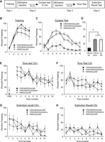
Activation of the BNST–vSUB pathway reduces context fear expression but not cued fear expression. (A) Schematic of the behavioral protocol. (B) Percentage freezing in response to the CS+ tone during training (±SEM). (C) Percentage freezing during each of the 10 min of context fear expression (±SEM). Animals received infusions of the hM3D(Gq) agonist J60 or saline 30 min prior to testing. Animals with both CRE and hM3D(Gq) viral infusions and J60 injection froze significantly less than the other two groups. (D) Percentage freezing averaged over the 10 min of context fear expression. The asterisk denotes a significant difference between groups (P < 0.05). (E,F) Percentage freezing (±SEM) to 10 CS+ tones (E) and 10 CS− tones (F) during the tone test. Animals received infusions of J60 or saline 30 min prior to testing. (G,H) Percentage freezing in response to 10 CS+ tones (G) and 10 CS− tones (H) 24 h after cued fear expression, testing extinction recall. Animals were tested drug-free for extinction recall.
Animals were habituated to the training environment for 10 min 24 h prior to training. All animals were then trained drug-free with six CS+ tones (CS+ = 2 kHz, 80 dB, 30 sec) coterminating with a footshock (US = 0.5-mA shock, 1 sec) interleaved with six CS− tones (CS− = white noise, 80 dB, 30 sec) that were not paired with the shock (Fig. 2B). The training session was 20 min. The first CS occurred after 5 min, and the ITI was random between 30 and 60 sec. A one-way repeated measures (RM) ANOVA revealed a significant effect of time (F(5,95) = 27.04; P < 0.001), with freezing increasing across the six CS+ tones but no effect of group (F(2,19) = 1.23; P = 0.31) or time × group interaction (F(10,95) = 0.53; P = 0.86).
Twenty-four hours later, animals were tested for context fear expression by placing them in the training context for 10 min (Fig. 2C,D). Thirty minutes prior to testing, animals received either a J60 or saline injection. A one-way RM-ANOVA revealed a significant difference between groups (F(2,19) = 5.39; P = 0.014) and a significant effect of time in minutes (F(9,171) = 5.43; P < 0.001) but no group × time interaction (F(18,171) = 1.26; P = 0.22). Tukey's HSD post-hoc tests revealed that the animals receiving both viral injections and J60 froze significantly less than animals receiving saline injections (P = 0.034) and animals with only hM3D(Gq) infusions and J60 injections (P = 0.021). Thus, activation of the vSUB–BNST pathway significantly reduces context fear expression.
Twenty-four hours after context fear expression, the animals were tested for cued fear expression. Thirty minutes prior to testing, the animals received either J60 or saline injections. There were no significant differences between groups in freezing in response to either the CS+ (Fig. 2E) or CS− (Fig. 2F). Specifically, for freezing in response to the CS+, a one-way RM-ANOVA revealed a significant effect of time (F(9,171) = 7.23; P < 0.001), with freezing decreasing across the 10 tones, but no effect of group (F(2,19) = 0.36; P = 0.7) or time × group interaction (F(18,171) = 0.96; P = 0.51). For freezing in response to the CS−, a one-way RM-ANOVA revealed a significant effect of time (F(9,171) = 4.52; P < 0.001), with freezing decreasing across the 10 tones, but no effect of group (F(2,19) = 1.71; P = 0.21) or time × group interaction (F(18,171) = 0.77; P = 0.74).
It is possible that activation of the BNST–vSUB pathway during fear expression affected fear extinction learning. To examine this, the animals were tested for fear extinction recall 24 h later, drug-free. Again, there were no significant differences between groups. For freezing in response to the CS+, a one-way RM-ANOVA revealed a significant effect of time (F(9,171) = 6.4; P < 0.001), with freezing decreasing across the 10 tones, but no effect of group (F(2,19) = 0.89; P = 0.43) or time × group interaction (F(18,171) = 0.7; P = 0.8) (Fig. 2G). For freezing in response to the CS−, a one-way RM-ANOVA revealed no significant effect of time (F(9,171) = 1.86; P = 0.06) and no effect of group (F(2,19) = 2.37; P = 0.12) or time × group interaction (F(18,171) = 0.92; P = 0.55) (Fig. 2H). In sum, activation of the vSUB–BNST pathway did not affect cued fear expression or cued extinction learning.
Together, these data suggest that activation of the vSUB–BNST pathway plays a specific role in mediating responses to environments rather than discrete cues in which the cue predicts the US with 100% contingency. The combined use of rgAAVs and DREADD technology allowed us to specifically activate the vSUB–BNST pathway. Here, we used the DREADD agonist J60 to activate hM3D(Gq) receptors. The more commonly used DREADD agonist clozapine-N-oxide (CNO) can be metabolized to clozapine, which can interact with both DREADDs as well as endogenous receptors (for review, see Mahler and Aston-Jones 2018). This is particularly problematic as clozapine itself can reduce context fear conditioning (Yavas et al. 2021). It should be noted that at higher concentrations (3 mg/kg), J60 administration can have nonspecific effects on behavior (Lawson et al. 2023). Here, the DREADD agonist J60 increased FOS expression in hM3D(Gq)-expressing neurons when compared with animals receiving saline. It also increased FOS expression throughout the vSUB in animals receiving both viral injections compared with animals receiving only one virus.
Here, we examined freezing in response to both a CS+ paired with an aversive shock and an unpaired CS−, given that lesions of the BNST reduce freezing in response to a CS− (Duvarci et al. 2009). However, we did not observe any effect on freezing in response to either the CS+ or CS− during activation of the vSUB–BNST pathway. Others have reported changes in neuronal activity within the BNST in response to both cued fear conditioning and expression (Haufler et al. 2013; Bjorni et al. 2020). As the BNST is a heterogeneous structure, our data suggest that inputs modulating CS responses in the BNST do not originate in the vSUB. Rather, the vSUB–BNST pathway appears to modulate defensive behaviors in aversive contexts. The BNST also processes cues that are poor predictors of aversive stimuli, as well as long duration cues (Goode et al. 2019). Further research should clarify whether the vSUB–BNST pathway modulates context fear expression only or any stimulus (cue or context) that is a poor predictor. More broadly, the vSUB–BNST pathway might contribute to shifting the balance between exploratory and defensive behaviors.
Acknowledgments
This work was supported by National Institutes of Health grant 1R15MH122969-01 to E.P.B., and Major Research Instrumentation grant 1828264 from the National Science Foundation Division of Biological Infrastructure.
Footnotes
-
Article is online at http://www.learnmem.org/cgi/doi/10.1101/lm.053797.123.
- Received May 26, 2023.
- Accepted August 3, 2023.
This article is distributed exclusively by Cold Spring Harbor Laboratory Press for the first 12 months after the full-issue publication date (see http://learnmem.cshlp.org/site/misc/terms.xhtml). After 12 months, it is available under a Creative Commons License (Attribution-NonCommercial 4.0 International), as described at http://creativecommons.org/licenses/by-nc/4.0/.



