


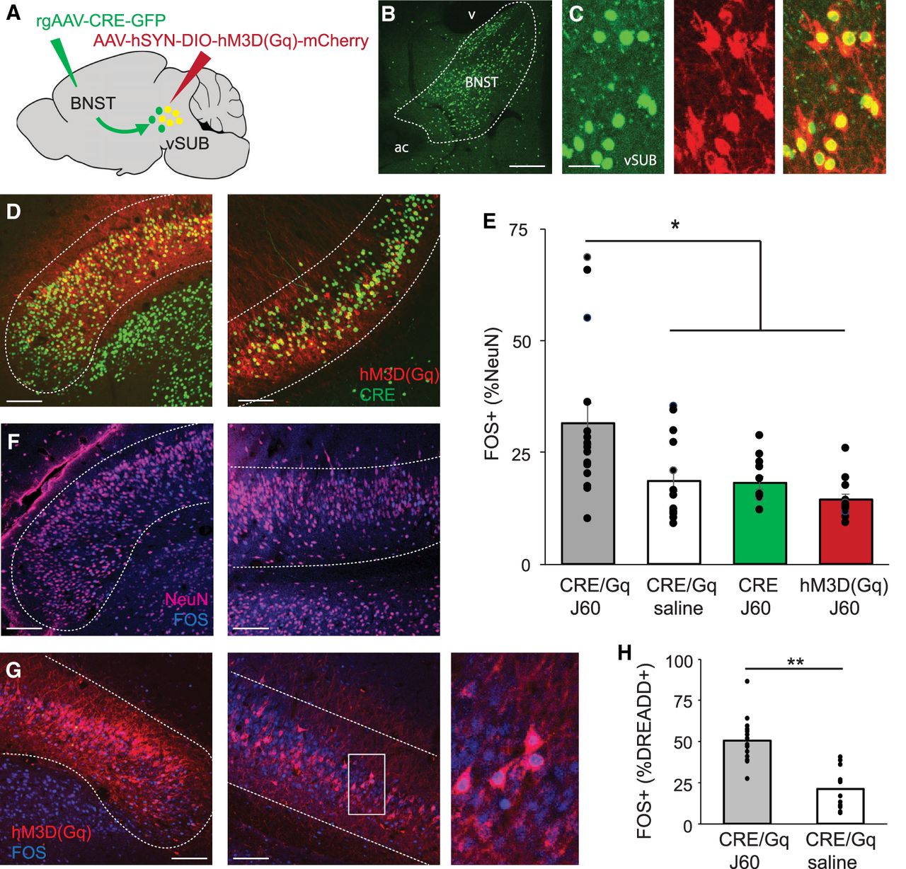
Cre-dependent hM3D(Gq) infusion into the vSUB specifically activates the vSUB–BNST pathway. (A) Schematic of dual-viral technique. (B) Example of CRE injection into the BNST. Scale bar, 100 µm. (C) Example of CRE (left), hM3D(Gq) (middle), and merged image (right) in the vSUB. Scale bar, 25 µm. (D) Extent of CRE and hM3D(Gq) expression in the vSUB. Dotted lines delineate the border of the vSUB. Scale bars, 100 µm. (E) FOS+ cells as a percentage of NeuN+ cells in the vSUB across four groups of animals. The asterisk represents a significant difference between animals receiving both viruses and the hM3D(Gq) agonist J60, animals receiving both viruses and saline injection, and animals receiving either the CRE virus alone or the hM3D(Gq) virus alone and J60 injections (P < 0.05). (F) Expression of FOS (blue) and NeuN (pink) throughout the vSUB in an example animal. Scale bars, 100 µm. (G) Expression of hM3D(Gq) and FOS (blue) in the vSUB. Scale bars, 100 µm. (H) The number of double-labeled FOS+/hM3D(Gq) cells as a percentage of hM3D(Gq)+ cells. Asterisks indicate a significant difference in FOS expression in animals receiving both viral infusions and injection of the hM3D(Gq) agonist J60 compared with animals receiving both viral infusions and injection of saline (P < 0.001).



