


Differential effects of emotional valence on mnemonic performance with greater hippocampal maturity
- 1Cognitive Neuroscience Program, Department of Psychology, Florida International University, Miami, Florida 33199, USA
- 2Clinical Science Program, Department of Psychology, Florida International University, Miami, Florida 33199, USA
- 3Center for Children and Families, Florida International University, Miami, Florida 33199, USA
- 4Department of Radiology and Medical Imaging, University of Virginia, Charlottesville, Virginia 22903, USA
- Corresponding author: amattfel{at}fiu.edu
Abstract
The hippocampal formation (HF) facilitates declarative memory, with subfields providing unique contributions to memory performance. Maturational differences across subfields facilitate a shift toward increased memory specificity, with peripuberty sitting at the inflection point. Peripuberty is also a sensitive period in the development of anxiety disorders. We believe HF development during puberty is critical to negative overgeneralization, a common feature of anxiety disorders. To investigate this claim, we examined the relationship between mnemonic generalization and a cross-sectional pubertal maturity index (PMI) derived from partial least squares correlation (PLSC) analyses of subfield volumes and structural connectivity from T1-weighted and diffusion-weighted scans, respectively. Participants aged 9–14 yr, from clinical and community sources, performed a recognition task with emotionally valent (positive, negative, and neutral) images. HF volumetric PMI was positively associated with generalization for negative images. Hippocampal–medial prefrontal cortex connectivity PMI evidenced a behavioral relationship similar to that of the HF volumetric approach. These findings reflect a novel developmentally related balance between generalization behavior supported by the hippocampus and its connections with other regions, with maturational differences in this balance potentially contributing to negative overgeneralization during peripuberty.
Memory increases in specificity, driven by maturation of key neurobiological substrates taking root in early childhood, with spatial pattern separation being observed as early as 18 mo (Lambert et al. 2015) and mnemonic object discrimination being observed as early as early as age 4 yr (Ngo et al. 2018; Canada et al. 2019). Memory specificity changes during another susceptible window around adolescence (Lavenex et al. 2007; Lee et al. 2014; Daugherty et al. 2017; Keresztes et al. 2018). This sensitive period of adolescent development before, during, and after puberty is referred to as peripuberty. Whether these specificity-supporting neurobiological mechanisms are similarly used across different stimulus valences (e.g., emotional vs. neutral) remains unknown. Around the same developmental window, the prevalence of anxiety disorders increases (Beesdo et al. 2009). Overgeneralization to threatening stimuli has been proposed as a possible etiological account for anxiety disorders (Dunsmoor and Paz 2015; Laufer et al. 2016). According to this theory, stimuli with similarities to originally conditioned fearful stimuli show increased aversive responding and show a shallow response decay gradient (Greenberg et al. 2013; Dymond et al. 2015; Laufer et al. 2016). In the current study, we aimed to build on this classical conditioning literature to show that similar mechanisms influence more episodic-like memory behavior within the context of an emotional mnemonic similarity task (McMakin et al. 2022) and examined how cross-sectional indices of neurobiological maturation around puberty are associated with behaviors. Together, understanding how developmental differences in memory specificity interact with emotional salience of stimuli may provide important insight into our understanding of negative overgeneralization, a characteristic symptom of anxiety where individuals generalize negative associations to similar events (Lissek et al. 2014).
A network of interconnected regions in the medial temporal lobe (MTL) and medial prefrontal cortex (mPFC) governs the specificity of declarative memories. The MTL is comprised of the rhinal cortices, the amygdala, and the hippocampal formation (HF) (Squire et al. 2004). Several distinct subfields comprise the HF—dentate gyrus [DG]; cornu ammonis [CA] 1, 2, and 3; and subiculum—with the different subfields playing important roles in the processes of pattern separation and completion. Pattern separation is the process thought to underlie discrimination, responsible for the orthogonalization of memories, whereas pattern completion relates to generalization (Yassa and Stark 2011). The sparse firing of the DG granule cells combined with the strong mossy fiber input to the CA3 can bias the system toward pattern separation under the right circumstances (Yassa and Stark 2011; Rolls 2013; Knierim and Neunuebel 2016). In contrast, weaker input to CA3 directly from layer II of the entorhinal cortex leads to pattern completion given the recurrent collateral architecture of the CA3, which is then propagated to the CA1 via Schaffer collaterals. While the HF is well suited for governing memory specificity, its functions are potently influenced by input arising from other subcortical and cortical regions. The amygdala contributes to the encoding of emotional salience (Mcgaugh and Ayala 2013) and the modulation of memory strength (McGaugh 2004), with damage selectively impairing gist or generalized memories (Adolphs et al. 2001, 2005), while the mPFC interacts with both the amygdala and HF (Colgin 2011; Xu and Sudhof 2013; Jin and Maren 2015; Sekeres et al. 2018). We contend that the balance in memory specificity arises from developmental differences across all these regions.
Notably, the HF and mPFC have prolonged developmental trajectories extending well into adulthood, with particularly rapid changes occurring around the onset of adolescence (Giedd et al. 1996; Lavenex and Banta Lavenex 2013; DeMaster et al. 2014; Avino et al. 2018), while the amygdala appears to mature earlier in life (Tottenham and Gabard-Durnam 2017). Examination of the HF alone has shown that developmental differences are associated with increased memory discriminability (Lee et al. 2014; Keresztes et al. 2018; Riggins et al. 2018). However, memory does not emerge from the independent contributions of individual brain regions but rather reflects the product of interacting networks (McIntosh 2000). Thus, similar to others, we contend that developmentally related increases in memory specificity reflect the composition of maturational changes across regions rather than the contribution of single regions alone (Keresztes et al. 2018). Specifically, we propose that connections between the AMY and HF will play a disproportionate role in the specificity of emotional stimuli; however, the mPFC, via the nucleus reuniens (RE) of the thalamus, has been shown to exert a powerful role on fear memory specificity (Xu and Sudhof 2013). Thus, we also examined the relation between generalization of emotional stimuli and the cross-sectional development of mPFC–HF anatomical connections.
To examine the role of MTL maturation during puberty on generalization of stimuli with emotional valence, we reanalyzed an existing data set (McMakin et al. 2022). In the original study, participants completed an emotional similarity task (Leal et al. 2014b) where they rated the valence of stimuli (negative, neutral, and positive) during a study session in the scanner. Participants returned to the scanner after 12 h (chosen within the original study to provide an interval suitable to contain a full night's sleep for the sleep condition within the study) for a surprise memory test where they made “old” or “new” judgments. Separate volumetric and connectivity-based HF and amygdala maturity were assessed using a partial least squares correlation (PLSC) analysis using the structural and diffusion-weighted scans (Keresztes et al. 2017, 2018). Behavioral measures of generalization were related to the multivariate measures of volume and connectivity. Here, negative and neutral images are given key focus, as the difference in emotional arousal was greatest between negative and neutral images in the original data set, with positive falling somewhere between the two (Leal et al. 2014b). Youth with anxiety are also known to experience wide generalization gradients to negative stimuli (Greenberg et al. 2013). Previous work using this data set found that the amygdala showed greater functional coupling with CA1 and dorsal mPFC during negative items that were later generalized (McMakin et al. 2022). We evaluated the hypothesis that emotional valence of stimuli differentially produced greater negative generalization with differences in hippocampal and amygdala maturity.
Results
Hippocampal maturation during puberty was related to increases in generalization
To test our hypothesis that valenced images differentially impacted mnemonic generalization across pubertal development, we
used a dimension reduction strategy known as PLSC to produce a singular index of hippocampal pubertal maturity (pubertal maturity
index [PMI]) and examined how the relationship between generalization performance was impacted as a function of PMI. Lure
generalization index (LGI) scores were calculated (Eq. 1) to assess an individual's tendency to behaviorally generalize emotional stimuli, with the idea that this emergent behavior
was dependent on pattern completion. As multiple models are presented in the current study, a Holm–Sidak-corrected alpha of
0.026 was used to assess significance.
 (1)
(1)
As predicted, when assessing generalization (e.g., LGI), we observed divergent patterns between negative and neutral stimuli (maturity × valence: b = 0.037, z = 3.074, P = 0.002). We found that enhanced generalization of negative images associated with greater HF-PMI [b = .029, CI (95) = (0.002, 0.055), t(33) = 2.228, P = 0.034], while neutral generalization remained steady or decreased slightly as a function of HF volumetric PMI [b = −0.011, CI (95) = (−0.037, 0.015), t(33) = −0.863, P = 0.395]. These same relationships were not observed in LGI for positive relative to neutral images (maturity × valence: b = 0.021, z = 1.747, P = 0.081) (Fig. 1B). For full results, please refer to Supplemental Table S1.
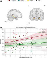
Differential relation between hippocampal maturity and memory performance across stimulus valence. (A) Hippocampal subfield volume estimates from the CA1, DG/CA2/CA3/CA4, subiculum, and entorhinal cortices (ERC) were used to produce a metric of hippocampal maturity as in Keresztes et al. (2017). (B) A significant interaction appeared for LGI performance (maturity × valence: b = 0.037, z = 3.074, P = 0.002), where negative LGI increases relative to neutral LGI as hippocampal maturity increases. This same pattern was not observed for positive relative to neutral LGI (maturity × valence: b = 0.021, z = 1.747, P = 0.081).
As rhinal cortex maturity increases, negative LGI performance and neutral LGI performance converge
To assess whether these relationships were unique to the hippocampus or were present in the MTL at large, we probed for similar associations between pubertal maturity and emotional valence in neighboring MTL regions such as the rhinal cortices and amygdala, two regions anatomically connected to the hippocampus (Suzuki and Amaral 1994) and important for memory and emotional processing (McGaugh 2004), with the rhinal cortices experiencing earlier development than both the amygdala and HF (Insausti et al. 2010). These regions were chosen a priori, as they provide critical contributions to hippocampal memory function but should be fully developed in a peripubertal age range. Rhinal PMI significantly interacted with negative and neutral lure generalization (LGI) (maturity × valence: b = −0.051, z = −2.637, P = 0.008) and positive versus neutral LGI (maturity × valence: b = −0.056, z = −2.902, P = 0.004) (Fig. 2A). As rhinal maturity increased, LGI decreased for negative images [b = −0.020, CI (95) = (−0.068, 0.029), t(33) = −0.831, P = 0.414] and positive images [b = −0.042, CI (95) = (−0.083, 0.002), t(33) = −2.157, P = 0.040] and increased for neutral images [b = 0.024, CI (95) = (−0.019, 0.068), t(33) = 1.146, P = 0.261]. For the amygdala, PMI did not appear to interact with valence to predict differences in negative versus neutral LGI (maturity × valence: b = 0.001, z = 0.087, P = 0.931) or positive versus neutral LGI (maturity × valence: b = −0.029, z = −1.733, P = 0.083) (Fig. 2B). For full results of analyses in this subsection, please refer to Supplemental Tables S2 and S3.
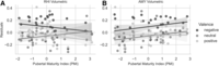
Rhinal cortex (RHI) PMI predicts reduced LDI and LGI for valenced information, but amygdala (AMY) PMI shows no differential relationship between PMI and valence with LDI and LGI performance. (A) RHI PMI predicted reductions in generalization for negative relative to neutral images (maturity × valence: b = −0.051, z = −2.637, P = 0.008) and for positive relative to neutral images (maturity × valence: b = −0.056, z = −2.902, P = 0.004). (B) Amygdala volumetric PMI did not differ between negative versus neutral stimuli for generalization (maturity × valence: b = 0.001, z = 0.087, P = 0.931) or positive versus neutral stimuli for generalization (maturity × valence: b = −0.029, z = −1.733, P = 0.083) behavior.
Hippocampal–mPFC anatomical connectivity PMI predicts increases in negative generalization
We next assessed whether nonvolumetric measures of PMI—in this case, diffusion-weighted connectivity—were related to changes in generalization with valence. We produced amygdala–hippocampal (AMY–HF) connectivity PMI (see the Materials and Methods) and examined the associations between this measure and image valence with LGI. When examining generalization (LGI), we did not observe a divergence in generalization with AMY–HF connectivity PMI for negative versus neutral images (maturity × valence: b = −0.003, z = −0.169, P = 0.866) or positive versus neutral images (maturity × valence: b = −0.012, z = −0.772, P = 0.440) (Fig. 3A). For full results, see Supplemental Table S4. We expanded this approach to other regions sharing anatomical connectivity with the hippocampus, creating pubertal maturity indices for hippocampal–rhinal (HF–RHI) and hippocampal–mPFC (HF–mPFC) connectivity. When examining generalization, we did not observe differences across our HF–RHI connectivity PMI for negative versus neutral images (maturity × valence: b = 0.028, z = 1.445, P = 0.148) or for positive versus neutral images (maturity × valence: b = −0.021, z = −1.062, P = 0.288) (Fig. 3B). For full results of these analyses, see Supplemental Table S5.
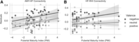
Association between connectivity PMI and generalization (LGI). (A) AMY–HF connectivity PMI showed no differential relationship with LGI across negative and neutral stimuli (maturity × valence: b = −0.003, z = −0.169, P = 0.866) or across positive and neutral stimuli (maturity × valence: b = −0.012, z = −0.772, P = 0.440). (B) Similarly, HF–RHI connectivity PMI showed no valence-related differential relationship with mnemonic performance in negative versus neutral LGI (maturity × valence: b = 0.028, z = 1.445, P = 0.148) or positive versus neutral LGI (maturity × valence: b = −0.021, z = −1.062, P = 0.288).
When examining the relationship between generalization and HF–mPFC anatomical connectivity PMI, a familiar pattern was evident. Generalization exhibited a significant relationship with valence in the opposite pattern for negative versus neutral images (maturity × valence: b = 0.027, z = 2.432, P = 0.015) and no relationship for positive versus neutral images (maturity × valence: b = 0.004, z = 0.316, P = 0.752) (Fig. 4). Greater HF–mPFC connectivity PMI was associated with greater negative image generalization [b = 0.023, CI (95) = (−0.002, 0.047), t(33) = 1.868, P = 0.072] and reduced neutral image generalization [b = −0.005, CI (95) = (−0.028, 0.019), t(33) = −0.415, P = 0.681]. These effects mirror the interactions observed in our HF volumetric PMI analyses (Fig. 1B). For full results of the analyses in this subsection, see Supplemental Table S6.
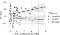
HF–mPFC connectivity PMI shows a pattern of results similar to that of volumetric hippocampal PMI in relation to mnemonic performance. There was a significant interaction between HF–mPFC connectivity PMI and valence-predicting LGI for negative versus neutral images (maturity × valence: b = 0.027, z = 2.432, P = 0.015), with no evidence of a relationship between positive versus neutral images (maturity × valence: b = 0.004, z = 0.316, P = 0.752), showing a pattern remarkably like the volumetric HF results.
Discussion
We examined the role of MTL pubertal maturity (defined both volumetrically and through diffusion-weighted connectivity) in generalization of stimuli with emotional valence. We found that greater volumetric pubertal maturity index (PMI) within the HF was related to greater mnemonic generalization (LGI) for negative images. This effect of volumetric PMI was constrained to the HF and was not present in the amygdala. When examining the same relations between behavior and a pubertal maturation index of the rhinal cortices, we observed the opposite pattern from the hippocampal formation. The divergence in the observed results may arise from differential connectivity and modulation by the amygdala across the hippocampus and rhinal cortices (Paz and Pare 2013). Alternatively, the disparate regions may independently generate mnemonic signals that are interpreted by downstream structures (Buzsaki 2010) to govern behavior depending on contextual or motivational differences. Using the same PLSC approach but using anatomical connectivity between the HF and other regions, a similar differential pattern of mnemonic generalization across stimulus valence was evident in the connectivity PMI between the HF and mPFC. Thus, puberty-related development of the HF, assessed both volumetrically and through its connections to the mPFC, produces differential patterns of generalization for negative compared with neutral images during a sensitive developmental period. Importantly, positive stimuli show similar but weaker effects, and their results fall between negative and neutral stimuli on all measures, likely arising from interactions between arousal and valence during this developmental window. Valence and arousal were previously assessed in older adults using the same task (Leal et al. 2014b). This finding may have implications for our understanding of negative overgeneralization, a core feature of anxiety, which increases in prevalence during this developmental window (Beesdo et al. 2009; Lissek et al. 2014). Specifically, maturational changes in how emotional valence drives generalization may contribute to increasing negative overgeneralization and anxiety among vulnerable youths.
Previous studies have found positive associations between age-related changes in the brain and mnemonic discrimination performance for neutral stimuli. For example, discrimination of neutral objects was positively associated with changes in DG/CA2/CA3/CA4 subfield volume across age (Lee et al. 2014). Similarly, greater volume of the DG/CA2/CA3/CA4 was associated with enhanced memory performance for neutral trivia information in early childhood (Riggins et al. 2018). When using a PLSC approach, a similar positive association between mnemonic discrimination of object images with HF maturity was shown (Keresztes et al. 2018). Our data corroborate these findings, showing that changes in HF pubertal maturity are associated with reductions in generalization of neutral images. The current study also extends the findings from prior work, showing generalization of negative images was positively associated with HF maturity. Together, our results suggest that generalization behavior is impacted differentially by emotional valence across pubertal development. Generalization of emotional memories is adaptive and common. Behaviorally, learning under conditions of threat (Starita et al. 2019) or following negative (Schechtman et al. 2010) or aversive (Resnik et al. 2011) feedback facilitates generalization of episodic memories. While the amygdala has long been known to facilitate consolidation (McGaugh 2004) and has recently been shown to prioritize declarative memories through its coordinated neural activity with the hippocampus (Manns and Bass 2016), damage to this region specifically impairs gist memories and leaves detailed memories spared (Adolphs et al. 2001, 2005). Neurons in the amygdala have specific tuning properties related to generalization (Resnik and Paz 2015), and specific populations in the lateral amygdala signal general versus cue-specific associations (Ghosh and Chattarji 2015). Our results provide a novel contribution to the potential mechanisms underlying generalization and suggest that the HF plays an important role in generalization of valenced information. These results support the notion that the generalization of emotional stimuli is supported by a broad network of regions (Asok et al. 2019).
Differences in development across the HF contribute to heterogeneity of behavior well into adulthood. The HF experiences a protracted postnatal development in primates, with histology indicating profound increases in the DG volume (Lavenex et al. 2007). This protracted development has also been observed in studies of the human hippocampus using structural MRI. Some studies have reported greater hippocampal volume from childhood through adolescence (Ostby et al. 2009), while others have localized these changes to the hippocampal body and reported concomitant decreases of volume in the head of the hippocampus (Gogtay et al. 2006; DeMaster et al. 2014). Focus on individual subfields using structural MRI have identified increasing DG/CA2/CA3/CA4 volume as participants approach adolescence (Lee et al. 2014; Daugherty et al. 2017). Stereological studies in macaques have similarly demonstrated that DG granule cell populations mature well beyond early life (Jabes et al. 2011). Notably, the CA3 appears to mature in lockstep with the DG (Jabes et al. 2011). Histological studies in humans have corroborated these findings, showing increases in postnatal volume across the HF marked by prolonged development of the DG and CA3, while the rhinal cortices exhibit a comparatively earlier maturational plateau (Insausti et al. 2010).
These differences in subfield volume and trajectories across development highlight the importance of using methods to simplify the input data when constructing models of HF development. Heterogeneity across the hippocampus is best captured by use of multivariate decomposition techniques (Keresztes et al. 2018). Our data demonstrate that findings such as those of Keresztes et al. (2018) are replicable across samples and different but related tasks using such techniques. Our data also demonstrate these techniques are sensitive to region-specific changes, as indicated by different mnemonic outcome predictions between different constituent regions of the MTL (HF, amygdala, and rhinal cortices).
In addition to multiple internal structures, the HF shares robust anatomical connections with the mPFC (Varela et al. 2014). The mPFC contributes to schema development and is sensitive to information congruent with previous experience (van Kesteren et al. 2010). Developmentally related differences in the detection of congruence between previous and current contexts could influence generalization and discrimination behavior. The mPFC also plays a key role in mnemonic control, influencing memory specificity and generalization at encoding and retrieval (Xu and Sudhof 2013). When examining connectivity between the HF and mPFC using our PLSC metric, we found results strikingly comparable with those in the initial analysis using only intra-HF volume.
The transition from childhood to adolescence (“peripuberty”) is a neurodevelopmental window when neural networks associated with generalization (Bowman and Zeithamova 2018) and emotional processing (Phelps and LeDoux 2005) undergo dynamic change. At the same time, disorders of emotion such as anxiety increase, putting youth at a higher risk for escalating mental health problems (e.g., depression) in later adolescence and adulthood (Pine et al. 1998). In later adulthood, studies have found that individuals with higher depressive symptoms in a community sample of adults show worse pattern separation performance with increasing symptom severity (Shelton and Kirwan 2013; Leal et al. 2014a). Other studies have found that individuals with deficits in mnemonic discrimination have increased shock expectancy during fear conditioning paradigms (Lange et al. 2017), indicating that a link between mnemonic performance and negative or fear generalization exists. The current study provides evidence that patterns of negative generalization appear to increase across HF development and may also be related to patterns of connectivity between the HF and mPFC. Findings that these patterns change during adolescent pubertal development indicate that the changes to emotional processing during puberty may play a key role in the development of anxiety disorders, and interventions to potentially address and improve outcomes for these disorders should likely be provided from a young age, as rates of anxiety disorders dramatically increase in this developmental window.
Our study has four important limitations. First, the sample was cross-sectional rather than longitudinal, and as such we cannot make claims that these changes in volume with puberty are developmental in nature (Raz and Lindenberger 2011). Second, HF subfields were defined using a consensus labeling approach. No harmonized protocol for HF segmentation exists, but one is currently in development (Olsen et al. 2019). Third, the limited sample size of the current study warrants caution of overinterpretation and the need for replication in a larger sample. Finally, the delay interval of the task may play a key role in understanding these findings, as longer delay intervals between task and study have been shown to reduce lure discrimination performance (Leal et al. 2019). Future studies may seek to examine the impact of interval duration on mnemonic generalization outcomes when paired with measures of hippocampal maturity.
Despite these limitations, our results both support and expand on previous findings in the literature. Puberty-related volumetric changes in the HF capture differences in generalization related to emotional salience. This difference in behavior appears unique to developmental changes in the hippocampus and may be related to changes in interregional connectivity between the HF and mPFC. Changes to the developmentally related balance between discrimination and generalization may support mechanisms of negative overgeneralization, a common feature of anxiety disorders often taking root during this developmental period.
Materials and Methods
Participants
The data reported in this study were originally acquired for McMakin et al. (2022), which recruited 52 peripubertal youths (age 9–14 yr) from a university pediatric anxiety specialty clinic and the Miami-Dade community at large to examine the neurobiological correlates of negative overgeneralization. A strategy of recruiting from anxiety clinics and the community was used to maximize variability in key dimensions of interest—generalization of emotional stimuli. Youths with anxiety are known to experience wide generalization gradients to negative stimuli in particular (Greenberg et al. 2013), which clinically appears as a tendency to pathologically extend fear from aversive contexts (e.g., house fire) to safe contexts with shared features (e.g., campfire). Participants recruited for the study were assessed for medical and psychiatric exclusionary criteria (e.g., current depressive episode, bipolar disorder, posttraumatic stress disorder, conduct disorder, oppositional defiant disorder, psychotic disorders, and obsessive–compulsive disorder) based on a screener assessing key symptoms associated with DSM-IV diagnoses and/or a parent-reported diagnosis. Following intake, three participants were ineligible for the study (left-handed) and another dropped out, leaving 48 volunteers to participate in the study session scan. Two participants were excluded from the test session scan due to excessive movement and an error in the experimental paradigm. A third participant failed to show up for their appointment, leaving 45 participants who completed the test session scan. Eleven participants were excluded following the test session—one failed to show up to their appointment, six were excluded for poor performance (hit rate for targets was 1.5 SD below the average performance), three were excluded for errors in triggering the onset of the scanner with the task, and one was removed for excessive motion during the scan (defined as >0.5 mm of framewise displacement for >30% of volumes)—leaving 34 participants (11.4 yr ± 2.0 yr, 16 female) in the final sample. All participants provided written informed consent (legal guardian) and assent and were compensated for their time.
Pubertal status
The pubertal development scale (PDS) (Petersen et al. 1988) was used to assess self-reported pubertal status. The scored measure captured two five-point scales corresponding to gonadal and adrenal hormone-related pubertal development and one composite measure capturing overall pubertal development, expressed as Tanner staging (1–5) (Shirtcliff et al. 2009). Subsequent analyses used the composite metric (PDS-Shirtcliff) to produce pubertal index scores using PLSC.
Anxiety
The pediatric anxiety rating scale (PARS) (The Research Units of Pediatric Pharmacology Anxiety Study Group 2002) was used to measure anxiety symptom severity based on clinician interview. The PARS-6 algorithm (Caporino et al. 2013) for scoring was used, providing a range of values between 0 and 30, with 30 indicating the most severe anxiety. The PARS was administered by a master's level clinician and a bachelor's level research assistant with clinical interview training. The parent and child were interviewed separately, and supervision was given by a trained clinical psychologist (D.L. McMakin). Parent and child ratings informed clinician ratings, with this clinician rating being used as the primary measure of anxiety severity.
Behavioral procedures and methods
Participants took part in an emotional similarity task (Supplemental Fig. S1). The task included an incidental encoding session inside of an MRI scanner, during which participants viewed scenes (2000 msec) and were instructed to endorse images as either negative, neutral, or positive. Stimuli were separated by a jittered interstimulus interval (2000–6000 msec), during which a white central fixation was presented on a black background. Each scene was presented once, totaling 145 images (48 negative, 47 neutral, and 50 positive). Participants returned 12 h later for a surprise memory test, with 17 of them performing the task in the morning postsleep, and the other 17 returning in the evening after 12 h of their normal routine. During the test session, which also took place within an MRI scanner, participants endorsed images as either “old” or “new.” A total of 284 images was presented: 48 targets (16 each of negative, neutral, and positive)—repetitions of the images presented during the incidental encoding session; 97 lures (32 negative, 32 neutral, and 33 positive)—images similar to but not exactly the same as an image shown during the incidental encoding session; and 139 foils (42 negative, 49 neutral, and 48 positive)—images never presented before and not sharing similarity to the original images. Participants indicated whether each image was either “old” (the subject recalls seeing that exact image during the study session) or “new.” Researchers instructed participants to endorse images as “old” only if they were the exact same as the image seen during the study session and to respond while the image was still on the screen. The timing of trial events was similar to the study session. Each image valence had a lure generalization index (LGI) calculated by subtracting the proportion of old responses when given a foil image from the proportion of old responses when given a lure image (Eq. 1).
Neuroimaging data collection and preprocessing
A 3T Siemens Magnetom Prisma scanner with a 32-channel head coil at the Center for Imaging Science at Florida International University was used to collect neuroimaging data. Diffusion-weighted images (1.7 mm isotropic) using a multiband sequence (slice acceleration factor = 3, 96 directions, seven b = 0 frames, and four b-values: six directions with 500 sec/mm2, 15 directions with 1000 sec/mm2, 15 directions with 2000 sec/mm2, and 60 directions with 3000 sec/mm2) in addition to a T1-weighted magnetization-prepared rapid gradient echo sequence (Mprage: TR = 2500 msec, TE = 2.9 msec, flip angle = 8°, FOV = 256 mm, 176 sagittal slices, and voxel size = 1 mm isotropic) were collected. For distortion correction, a field map opposite of the phase encode direction of the dMRI acquisition was acquired.
Each participant's T1-weighted structural scan was processed through FreeSurfer's (version 6.0.0; Fischl 2012) “recon-all” algorithm to obtain cortical surface reconstruction and cortical/subcortical segmentations. Each participant's diffusion-weighted scan was registered to FreeSurfer structural space using boundary-based registration, with the reference image being the first acquired b = 0 frame. The FreeSurfer parcellation and segmentation file (aparc + aseg) was then binarized and transformed into diffusion space using FreeSurfer's “ApplyVolTransform” tool and was then binarized and dilated by 1 mm to include edge voxels to function as a brain mask. Susceptibility distortion correction was then performed using FSL Topup, followed by eddy current correction using FSL Eddy on the diffusion data masked by the dMRI space brain mask. FSL's Bedpostx (Jbabdi et al. 2012) was then used on the preprocessed data to model crossing fibers within each brain voxel. The results of Bedpostx were the basis of all subsequent probabilistic tractography-based analyses.
Delineating amygdala subregions
The amygdala is comprised of several nuclei, each having unique anatomical connections to cortical and subcortical targets. We used probabilistic tractography combined with a novel method of k-means clustering analysis to identify amygdala subregions. Probabilistic tractography was computed from bilateral masks of the amygdala to 24 ipsilateral cortical and subcortical targets while avoiding the ventricles. This resulted in separate files (one for each target) containing the total number of random walks completed from each voxel in the amygdala mask to that specific target. Each file was then vectorized and included in an n × m array, with n being the number of voxels in the left or right amygdala masks, and m being the number of amygdala targets. The array was then subjected to a k-means clustering using the “scikit-learn” (0.23.2) (Pedregosa et al. 2011) module in Python 3.8.3 with a limit of four clusters, with voxels serving as samples and targets serving as features. A k-means cluster value was then assigned to each voxel in the amygdala masks based on their connectivity across the targets (also known as features), which were then coerced back into their three-dimensional anatomical representations. Each cluster's volume estimates were then extracted while also controlling for overall intracranial volume (Supplemental Fig. S2).
Delineating hippocampal and cortical regions of interest (ROIs)
A consensus labeling approach was used to segment the hippocampal subfields (bilateral DG/CA2/CA3/CA4, CA1, and subiculum) and the posterolateral and anteromedial entorhinal cortices. First, manual segmentations (Yassa and Stark 2009; Yushkevich et al. 2015) were applied to an atlas set of 19 T1 Mprage scans and their corresponding T2-FSE scans (oblique orientation perpendicular to the long axis of the hippocampus; 0.47 mm2 in-plane, 2.0-mm slice thickness). Weighted consensus labeling from the atlas to an unlabeled T1 was accomplished by normalizing the atlas set to the unlabeled subject and applying multiatlas segmentation with joint label fusion (Wang and Yushkevich 2013). This approach capitalizes on both label and intensity information and has been used in several recent publications to segment hippocampal subfields (Fig. 3A; Sinha et al. 2018; Brown et al. 2019). These hippocampal subfield segmentations were then visually inspected by A. Kimbler. Within the sample of subjects, no correction was needed. Cortical ROIs (e.g., perirhinal cortex, parahippocampal cortex, amygdala, superior frontal cortex, caudal and rostral anterior cingulate cortices, and medial orbitofrontal cortex) were created by binarizing FreeSurfer segmentations.
Measures of regional connectivity
To examine the structural connectivity between regions, probabilistic tractography was conducted using FSL's Probtrackx (Behrens et al. 2003, 2007) with 25,000 streamline samples (step length = 0.5, curvature threshold = 0.2, maximum steps = 2000) in each seed voxel to produce a connectivity distribution between each seed and target region while avoiding paths through the ventricles. A list of the connections examined appears in Table 1.
Seeds to target regions for connectivity analyses
Regional volumetric maturity estimates using PLSC
Partial least squares correlation (PLSC) was used to produce a singular maturity value associated with pubertal development for each of the following regions: HF (DG/CA2/CA3/CA4, CA1, subiculum, and posterolateral and anteromedial entorhinal cortices), rhinal (RHI) cortices (perirhinal cortex and parahippocampal cortex), and amygdala (AMY) (resulting k-means clusters were determined by probabilistic tractography). For each region, estimates of subregion volume were extracted and were corrected for intercranial volume and age by multiplying each volume estimate by the ratio of age-predicted whole-brain volume to actual whole-brain volume obtained via FreeSurfer. A measure of development was entered along with subregion volume estimates into this PLSC analysis. Previous studies have used age as a measure of development (Keresztes et al. 2017), but given the nature of our sample and the strong effects of pubertal status on development within our age range, we chose to forgo age and use the PDS-Shirtcliff in construction of maturity scores. A correlation matrix was produced by correlating PDS-Shirtcliff and volumetric measures for each subregion for a given ROI. The resultant matrix was then decomposed via singular value decomposition (SVD). This process produced a left singular vector of PDS weights (U), a diagonal matrix of singular values (S), and a right singular vector of ROI weights (V). Resultant weights for each ROI (V) were then multiplied by each subject's vector of ROI volumes to produce a singular regional maturity score. This process was completed for the HF, RHI, and AMY to produce hippocampal, amygdala, and rhinal PMI, respectively. This process is described in detail in Keresztes et al. (2017). This PLSC analysis was conducted using NumPy (1.19.4) (Harris et al. 2020) in Python 3.8.3.
Connectivity maturity estimates using PLSC
PLSC was also used to compute connectivity maturity metrics between regions. A correlation matrix was constructed using PDS-Shirtcliff and the median number of random walks between the seed and target regions. This matrix was decomposed via SVD, and the resultant connectivity weights were multiplied by the median connection strength to produce a connectivity maturity score. This was conducted for all connections outlined in Table 1 (amygdala to HF, HF to rhinal cortices, and HF to mPFC). These formed the basis of our AMY–HF connectivity, HF–RHI connectivity, and HF–mPFC connectivity PMI scores, respectively.
Statistical analyses
The “statsmodels” (0.12.1) package (Seabold and Perktold 2010) in Python 3.8.3 was used to conduct linear mixed-effects modeling of the effects of maturity and valence on lure generalization outcomes while controlling for age, anxiety, gender, and experimental condition of participants. The “statsmodels” package was also used to conduct simple effects analyses.
Competing interest statement
The authors declare no competing interests.
Acknowledgments
We acknowledge the technicians at the Center for Imaging Sciences at Florida International University, project staff, and participant families for their assistance. This research was supported in part by an R01 from the National Institutes of Mental Health (NIMH) to D.L.M. and A.T.M. (MH116005).
Footnotes
-
[Supplemental material is available for this article.]
-
Article is online at http://www.learnmem.org/cgi/doi/10.1101/lm.053628.122.
- Received July 8, 2022.
- Accepted February 13, 2023.
This article is distributed exclusively by Cold Spring Harbor Laboratory Press for the first 12 months after the full-issue publication date (see http://learnmem.cshlp.org/site/misc/terms.xhtml). After 12 months, it is available under a Creative Commons License (Attribution-NonCommercial 4.0 International), as described at http://creativecommons.org/licenses/by-nc/4.0/.



