


On the participation of adenosinergic receptors in the reconsolidation of spatial long-term memory in male rats
- Anne Karine Bosetto Fiebrantz1,2,
- Luana Felski Leite1,
- Eduarda Dal Pisol Schwab1,
- Juliana Sartori Bonini3 and
- Weber Cláudio da Silva1,2
- 1Laboratório de Neuropsicofarmacologia, Departamento de Farmácia, Universidade Estadual do Centro-Oeste, Guarapuava, Paraná 85040-167, Brasil
- 2Programa de Pós-Graduação em Ciências Biológicas: Fisiologia, Departamento de Fisiologia, Instituto de Ciências Básicas da Saúde, Universidade Federal do Rio Grande do Sul, Porto Alegre, Rio Grande do Sul 90035-003, Brasil
- 3Laboratório de Neurociências e Comportamento, Departamento de Farmácia, Universidade Estadual do Centro-Oeste, Guarapuava, Paraná 85040-167, Brasil
- Corresponding author: wwwclaudion{at}gmail.com
Abstract
To date, there is insufficient evidence to explain the role of adenosinergic receptors in the reconsolidation of long-term spatial memory. In this work, the role of the adenosinergic receptor family (A1, A2A, A2B, and A3) in this process has been elucidated. It was demonstrated that when infused bilaterally into the hippocampal CA1 region immediately after an early nonreinforced test session performed 24 h posttraining in the Morris water maze task, adenosine can cause anterograde amnesia for recent and late long-term spatial memory. This effect on spatial memory reconsolidation was blocked by A1 or A3 receptor antagonists and mimicked by A1 plus A3 receptor agonists, showing that this effect occurs through A1 and A3 receptors simultaneously. The A3 receptor alone participates only in the reconsolidation of late long-term spatial memory. When the memory to be reconsolidated was delayed (reactivation 5 d posttraining), the amnesic effect of adenosine became transient and did not occur in a test performed 5 d after the reactivation of the mnemonic trace. Finally, it has been shown that the amnesic effect of adenosine on spatial memory reconsolidation depends on the occurrence of protein degradation and that the amnesic effect of inhibition of protein synthesis on spatial memory reconsolidation is dependent on the activation of A3 receptors.
Adenosine is an essential substance present in all living cells. In mammals, the extracellular concentration of adenosine is directly linked to the cellular energy load, where fine-tuning occurs between the concentration of extracellular adenosine and intracellular adenosine. Adenosine therefore plays an important role in homeostasis. High energy consumption in the cell at rates higher than the production of energy leads to increased intracellular concentration of adenosine, indicating that ATP is being hydrolyzed to ADP, which is converted to AMP and, finally, to adenosine (Latini and Pedata 2001).
Various regions of the brain are known for presenting a high density of adenosine receptors (Sebastião and Ribeiro 2009), which control neuronal excitability and, particularly, the release of neurotransmitters. Adenosine is closely linked to the secretion of neurotransmitters and, consequently, to the frequency and intensity of neuronal firing (Cunha 2005). Therefore, adenosine acts as a “flow controller” of brain information and not as a transmitter of information itself.
To date, four types of membrane-bounded adenosine receptors have been described, named A1, A2A, A2B, and A3 receptors. These receptors have been cloned in different species and are characterized as transmembrane, metabotropic, and G-protein-coupled (Ralevic and Burnstock 1998; Cunha 2005).
The A1 receptor is highly expressed in the cerebral cortex, cerebellum, hippocampus, and dorsal horn of the spinal cord (Sebastião and Ribeiro 2009). It is coupled to a Gi, where A1 receptor activation is associated with inhibition of adenylate cyclase, activation of K+ currents, and in some cases activation of phospholipase C.
A2 receptor stimulation results in adenylate cyclase activation, leading to cAMP elevation. A2 receptors are more widely distributed and bind adenosine with less affinity than A1 receptors. A2 receptors are subdivided into A2A and A2B receptors based on their high and low affinity to adenosine, respectively (Livingston et al. 2004). A2A receptors are expressed in the striatum, caudate putamen, and nucleus accumbens and at lower levels in glial cells (Sebastião and Ribeiro 2009; Boison et al. 2012). They have been shown to modulate the neurotransmission of γ-aminobutyric acid (GABA), glutamate, glycine, acetylcholine, noradrenaline, and serotonin in different brain regions (Cunha 2005). A2B receptors are highly expressed in the gastrointestinal tract, bladder, lungs, and mast cells and down-expressed in the brain (Sebastião and Ribeiro 2009). The A2B receptor, although structurally related to the A2A receptor and capable of activating adenylate cyclase, is functionally very different. It has been postulated that this subtype might use signal transduction systems differently from adenylate cyclase due to these functional differences (Livingston et al. 2004).
The A3 receptor is broadly distributed in the kidneys, testis, lungs, mast cells, eosinophils, neutrophils, heart, and cerebral cortex, with an apparent moderate distribution in the cerebellum and hippocampus and low expression in other brain areas (Fredholm et al. 2000; Livingston et al. 2004; Huang et al. 2005; Sebastião and Ribeiro 2009). Just as in the A1 receptor, the stimulation of the A3 receptor results in the inhibition of adenylate cyclase.
The presence of A1, A2A, A2B, and A3 in the hippocampus and their interactions are well known (Stockwell et al. 2017; León-Navarro et al. 2019; Lillo et al. 2020). Adenosine increases synaptic facilitation in the in vitro rat hippocampus (Dunwiddie and Haas 1985). The interaction between the A1 and A2A receptors, for example, can produce opposite effects on glutamate release through the interaction with metabotropic glutamate receptors (León-Navarro et al. 2019).
Adenosine is also involved in learning and memory (Chen 2014). However, the actual role of this modulatory neurotransmitter during information storage remains mainly unclear, with few studies on the effect of adenosine on memory consolidation and long-term potentiation (LTP) (de Mendonça and Ribeiro 2001). In several studies, it has been indicated that exogenous adenosine (or its analogs) acts on A1 receptors, inhibiting LTP in hippocampal slices (Arai et al. 1990; de Mendonça and Ribeiro 1990). In fact, several A1 receptor agonists have been reported to decrease inhibitory learning in mice (Normile and Barraco 1991; Zarrindast and Shafaghi 1994). Because the activation of astrocytes in the hippocampus decreases fear memory consolidation via A1 receptors (Li et al. 2020), adenosine A1 receptor blockage in the posterior cingulate cortex facilitates memory in rats (Pereira et al. 2002). In addition, A1 adenosine receptors play a role in the memory impairment caused by sleep deprivation in tasks that involve the striatum through modulation of the cAMP/PKA pathway (Oliveira et al. 2019). A2A receptors, which are localized postsynaptically at synapses between hippocampal mossy fibers and CA3 pyramidal cells, are essential for forming long-term potentiation (LTP) of NMDA EPSCs, induced by short bursts of mossy fiber stimulation (Rebola et al. 2008). In addition, aged rats present enhanced A2AR-mediated modulation of LTP (Costenla et al. 2011). It has also been shown that adenosine A2A receptors control fear memory acquisition and consolidation (Simões et al. 2016) and that they are necessary and sufficient to trigger memory impairment in adult mice (Pagnussat et al. 2015). Neuronal adenosine A2A receptors interplay with mGluR5, and NMDA receptors underlie age-related shift in LTD (Temido-Ferreira et al. 2020). Adenosine A2A receptor blockade prevents synaptotoxicity and memory dysfunction caused by β-amyloid peptides via the p38 mitogen-activated protein kinase pathway in mice and rats (Canas et al. 2009). Selective inactivation of adenosine A2A receptors in striatal neurons enhances working memory and reversal learning in mice (Wei et al. 2011). Optogenetic activation of intracellular adenosine A2A receptor signaling in the hippocampus has been shown to be sufficient to trigger CREB phosphorylation and impair spatial recognition memory in mice (Li et al. 2015). Astrocytic adenosine receptor A2A and Gs-coupled signaling regulate memory in mice, reducing long-term memory without affecting learning (Orr et al. 2015). Also, caffeine affects synaptic plasticity (Costenla et al. 2010). Caffeine-facilitated synaptic transmission has been proven to act on A1 receptors and decrease the amplitude of LTP acting on A2A receptors in Schaffer fibers–CA1 pyramid synapses of mouse hippocampal slices (Lopes et al. 2019). Caffeine and the selective A2A adenosine receptor antagonist SCH 58261 facilitate retention when administered immediately after training in mice (Kopf et al. 1999). Caffeine acts through neuronal adenosine A2A receptors to prevent mood and memory dysfunction triggered by chronic stress in adult mice (Kaster et al. 2015). In humans, caffeine has been shown to enhance the consolidation of long-term memories (Borota et al. 2014). Regarding the A3 receptor, treatment with CF102, an A3 receptor agonist, significantly prevented the early and late memory deficits and object recognition memory deficits caused by intracerebroventricular injections of streptozotocin in male mice (Andrade et al. 2019). Adenosine A3 receptor stimulation may ameliorate spatial memory and long-term memory impairments in terms of cholinergic and glutamatergic deficits induced by scopolamine and MK-801, respectively (Rubaj et al. 2003). Activation of hippocampal adenosine A3 receptors, when brain concentrations of adenosine are elevated, produces a desensitization of A1 receptor-mediated responses in the rat hippocampus (Dunwiddie et al. 1997). On the other hand, to date, there are no published studies regarding the effect of adenosine on memory reconsolidation.
Reconsolidation is an important factor in memory maintenance (Nader 2003; Alberini 2005). It originates in the labilization of memories caused by nonreinforced retrieval. Reconsolidation is a gene transcription- and protein synthesis-dependent mechanism, without which memory traces become progressively weaker (Morris et al. 2006; Da Silva et al. 2008; Wang and Morris 2010; Alberini 2011; Cláudio da Silva and Sartori Bonini 2022).
The reconsolidation process occurs only through the absence of reinforcing stimulus during learning. In most cases, it occurs promptly the first few times that memories are retrieved (Milekic and Alberini 2002). In addition, if the reinforcing stimulus is present during evocation, the process of labilization, followed by reconsolidation, is impaired. Instead, only a reinforcement of the consolidated mnemonic trace occurs. This is a subtle but important distinction between the consolidation and reconsolidation processes (McKenzie and Eichenbaum 2011).
Reconsolidation is considered to take place mainly as a memory-updating process (Lee et al. 2017), the biochemical mechanisms of which are not fully understood (Tronson and Taylor 2007). For example, it has already been shown that memory reconsolidation requires the activation of L-subtype voltage-dependent calcium channels (L-VDCCs) (Da Silva et al. 2013), protein kinase A (Kemenes et al. 2006; Zhang et al. 2022), protein kinase C (Bonini et al. 2007), and extracellular signal-regulated kinase subtypes 1 and 2 (ERK1/2) (Krawczyk et al. 2019). It has also been proven that reconsolidation requires the activation of transcription factors to occur (de la Fuente et al. 2015; Gonzalez et al. 2019).
Here, analysis was made of the effect of the intra-CA1 infusion of adenosine and other adenosinergic agents, including adenosine receptor subtype-selective antagonists and agonists, on the reconsolidation of spatial memory. To that end, the highly validated, hippocampal-dependent Morris water maze task was used (Bures et al. 1997; Redish and Touretzky 1998; de Hoz et al. 2004; Schimanski and Nguyen 2004).
Results
Adenosine impairs spatial memory reconsolidation
To investigate whether adenosine affects spatial memory reconsolidation, rats trained for 5 d in the spatial version of the Morris water maze (MWM) were subjected to a probe test without the escape platform 24 h after the final training session. The rats received an intrahippocampal bilateral infusion of either vehicle (0.9% saline) or adenosine (0.1, 1.0, 10, or 100 nmol/side) immediately after the first probe test. The rats were then tested 24 h postinfusion (for evaluating early reconsolidated memory) and 5 d postinfusion (for late reconsolidated memory).
Initially, it was verified that the rats learned the task over the training days, which was evidenced by the reduction in the average latency time during the training, and that the rats consolidated a recent long-term memory for the task, as evidenced by the time spent in the target quadrant (Fig. 1) and the number of crossings on the local platform (Supplemental Fig. S1) in the test 24 h posttraining.
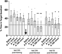
Intrahippocampal infusion of adenosine immediately after nonreinforced retrieval hindered recent and late spatial memory retention, as measured 24 h and 5 d after infusion. Male Wistar rats with infusion cannulas implanted into the CA1 region of the dorsal hippocampus were trained for 5 d in the spatial version of the MWM. Twenty-four hours after the last training session, the animals were randomly assigned to one out of five experimental groups and subjected to a 60-sec probe test in the absence of the escape platform. Immediately after this first test, the animals received intrahippocampal bilateral infusions (black arrow) of either saline solution (saline) or adenosine (A 0.1, 1, 10, or 100 nmol). Memory retention was assessed in successive 60-sec probe tests carried out either 24 h or 5 d after the first test. Data are expressed as means (±SD) of the percentage of swimming time spent in the target quadrant. (Dots) Individual value for each rat, (dashed line) theoretical mean of 25% of test time spent in the target quadrant at random. (*) P < 0.05, (**) P < 0.01 versus respective saline groups; one-way ANOVA: test 24 h after the first test, F(4,48) = 5.933; test 5 d after the first test, F(4,48) = 3.321; Dunnett's posttest; n = 7–12 per group.
It was observed that when adenosine was infused at a dose of 10 nmol/side or 100 nmol/side immediately after the first probe test, there was a reduction in the time spent in the target quadrant in the tests run 24 h and 5 d after the first tests. These results show that reconsolidations of both early and late long-term memory were impaired in these groups and that this effect is dose-dependent (Fig. 1; Supplemental Fig. S1).
The observed effect was attributed to the reconsolidation of spatial memory, as there were no significant differences in all the tests in the time spent in the target quadrant (Fig. 2A) or in the number of crossings on the local platform (Supplemental Fig. S2A) compared with the saline group when adenosine was infused at 10 nmol/side 24 h posttraining in the absence of an immediately preceding probe test. This result precludes the possibility that the previous effects verified after the bilateral infusion of adenosine (Fig. 2) might be independent of the fact that infusion was performed immediately after a reactivation session test without reinforcement (the test 24 h posttraining) or due to some long-term alteration triggered by adenosine, which has been observed in any postinfusion test. Additionally, there was no significant difference in the time spent in the target quadrant (Fig. 2B) or in the number of crossings on the local platform (Supplemental Fig. S2B) between the saline and adenosine groups infused immediately after a reactivation test with reinforcement (test 24 h posttraining with the platform in the same location as it was during training days). This result restricts the effects related to the bilateral hippocampal infusion of adenosine to the absence of reinforcement (escape platform) during the reactivation test session (24 h posttraining). The absence of reinforcement is admittedly required for triggering the labilization process, which precedes the reconsolidation process. Moreover, there was no significant difference in the time spent in the target quadrant (Fig. 2C) or in the number of crossings on the local platform (Supplemental Fig. S2C) between the saline and adenosine infused 3 h after the first probe test in all the performed tests, indicating that the amnesic effect of adenosine on spatial memory reconsolidation is time-dependent. Finally, there was no significant difference in the time spent in the target quadrant (Fig. 2D) or in the number of crossings on the local platform (Supplemental Fig. S2D) of the saline group compared with when adenosine was infused immediately after the first probe test into the occipital cortex, which is 1.0 mm above the pyramidal cell layer of the dorsal CA1 region, using coordinates (−4.2 anterior, ±3.0 lateral, −1.0 ventral from bregma) obtained from the atlas of Paxinos and Watson (2006) as in all the tests. This result proves that the effects caused by bilateral hippocampal infusion of adenosine on retrieval of either early or late long-term spatial memory are structure-dependent. Therefore, the aforementioned effects were observed only when adenosine was infused immediately after the nonreinforced reactivation test (first test 24 h posttraining) into the hippocampal CA1 region.
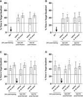
(A) Intrahippocampal infusion of adenosine after the final day of training does not affect early and late spatial memory retention, as measured 24 h and 5 d after infusion. Male Wistar rats with infusion cannulas implanted into the CA1 region of the dorsal hippocampus were trained for 5 d in the spatial version of the MWM. Twenty-four hours after the last training session, the animals received intrahippocampal bilateral infusions of either saline solution (saline) or 10 nmol of adenosine (A 10 nmol). Memory retention was assessed in successive 60-sec probe tests carried out either 24 h or 5 d after infusion (INF; black arrow). (Dots) Individual value for each rat, (dashed line) theoretical mean of 25% of test time spent at random in the target quadrant. Data are expressed as means (±SD) of the percentage of swimming time spent in the target quadrant. There was no difference between the saline and adenosine groups; Student's t-test: test 24 h after the first test, t(19) = 2.065; test 5 d after the first test, t(19) = 1.397; n = 10–11 per group. (B) Intrahippocampal infusion of adenosine immediately after reinforced retrieval does not affect early and late spatial memory retention, as measured 24 h and 5 d after the first test. Male Wistar rats with infusion cannulas implanted into the CA1 region of the dorsal hippocampus were trained for 5 d in the spatial version of the MWM. Twenty-four hours after the last training session, the animals were subjected to a 60-sec probe test in the presence of the escape platform (R). Immediately after R, the animals received intrahippocampal bilateral infusions (black arrow) of either saline solution (saline) or 10 nmol of adenosine (A 10 nmol). Memory retention was assessed in successive 60-sec probe tests carried out either 24 h or 5 d after R. (Dots) Individual value for each rat, (dashed line) theoretical mean of 25% of test time spent at random in the target quadrant. Data are expressed as means (±SD) of the percentage of swimming time spent in the target quadrant. There was no difference between the saline and adenosine groups; Student's t-test: test 24 h after the first test, t(16) = 0.3869; test 5 d after the first test, t(16) = 0.4349; n = 6–12 per group. (C) Intrahippocampal infusion of adenosine 3 h after nonreinforced retrieval does not affect early and late spatial memory retention, as measured 24 h and 5 d after the first test. Male Wistar rats with infusion cannulas implanted into the CA1 region of the dorsal hippocampus were trained for 5 d in the spatial version of the MWM. Twenty-four hours after the last training session, the animals were subjected to a 60-sec probe test in the absence of the escape platform. Three hours after this test, the animals received intrahippocampal bilateral infusions (black arrow) of either saline solution (saline) or 10 nmol of adenosine (A 10 nmol). Memory retention was assessed in successive 60-sec probe tests carried out either 24 h or 5 d after the first test. (Dots) Individual value for each rat, (dashed line) theoretical mean of 25% of test time spent at random in the target quadrant. Data are expressed as means (±SD) of the percentage of swimming time spent in the target quadrant. There was no difference between the saline and adenosine groups; Student's t-test: test 24 h after the first test, t(12) = 1.923; test 5 d after the first test, t(12) = 0.6724; n = 6–8 per group. (D) Intramediolateral occipital cortex infusion of adenosine immediately after nonreinforced retrieval does not affect early and late spatial memory retention, as measured 24 h and 5 d after the first test. Male Wistar rats with infusion cannulas implanted into the mediolateral occipital cortex were trained for 5 d in the spatial version of the MWM. Twenty-four hours after the last training session, the animals were subjected to a 60-sec probe test in the absence of the escape platform. Immediately after this test, the animals received intramediolateral occipital cortex bilateral infusions (black arrow) of either saline solution (saline) or 10 nmol of adenosine (A 10 nmol). Memory retention was assessed in successive 60-sec probe tests carried out either 24 h or 5 d after the first test. (Dots) Individual value for each rat, (dashed line) theoretical mean of 25% of test time spent at random in the target quadrant. Data are expressed as means (±SD) of the percentage of swimming time spent in the target quadrant. There was no difference between the saline and adenosine groups; Student's t-test: test 24 h after the first test, t(11) = 2.170; test 5 d after the first test, t(11) = 0.6221; n = 6–7 per group.
Adenosine impairment on spatial memory reconsolidation is blocked by A1 and A3 receptor antagonists
To explore the receptor(s) by which adenosine exerts its amnesic effect on the reconsolidation of early and late spatial memory, rats trained for 5 d in the spatial version of the MWM were subjected to a probe test without the escape platform 24 h after the final training session. Immediately after the first probe test, the rats received an intrahippocampal bilateral infusion of either vehicle (0.9% saline), adenosine (10 nmol/side), or adenosine plus each one of the adenosinergic receptor antagonists. The rats were then tested 24 h postinfusion (for evaluating early reconsolidated memory) and 5 d postinfusion (for late reconsolidated memory) (Fig. 3; Supplemental Fig. S3).
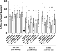
The amnesic effect induced by the intrahippocampal infusion of adenosine is blocked by A1 and A3 but not by A2A or A2B receptor antagonists. Male Wistar rats with infusion cannulas implanted into the CA1 region of the dorsal hippocampus were trained for 5 d in the spatial version of the MWM. Twenty-four hours after the final training session, the animals were subjected to a 60-sec probe test in the absence of the escape platform. Immediately after this first test, the animals received intrahippocampal bilateral infusions (black arrow) of either saline solution (saline), 10 nmol of adenosine (A 10 nmol), or 10 nmol of adenosine plus A1 (A + antag A1), A2A (A + antag A2A), A2B (A + antag A2B), or A3 (A + antag A3) receptor antagonist (50 nmol for each antagonist). Memory retention was assessed in successive 60-sec probe tests carried out either 24 h or 5 d after the first test. Data are expressed as means (±SD) of the percentage of swimming time spent in the target quadrant. (Dots) Individual value for each rat, (dashed line) theoretical mean of 25% of test time spent in the target quadrant at random. (*) P < 0.05, (**) P < 0.01 versus respective saline groups; one-way ANOVA: test 24 h after the first test, F(5,71) = 4.733; test 5 d after the first test, F(5,71) = 3.895; Dunnett's posttest; n = 9–18 per group.
It was observed that when adenosine was coinfused with A1 or A3 receptor antagonists, its amnesic effect on both early and late spatial memory reconsolidation was blocked, as there was no significant difference in the time spent in the target quadrant (Fig. 3) or in the number of crossings on the local platform (Supplemental Fig. S3) compared with the saline group. Likewise, there was an amnesic effect of adenosine on spatial memory reconsolidation when adenosine was coinfused with A2A or A2B receptor antagonists. These results indicate that adenosine exerts its amnesic effect through its action on A1 and A3 receptors.
Adenosine receptors do not participate individually in the spatial memory reconsolidation process
To investigate whether adenosinergic receptors participate individually in the reconsolidation of early and late spatial memory, rats trained for 5 d in the spatial version of the MWM were subjected to a probe test without the escape platform 24 h after the last training session. The rats received an intrahippocampal bilateral infusion of either vehicle (0.9% saline) or adenosinergic receptor antagonists (50 nmol/side) immediately after this first probe test. The rats were then tested 24 h postinfusion (for evaluating early reconsolidated memory) and 5 d postinfusion (for late reconsolidated memory) (Fig. 4; Supplemental Fig. S4).
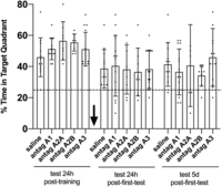
Antagonists of A1, A2A, A2B, and A3 receptors have no effect on spatial memory reconsolidation. Male Wistar rats with infusion cannulas implanted into the CA1 region of the dorsal hippocampus were trained for 5 d in the spatial version of the MWM. Twenty-four hours after the last training session, the animals were subjected to a 60-sec probe test in the absence of the escape platform. Immediately after this first test, the animals received intrahippocampal bilateral infusions (black arrow) of either saline solution (saline) or A1 (antag A1), A2A (antag A2A), A2B (antag A2B), or A3 (antag A3) receptor antagonist (50 nmol for each antagonist). Memory retention was assessed in successive 60-sec probe tests carried out either 24 h or 5 d after the first test. Data are expressed as means (±SD) of the percentage of swimming time spent in the target quadrant. (Dots) Individual value for each rat, (dashed line) theoretical mean of 25% of test time spent in the target quadrant at random. There was no difference between the respective saline groups regarding distinct drug infusion; one-way ANOVA: test 24 h after the first test, F(4,53) = 0.09668; test 5 d after the first test, F(4,53) = 0.8682; Dunnett's posttest; n = 8–15 per group.
It was verified that none of the antagonists caused any effect per se on both early and late spatial memory reconsolidation, as there was no significant difference in the time spent in the target quadrant (Fig. 4) or in the number of crossings on the local platform (Supplemental Fig. S4) compared with the saline group. These results imply that none of the adenosinergic receptors participate individually in the spatial memory reconsolidation process.
The amnesic effect of adenosine on spatial memory reconsolidation is mimicked by coactivation of receptors A1 and A3
To examine which adenosinergic receptor mimics the adenosine effect on the reconsolidation of early and late spatial memory, rats trained for 5 d in the spatial version of the MWM were subjected to a probe test without the escape platform 24 h after the final training session. The rats received an intrahippocampal bilateral infusion of either vehicle (0.9% saline) or adenosinergic receptor agonists (10 nmol/side) immediately after this first probe test. The rats were then tested 24 h postinfusion (for evaluating early reconsolidated memory) and 5 d postinfusion (for late reconsolidated memory) (Fig. 5; Supplemental Fig. S5).
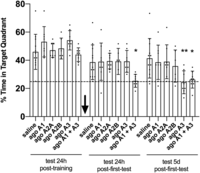
The amnesic effect of adenosine is mimicked by A1 and A3 receptor agonists infused together. Male Wistar rats with infusion cannulas implanted into the CA1 region of the dorsal hippocampus were trained for 5 d in the spatial version of the MWM. Twenty-four hours after the final training session, the animals were subjected to a 60-sec probe test in the absence of the escape platform. Immediately after this first test, the animals received intrahippocampal bilateral infusions (black arrow) of either saline solution (saline) or A1 (ago A1), A2A (ago A2A), A2B (ago A2B), A3 (ago A3), or A1 plus A3 (ago A1 + A3) receptor agonists (10 nmol for each agonist). Memory retention was assessed in successive 60-sec probe tests carried out either 24 h or 5 d after the first test. Data are expressed as means (±SD) of the percentage of swimming time spent in the target quadrant. (Dots) Individual value for each rat, (dashed line) theoretical mean of 25% of test time spent in the target quadrant at random. (*) P < 0.05, (**) P < 0.01 versus respective saline groups; one-way ANOVA: test 24 h after the first test, F(5,44) = 2.690; test 5 d after the first test, F(5,44) = 3.513; Dunnett's posttest; n = 6–12 per group.
A significant difference was observed in the time spent in the target quadrant (Fig. 5) and in the number of crossings on the local platform (Supplemental Fig. S5) when A1 and A3 receptors were simultaneously activated compared with the saline group, similar to that which occurred with adenosine. Adenosine therefore might act on receptors A1 and A3 to impair early and late spatial memory reconsolidation. Notably, only the receptor A3 activation is sufficient for impairing late, but not early, spatial memory reconsolidation.
Adenosine and A1 and A3 receptor agonists impair early spatial memory reconsolidation when this reconsolidation is late
To investigate whether both adenosine and the A1 and A3 receptor agonists impair spatial memory reconsolidation when this reconsolidation occurs late, rats trained for 5 d in the spatial version of the MWM were subjected to a probe test without the escape platform 5 d after the last training session. The rats received an intrahippocampal bilateral infusion of either vehicle (0.9% saline), adenosine (10 nmol/side), or adenosinergic receptor agonists (10 nmol/side) immediately after this first probe test. The rats were then tested 24 h postinfusion (for evaluating early reconsolidated memory) and 5 d postinfusion (for late reconsolidated memory) (Fig. 6A; Supplemental Fig. S6A).
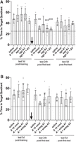
(A) Intrahippocampal infusion of adenosine or A3 receptor agonist immediately after a late nonreinforced retrieval hinders recent but not late spatial memory retention, as measured 24 h and 5 d after infusion. Male Wistar rats with infusion cannulas implanted into the CA1 region of the dorsal hippocampus were trained for 5 d in the spatial version of the MWM. Five days after the last training session, the animals were subjected to a 60-sec probe test in the absence of the escape platform. Immediately after this first test, the animals received intrahippocampal bilateral infusions (black arrow) of either saline solution (saline), 10 nmol of adenosine (A 10 nmol), A1 (ago A1), A3 (ago A3), or A1 plus A3 (ago A1 + A3) receptor agonists (10 nmol for each agonist). Memory retention was assessed in successive 60-sec probe tests carried out either 24 h or 5 d after the first test. Data are expressed as means (±SD) of the percentage of swimming time spent in the target quadrant. (Dots) Individual value for each rat, (dashed line) theoretical mean of 25% of test time spent in the target quadrant at random. (**) P < 0.01, (***) P < 0.001 versus respective saline groups; one-way ANOVA: test 24 h after the first test, F(4,34) = 6.255; test 5 d after the first test, F(4,34) = 1.142; Dunnett's posttest; n = 7–10 per group. (B) The amnesic effect on late reconsolidation induced by the intrahippocampal infusion of adenosine is blocked by A1 and A3 receptor antagonists. Male Wistar rats with infusion cannulas implanted into the CA1 region of the dorsal hippocampus were trained for 5 d in the spatial version of the MWM. Five days after the final training session, the animals were subjected to a 60-sec probe test in the absence of the escape platform. Immediately after this first test, the animals received intrahippocampal bilateral infusions (black arrow) of either saline solution (saline), 10 nmol of adenosine (A 10 nmol), or 10 nmol of adenosine plus A1 (A + antag A1) or A3 (A + antag A3) receptor antagonist (50 nmol for each antagonist). Memory retention was assessed in successive 60-sec probe tests carried out either 24 h or 5 d after the first test. Data are expressed as means (±SD) of the percentage of swimming time spent in the target quadrant. (Dots) Individual value for each rat, (dashed line) theoretical mean of 25% of test time spent in the target quadrant at random. (*) P < 0.05 versus respective saline groups; one-way ANOVA: test 24 h after the first test, F(3,38) = 3.247; test 5 d after the first test, F(3,38) = 0.4823; Dunnett's posttest; n = 7–18 per group.
Both the adenosine and coinfused A1 and A3 receptor agonists impaired the reconsolidation of spatial memory tested 24 h posttest, but this effect did not endure until 5 d posttest. This result is in line with the fact that the effect of adenosine on recent spatial memory was blocked by A1 and A3 receptor antagonists (Fig. 6B; Supplemental Fig. S6B).
The amnesic effect of adenosine on spatial memory reconsolidation is blocked by proteasome inhibition
To investigate whether the impairment of spatial memory reconsolidation by adenosine depends on protein turnover, rats trained for 5 d in the spatial version of the MWM were subjected to a probe test without the escape platform 24 h after the last training session. The rats received an intrahippocampal bilateral infusion of either vehicle (0.9% saline), adenosine (10 nmol/side), lactacystin (200 pmol/side), or adenosine plus lactacystin immediately after this first probe test. The rats were then tested 24 h postinfusion (for evaluating early reconsolidated memory) and 5 d postinfusion (for late reconsolidated memory) (Fig. 7; Supplemental Fig. S7).
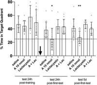
The amnesic effect on early reconsolidation induced by the intrahippocampal infusion of adenosine is blocked by the proteasome inhibitor lactacystin. Male Wistar rats with infusion cannulas implanted into the CA1 region of the dorsal hippocampus were trained for 5 d in the spatial version of the MWM. Twenty-four hours after the final training session, the animals were subjected to a 60-sec probe test in the absence of the escape platform. Immediately after this first test, the animals received intrahippocampal bilateral infusions (black arrow) of either saline solution (saline), 10 nmol of adenosine (A 10 nmol), 200 pmol of lactacystin (Lactacystin), or 10 nmol of adenosine plus 200 pmol of lactacystin (A + Lac). Memory retention was assessed in successive 60-sec probe tests carried out either 24 h or 5 d after the first test. Data are expressed as means (±SD) of the percentage of swimming time spent in the target quadrant. (Dots) Individual value for each rat, (dashed line) theoretical mean of 25% of test time spent in the target quadrant at random. (*) P < 0.05, (**) P < 0.01 versus respective saline groups; one-way ANOVA: test 24 h after the first test, F(3,40) = 5.464; test 5 d after the first test, F(3,40) = 6.879; Dunnett's posttest; n = 7–14 per group.
It was observed that when protein degradation is blocked, the amnesic effect of adenosine on spatial memory reconsolidation disappears, indicating that the action of A1 and A3 receptors somehow implies a lower protein input in the reconsolidation process. If this is indeed the case, the already known effect of blocking protein synthesis on spatial memory reconsolidation (Morris et al. 2006; Myskiw et al. 2008; Mac Callum et al. 2014) can be reversed by blocking one of these A1 or A3 receptors. Next, it was decided to test this hypothesis for the A3 receptor antagonist.
The amnesic effect of protein synthesis inhibition on spatial memory reconsolidation is blocked by inhibiting the adenosinergic A3 receptor
To establish whether the impairment of spatial memory reconsolidation caused by inhibition of protein synthesis can be reversed by blocking the adenosinergic A3 receptor, rats trained for 5 d in the spatial version of the MWM were subjected to a probe test without the escape platform 24 h after the final training session. The rats received an intrahippocampal bilateral infusion of either vehicle (0.9% saline), A3 receptor antagonist (50 nmol/side), rapamycin (5 μg/side), or A3 receptor antagonist plus rapamycin immediately after this first probe test. The rats were then tested 24 h postinfusion (for evaluating early reconsolidated memory) and 5 d postinfusion (for late reconsolidated memory) (Fig. 8; Supplemental Fig. S8).
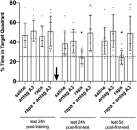
The amnesic effect on early reconsolidation induced by the intrahippocampal infusion of rapamycin is blocked by the adenosine A3 receptor antagonist. Male Wistar rats with infusion cannulas implanted into the CA1 region of the dorsal hippocampus were trained for 5 d in the spatial version of the MWM. Twenty-four hours after the final training session, the animals were subjected to a 60-sec probe test in the absence of the escape platform. Immediately after this first test, the animals received intrahippocampal bilateral infusions (black arrow) of either saline solution (saline), 50 nmol of adenosine A3 receptor antagonist (antag A3), 5 µg of rapamycin (rapa), or A3 receptor antagonist plus 5 µg of rapamycin (rapa + antag A3). Memory retention was assessed in successive 60-sec probe tests carried out either 24 h or 5 d after the first test. Data are expressed as means (±SD) of the percentage of swimming time spent in the target quadrant. (Dots) Individual value for each rat, (dashed line) theoretical mean of 25% of test time spent in the target quadrant at random. (*) P < 0.05 versus respective saline groups; one-way ANOVA: test 24 h after the first test, F(3,50) = 7.467; test 5 d after the first test, F(3,50) = 8.137; Dunnett's posttest; n = 11–18 per group.
As predicted previously, the blockade of the A3 receptor was sufficient to reverse the amnesic effect on reconsolidation caused by the inhibition of protein synthesis by rapamycin. This result implies that endogenous adenosine, acting via A3 receptors, is required for the inhibition of protein synthesis to affect protein turnover in a way that compromises the protein input required for spatial memory reconsolidation.
Discussion
The results presented indicate that adenosine and adenosinergic receptor A1 plus A3 agonists administered in the dorsal hippocampus immediately after a probe test in the absence of the escape platform and performed 24 h posttraining impair spatial memory retention during subsequent probe tests carried out 24 h and 5 d later. The amnesic effect of adenosine and A1 plus A3 receptor agonists is contingent on the nonreinforced reactivation of the mnemonic trace. Also, the A3 receptor agonist is sufficient to inhibit the reconsolidation of late spatial memory, as tested 5 d after the first test, when this first test was performed 24 h posttraining. In addition, when the first probe test was conducted 5 d posttraining, the amnesic effect of adenosine and A1 plus A3 receptor agonists was transitory, not extending until a test performed 5 d after the first test.
The fact that the A3 receptor agonist only caused a verifiable effect on the reconsolidation of late but not early spatial long-term memory points out a difference in the level of biochemical mechanisms between these two types of long-term memories—early versus late. The first time a distinction in the biochemical level was found between these two types of long-term memory, it was attributed to the activation of CaMKII, which needs to be activated immediately after an unreinforced test in the MWM task for a reconsolidation to occur that generates a stable memory in the late-term (5 d posttest without reinforcement) (Da Silva et al. 2013). These results—together with the fact that adenosine is a critical factor in neuronal metabolic regulation and that in these regulatory mechanisms, intracellular calcium levels are also a key factor (Llorente-Folch et al. 2015)—suggest an interesting line of investigation. This new approach may lead to the discovery of a regulatory link between the extracellular/intracellular adenosine ratio and calcium-dependent signal transduction pathways, as well as the participation of this link in mnemonic processes, such as the reconsolidation of long-term memories and how they differ in determining the persistence of the mnemonic trace upon reconsolidation.
Although our data showed significant change only concerning late long-term spatial memory when the A3 receptor was activated, it is unusual that these results are related to A3 receptors. A3 receptors are described in detail in the literature, confirmed by our literature review, where we found several studies in which the function of adenosinergic receptors is described through pharmacological administrations only in A1 receptors and the A2 family (Kopf et al. 1999; Huang et al. 2005; Dall'Igna et al. 2007; Mioranzza et al. 2011; Harvey et al. 2012; Sachdeva and Gupta 2013; Bortolotto et al. 2015; Liu et al. 2018), with A3 receptors being described the minority of times (Zhou et al. 1992; Corodimas and Tomita 2001; Rubaj et al. 2003).
A1 and A3 receptors are coupled to an inhibitory G protein (Ralevic and Burnstock 1998), which inhibits membrane adenylyl cyclase when activated, thereby decreasing the intracellular concentration of the cAMP second messenger. This leads to lower activation of PKA and, subsequently, lower activation of the transcription of immediate and late genes related to long-lasting synaptic plasticity (Lamprecht 1999; Alberini and Kandel 2014; Cláudio da Silva and Sartori Bonini 2022). These factors converge to a decrease in the synthesis rate of new proteins, which entails a consequent impairment in long-term memory retention (Izquierdo et al. 2006; Morris et al. 2006; Rosenberg et al. 2014; Roesler 2017). Corroborating this successive description of events, it was verified that the amnesic effect of adenosine on spatial memory reconsolidation disappears if there is no protein degradation. In the absence of protein degradation, a decrease in the rate of new protein synthesis does not translate into a lower protein input required for the reconsolidation process (Lee et al. 2008, 2012; Choi et al. 2010). Furthermore, we also discovered that the amnesic effect caused by inhibition of protein synthesis during spatial memory reconsolidation is dependent on the activation of A3 adenosinergic receptors, implying this receptor's participation in the regulation of the protein turnover process.
Another interesting result obtained was the observation of the fact that the amnesic effect of adenosine on spatial memory reconsolidation becomes transient when the reconsolidation takes place on a delayed memory. To explain this outcome, we propose the following hypothesis. When the memory to be reconsolidated is still recent, it has much of its engram allocated in the hippocampus. Thus, immediately after the labilization resulting from retrieval without reinforcement, when reconsolidation is blocked and the underlying engram is not reconstituted, lasting amnesia takes place as a consequence. When the memory to be reconsolidated is late, a considerable part of the engram has already migrated to other locations in the cerebral cortex (Bontempi et al. 1999; Dudai 2004; Alberini 2011). This engram, or access to this engram, is sent to the hippocampus during evocation, as described in studies showing that even a hippocampus-independent memory becomes transiently hippocampus-dependent again when reactivated (Debiec et al. 2002; Einarsson et al. 2015). If there is no reconsolidation after labilization, again, this engram is lost in the hippocampus, explaining the amnesia observed the following day. However, there is now a backup copy of this engram outside the hippocampus, which can be reaccessed. This reaccessing takes time, which explains the return of the ability to recall the mnemonic trace corresponding to the engram only several days after the reconsolidation of the remote memory has been blocked.
Finally, several studies have described the actions of adenosine as inhibitory and neuroprotective due to its extracellular concentrations increasing under conditions of cellular stress (Jacobson and Gao 2006; Rahman 2009; Sebastião and Ribeiro 2009; Gomes et al. 2011) as it aims to protect against any cellular damage common in these situations. This role of adenosine, along with its physiological action on the reconsolidation of long-term spatial memory, also allows connecting scenarios in which there is cellular stress with subsequent mnemonic performance alteration. Among these scenarios, excitotoxicity (Popoli et al. 2003; Melani et al. 2014; Serpa et al. 2015), neuroinflammation (Shakya et al. 2019; Martí Navia et al. 2020), or even the onset of neurodegenerative processes involving the accumulation of β-amyloid plaques (Castillo et al. 2021; Lopes et al. 2021; Trinh et al. 2022) and neurofibrillary tangles (Angulo et al. 2003; Launay et al. 2023) are described. The adenosinergic system certainly participates in the background of all these processes, implying that a greater understanding of the role of its receptors in physiological and pathophysiological processes serves as an important source of insights for new therapeutic approaches.
Materials and Methods
Subjects
All experiments were conducted blindly for the animals’ treatment conditions according to the National Institutes of Health guidelines for animal care and use and were approved by the Animal Care and Ethical Committee of the University of Center-West of Paraná. Furthermore, all methods were in accordance with “Animal Research: Reporting of in Vivo Experiments” (ARRIVE) guidelines (https://arriveguidelines.org) for the reporting of animal experiments. Three-month-old male Wistar rats weighing 220–280 g and raised in our animal facilities were used in the experiments. The animals were housed four or five in a cage and maintained at 21°C–23°C under a 12-h light/12-h dark cycle (lights on at 7:00 a.m.) with free access to food and water.
Experimental design
The effect of intrahippocampal infusion of either adenosine (10 nmol/side) or adenosinergic agonists (10 nmol/side) or antagonists (50 nmol/side) on early and late reconsolidation of early and late long-term spatial memory in rats was evaluated. First, the rats underwent stereotaxic surgery to implant guide cannulas, and after 3 d of recovery, the rats were randomly divided into the following experimental groups: dose curve of adenosine, adenosine plus antagonists, antagonists, agonists, adenosine plus lactacystin, and finally A3 receptor antagonist plus rapamycin. For the early reconsolidation experiments, they were trained in the Morris water maze (MWM) task for five consecutive days (eight trials per day). They then received an intrahippocampal bilateral infusion of the drug(s) under scrutiny immediately after the first test performed 24 h after the final training day. The rats were then retested after saline/drug infusion after 24 h (early long-term spatial memory) or after 5 d (late long-term spatial memories). For the late reconsolidation experiments, the rats were trained and tested as mentioned earlier, but the first test was performed 5 d after the final training day. There were also four experimental control groups (infusion, reinforcement, temporal, and structure controls) with other sets of rats.
Stereotaxic surgery for cannula implants
To implant the rats with indwelling cannulas, they were deeply anesthetized with 75 mg/kg ketamine (König) plus 10 mg/kg xylazine (Coopers), and 27-gauge 9.0-mm guide cannulas were stereotaxically directed toward the pyramidal cell layer of the dorsal CA1 region using coordinates (−4.2 anterior, ±3.0 lateral, −2.0 ventral from bregma) taken from the atlas of Paxinos and Watson (2006). The animals were allowed to recover from surgery for 3 d before being subjected to any other procedure.
Microinfusion procedures
At the drug delivery time, 30-gauge, 10.0-mm infusion needles (extending 1.0 mm beyond guide cannulas) were tightly fitted into the guides. Infusions (1.0 µL per side) were carried out for 60 sec, and the infusion cannulas were left in place for 30 additional seconds to minimize backflow. Cannula placement was verified postmortem. Two hours to 4 h after the final behavioral test, 1.0 µL of a 4% (m/v) methylene-blue solution was infused as described earlier, and the extension of the dye 30 min after that was taken as indicative of the presumable diffusion of the vehicle or drug previously given to each animal. Only data from animals with correct implants were included in the statistical analysis (Fig. 9A–D).
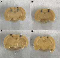
Images showing the cannula placement and the infusion extension (1.0 µL of a 4% [m/v] methylene-blue solution) in both structures: the hippocampus and occipital cortex. (A,B) Hippocampal infusions (two rats shown). (C,D) Occipital cortex (two rats shown).
Drugs
Adenosine was purchased from Sigma (A9251-5G). CPA (A1 receptor agonist; Tocris 1702), PSB 0777 (A2A receptor agonist; Tocris 4334), BAY 60-6583 (A2B receptor agonist; Tocris 4472), HEMADO (A3 receptor agonist; Tocris 1579), DPCPX (A1 receptor antagonist; Tocris 439), SCH 442416 (A2A receptor antagonist; Tocris 2463), PSB 603 (A2B receptor antagonist; Tocris 3198), MRS 3777 (A3 receptor antagonist; Tocris 2403), lactacystin (Tocris 2267), and rapamycin (Tocris 1292) were obtained from Tocris. Adenosinergic agonists and antagonists were first dissolved in DMSO and stored frozen at −20°C until the moment of use, when they were diluted with saline to working concentration. Adenosine, lactacystin, and rapamycin were first dissolved in saline solution and stored frozen at −20°C until the moment of use, when they were diluted with saline to working concentration.
Morris water maze task
The water maze was a dark-blue circular pool (180 cm in diameter) conceptually divided into four equal imaginary quadrants for data analysis. The water temperature was 21°C–23°C. There was a black circular platform (12 cm in diameter) 2 cm beneath the surface of the water and hidden from the rat's view. It had a rough surface, allowing the rats to climb onto it easily once detected. The swimming path of the animals was recorded by using a video camera mounted above the center of the pool and analyzed using a video tracking and analysis system. The water maze was located in a white, well-lit room with several posters and other distal visual stimuli hanging on the walls to provide spatial cues. The rats were handled 5 min per day for 3 d before training. Training using the spaced training protocol was carried out for five successive days (Da Silva et al. 2008). On each day, the rats received eight consecutive training trials, during which the hidden platform was kept in a constant location. A different starting location was used for each trial, which consisted of a swim followed by a 30-sec platform sit. Any rat that did not find the platform within 60 sec was guided to it by the experimenter. To evaluate the effect of the drugs given after memory reactivation, the rats were trained for 5 d, as mentioned earlier, before being subjected to the first probe test without the escape platform either 24 h or 5 d after the final training session. Immediately after this test session, the rats received intra-CA1 infusions of the drug under scrutiny or vehicle. Memory retention was evaluated in successive probe tests carried out at 24 h or 5 d after the first test.
Data analysis
The Shapiro–Wilk normality test was carried out, and the behavioral data are presented as mean and standard deviation. As appropriate, data were analyzed by either two-tailed Student's t-test or one-way ANOVA followed by Dunnett's posthoc tests. Values <0.05 were considered significant. All the raw data are shown in Supplemental Data Set S1 (percentage of time spent in target quadrant) and Supplemental Data Set S2 (number of crossings on the local platform).
Acknowledgments
This work was supported by fellowships from the Brazilian government agency Coordination for the Improvement of Higher Education Personell (CAPES) and financial support from the nongovernmental organization Association for Research, Studies, and Support for Alzheimer's Patients (AEPAPA), who had no participation in the design, analysis, or reporting of the study.
Footnotes
-
[Supplemental material is available for this article.]
-
Article is online at http://www.learnmem.org/cgi/doi/10.1101/lm.053785.123.
- Received April 22, 2023.
- Accepted August 9, 2023.
This article is distributed exclusively by Cold Spring Harbor Laboratory Press for the first 12 months after the full-issue publication date (see http://learnmem.cshlp.org/site/misc/terms.xhtml). After 12 months, it is available under a Creative Commons License (Attribution-NonCommercial 4.0 International), as described at http://creativecommons.org/licenses/by-nc/4.0/.



