


Sleep strengthens resting-state functional communication between brain areas involved in the consolidation of problem-solving skills
- Nicholas H. van den Berg1,
- Dylan Smith1,
- Zhuo Fang1,2,3,
- Alyssa Pozzobon1,
- Balmeet Toor1,
- Julia Al-Kuwatli1,
- Laura Ray1 and
- Stuart M. Fogel1,2,3
- 1School of Psychology, University of Ottawa, Ottawa, Ontario K1N 6N5, Canada
- 2The Royal's Institute of Mental Health Research, University of Ottawa, Ottawa, Ontario K1Z 7K4, Canada
- 3University of Ottawa Brain and Mind Institute, Ottawa, Ontario K1H 8M5, Canada
- Corresponding author: sfogel{at}uottawa.ca
Abstract
Sleep consolidates procedural memory for motor skills, and this process is associated with strengthened functional connectivity in hippocampal–striatal–cortical areas. It is unknown whether similar processes occur for procedural memory that requires cognitive strategies needed for problem-solving. It is also unclear whether a full night of sleep is indeed necessary for consolidation to occur, compared with a daytime nap. We examined how resting-state functional connectivity within the hippocampal–striatal–cortical network differs after offline consolidation intervals of sleep, nap, or wake. Resting-state fMRI data were acquired immediately before and after training on a procedural problem-solving task that requires the acquisition of a novel cognitive strategy and immediately prior to the retest period (i.e., following the consolidation interval). ROI to ROI and seed to whole-brain functional connectivity analyses both specifically and consistently demonstrated strengthened hippocampal–prefrontal functional connectivity following a period of sleep versus wake. These results were associated with task-related gains in behavioral performance. Changes in functional communication were also observed between groups using the striatum as a seed. Here, we demonstrate that at the behavioral level, procedural strategies benefit from both a nap and a night of sleep. However, a full night of sleep is associated with enhanced functional communication between regions that support problem-solving skills.
Proficiency at solving cognitively complex procedural problems does not materialize instantly. Rather, this type of skill learning develops gradually, usually requiring trial and error, repeated practice, and, for optimal performance, an offline consolidation period of sleep (Fogel et al. 2015). Sleep is indeed known to facilitate creativity and to act as a cognitive catalyst for realizing solutions to cognitively complex problems (Wagner et al. 2004; Sio et al. 2013; Monaghan et al. 2015). Sleep is known to preferentially consolidate this type of memory over and above memory of the simple motor movements typically required to execute the solution (Van Hedger et al. 2015; van den Berg et al. 2019; Conte et al. 2020).
Functional magnetic resonance imaging (fMRI) techniques can further elucidate how the sleeping brain actively consolidates newly formed memory traces. For example, sleep enhances consolidation of procedural memory traces for motor skills (Debas et al. 2010; Albouy et al. 2013c). Brain areas such as the striatum, hippocampus, motor cortex, and prefrontal cortex are recruited during learning. They exhibit subsequent sleep-dependent changes in activity, and these changes are associated with offline gains in performance (Debas et al. 2010; Albouy et al. 2013c).
Specifically, striatal activity in both the putamen (Albouy et al. 2013c; Barakat et al. 2013; Boutin et al. 2018) and the caudate (Cousins et al. 2016; van den Berg et al. 2021) is associated with sleep-related consolidation of procedural memories. During sleep itself, the putamen is reactivated when sleep spindles occur. Both sleep spindle characteristics and the extent of this reactivation are associated with improved motor sequence memory performance upon awakening (Fogel et al. 2017). In addition, in part because of recent findings from sleep studies, the hippocampus is becoming increasingly recognized as a central hub for procedural memory consolidation (Schendan et al. 2003; Albouy et al. 2013a,b; Sawangjit et al. 2018; Schapiro et al. 2019). Sleep-related consolidation of both simple motor sequences (Fogel et al. 2014) and procedural problem-solving skills involving cognitive strategies (van den Berg et al. 2021) is associated with increased hippocampal activity compared with an equivalent period of wake.
Importantly, these brain areas (i.e., the hippocampus and striatum) do not act in isolation when memory consolidation occurs. Rather, each has functionally distinct roles as the consolidation process unfolds over a period of sleep. Specifically, a competitive interaction exists between the hippocampus and striatum during learning and consolidation periods of simple motor sequences (Albouy et al. 2008, 2013c). The hippocampus is involved in consolidation of the spatial aspect and is sleep-dependent (Albouy et al. 2013a, 2015). The striatum (i.e., the putamen) is involved in consolidating the motor aspect, and although it does so regardless of sleep or wake (Albouy et al. 2013a), a period of sleep can modulate its involvement through interactions with the hippocampus (Albouy et al. 2013b). In addition, the strength of reactivation of the putamen at night correlates with the magnitude of offline gains in motor sequence performance (Fogel et al. 2017). Most recently, we identified an analogous process for the hippocampus and the caudate for procedural skills that involve the acquisition of novel cognitive strategies necessary for problem-solving. Specifically, activation of the caudate increased after a period of sleep (vs. wake), whereas activation of the hippocampus decreased (van den Berg et al. 2021). Thus, depending on the exact nature of the memory type, dissociable domain-specific areas (i.e., the putamen or caudate) interact with the hippocampus over the course of sleep-dependent memory consolidation. One of the aims of the current study is to determine whether a similar functional dissociation is observed for sleep-related changes in hippocampal–striatal functional connectivity over the course of the consolidation of novel cognitive strategies when comparing retention intervals of nocturnal sleep, a daytime nap, or daytime wakefulness.
Prefrontal cortical areas are also thought to interact with hippocampal and striatal areas (Albouy et al. 2013b). The orbitofrontal cortex (OFC), in particular, interacts with the hippocampus when performing cognitively demanding skills (Wikenheiser and Schoenbaum 2016), specifically when encoding memories that involve reward learning (Knudsen et al. 2020), trial and error learning (Hornak et al. 2002), and goal-directed strategy switching (Young and Shapiro 2011). Functional connectivity between these two areas is also thought to optimize complex and abstract reinforcement learning, even in the absence of direct physical cues (Wang et al. 2020). Similarly, the striatum and OFC are thought to work cooperatively; indeed, they are structurally connected to one another (Haber et al. 1995). Excision of either the OFC or the dorsolateral prefrontal cortex (dlPFC) impairs trial and error reinforcement learning (Hornak et al. 2002), and the dlPFC is recruited alongside both the hippocampus and striatum during the insight moment of creative problem-solving (Tik et al. 2018). Together, these studies support the developing hypothesis that the hippocampus and striatum interact, possibly via the prefrontal cortex, to consolidate procedural memories (Albouy et al. 2013b). The caudate and the prefrontal cortex might be especially important for the consolidation of procedural memories involving complex problem-solving skills; however, more research is required to support this speculation.
Given these complex interactions between brain areas, resting-state functional connectivity approaches provide a useful way to assess system-level memory consolidation. Assessing the functional connectivity among these brain regions has further revealed the transformation process that occurs over the course of sleep-dependent memory consolidation (Vahdat et al. 2017; Fang et al. 2021; Samanta et al. 2021). For example, taking a short daytime nap (as compared with remaining awake) after learning a novel motor procedural skill enhances both offline gains in performance and the functional connectivity between the putamen and motor cortical areas (Fang et al. 2021). In addition, simultaneous EEG–fMRI sleep recording studies have shown that functional connectivity increases only during periods of NREM sleep, but not wake, in subcortical areas involved in the consolidation of procedural memories (Vahdat et al. 2017). Moreover, the putamen was found to be a central hub for this increased connectivity that occurred exclusively during postlearning sleep, and the strength of this connectivity was related to offline gains in performance (Vahdat et al. 2017). Thus, changes in functional connectivity can provide insight into how sleep is involved in the enhancement and reorganization process that supports system-level memory consolidation.
Our understanding of the alterations of functional connectivity that reflect sleep-related procedural memory consolidation is currently limited to cognitively simple motor skills. Importantly, task complexity moderates the benefit of sleep for procedural memory consolidation (Kuriyama et al. 2004; Blischke and Malangré 2017), and sleep preferentially enhances memory for cognitive strategies over and above the motor skills needed to acquire them (Van Hedger et al. 2015; van den Berg et al. 2019; Conte et al. 2020). At the neural level, sleep-related consolidation of procedural problem-solving skills is associated with changes in both brain areas involved in motor skills (e.g., motor cortex and cerebellum) and areas involved in higher-order cognitive skills (e.g., caudate, hippocampus, and prefrontal cortex) (van den Berg et al. 2021). However, it is not known how a period of sleep versus wake may impact the evolution of functional connectivity over the course of the consolidation of novel cognitive strategies that are involved in problem-solving or whether such sleep-related changes in functional connectivity might be correlated with the extent of offline improvements in performance. Examining functional connectivity, specifically in these hippocampal–striatal–cortical areas, can provide valuable insight into the processes that enhance memory consolidation, which unfold preferentially over the course of sleep.
Finally, it is not clear how much sleep is needed for optimal memory consolidation to occur. Some studies suggest that a nap is as good as a night of sleep for consolidating memory (Mednick et al. 2003; Doyon et al. 2009). Our recent work has shown that a consolidation interval containing either a full night of sleep or a 90-min daytime nap does benefit problem-solving skills. However, at the neural system level, task-related cerebral activation of critical areas was greatest following a full night of sleep (van den Berg et al. 2021). It remains to be investigated whether changes in resting-state functional connectivity might elucidate the unique benefits of a nap versus a night of sleep for memory consolidation.
Here, we examined changes in resting-state functional connectivity following a consolidation interval of either a sleep, nap, or equivalent period of wakefulness among brain areas required for problem-solving skills. Regions of interest (ROIs) that showed sleep-related, task-specific activation from the same experiment (van den Berg et al. 2021), as well as a similar study using motor sequence learning (Vahdat et al. 2017), were used as seed regions for the fMRI analyses in the present study.
Specifically, we examined (1) how sleep impacts memory consolidation for a novel problem-solving skill; (2) how functional communication between the hippocampus, striatum (i.e., caudate, putamen), motor cortex, and prefrontal cortex (i.e., OFC and dlPFC) evolves after acquiring a novel cognitive procedural strategy; (3) whether changes in functional connectivity between these ROIs differ depending on whether a night of sleep or a daytime nap compared with a day of wakefulness occurs during the consolidation interval; and (4) whether the changes in functional connectivity are correlated with offline gains in performance for problem-solving skills in sleep versus nap versus wake groups.
Results
Behavioral performance
Briefly, performance improved over the course of the training session; a block by trial ANOVA revealed a significant improvement in speed (F(7,413) = 61.52, P < 0.0001, η2 = 0.51), accuracy (F(7,413) = 12.65, P < 0.0001, η2 = 0.17), and speed–accuracy trade-off (SATO; F(7,413) = 123.33, P < 0.0001, η2 = 0.68). Detailed supplemental results are discussed in a related study (van den Berg et al. 2021).
Next, following the interval of sleep, nap, or wake, a one-way ANOVA showed significant differences between groups on percent task improvement for speed (F(2,57) = 51.35, P < 0.0001, η2 = 0.64) and accuracy (F(2,57) = 3.48, P = 0.038, η2 = 0.11) but not for SATO (F(2,57) = 0.79, P = 0.459, η2 = 0.03). Follow-up t-tests indicated that the sleep group outperformed the wake group on speed (t(38) = 6.69, P < 0.0001, d = 2.12) and accuracy (t(38) = 2.08, P = 0.044, d = 0.66). The nap group also outperformed the wake group on speed (t(38) = 8.86, P < 0.0001, d = 2.80) and accuracy (t(38) = 2.36, P = 0.024, d = 0.75). Finally and surprisingly, the nap group improved more than the sleep group on speed (t(38) = 4.11, P < 0.001, d = 1.30). Thus, either a full night of sleep or a daytime nap benefitted task performance in terms of speed and accuracy more than a period of wakefulness (Fig. 1).
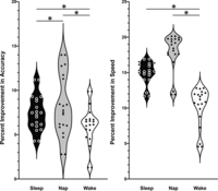
Behavioral results. Offline gains in ToH performance (expressed as percent improvement in speed and accuracy) from training to retest for sleep, nap, and wake groups.
Resting-state functional connectivity results
ROI to ROI results
To test our main research question using a hypothesis-driven a priori set of six ROIs, a 3 × 2 group (sleep, nap, and wake) by session (rest 2 and rest 3) ANOVA revealed differences in functional connectivity from rest 2 to rest 3 as a function of group (F(4,112) = 3.17, PFDR = 0.049). To further investigate the between-group differences from rest 2 to rest 3, follow-up 2 (group) × 2 (session) ANOVAs revealed that this connectivity difference was specific to the sleep versus wake comparison (F(1,38) = 8.11, PFDR = 0.042) and not the nap versus wake (all PFDR > 0.550) or sleep versus nap (all PFDR > 0.590) comparisons. Independent t-tests revealed that connectivity increased between the hippocampus and the inferior OFC from rest 2 to rest 3 in the sleep versus wake group (t(38) = 2.85, PFDR = 0.035) (Fig. 2). Individual hippocampal–orbitofrontal connectivity values at rest 2 and at rest 3 separately, for each participant in each group, are descriptively shown in Figure 3.
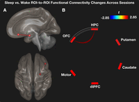
ROI to ROI results using six a priori ROIs. (A) The hippocampus showed strengthened functional connectivity to the inferior OFC following a period of sleep versus wake. (B) Significant edge between ROIs reflects independent t-test value. (HPC) Hippocampus, (dlPFC) dorsolateral prefrontal cortex, (OFC) orbitofrontal cortex.
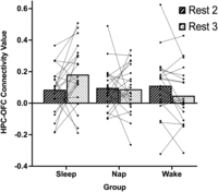
Individual and group mean hippocampal–orbitofrontal resting-state functional connectivity values at both rest 2 and rest 3 for the sleep, nap, and wake groups. The sleep group showed increased functional connectivity versus wake, the nap group showed stabilization, and the wake group showed decreased functional connectivity versus the sleep group.
Seed to whole-brain results
In order to confirm the ROI to ROI results in a less constrained and more exploratory analysis approach, a series of 3 × 2 group (sleep, nap, and wake) by session (rest 2 and rest 3) ANOVAs using the six ROIs as seeds revealed significant changes in connectivity as a function of group between the hippocampus and bilateral supplementary motor area (SMA; F(2,57) = 12.59, PFDR = 0.046, k = 92) (Fig. 4A) and between the putamen and the left anterior cerebellum (F(2,57) = 18.39, PFDR = 0.040, k = 95) (Fig. 4B). Planned comparison follow-up 2 × 2 group by session ANOVAs showed that these omnibus effects in both cases were specific to the sleep versus nap comparison, whereby functional connectivity among these areas was greater in the sleep group (Table 1).
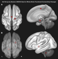
Seed to whole-brain analyses using a 3 × 2 group (sleep, nap, and wake) by session (rest 2 and rest 3) ANOVA demonstrated changes in functional connectivity between the hippocampus and SMA (A) and between the putamen and anterior cerebellum (B). Both omnibus effects were specific to the sleep versus nap comparison, as suggested by follow-up 2 × 2 group by session ANOVAs (Table 1). Spheres represent seeds.
Seed to whole-brain analyses between groups at postinterval (rest 3 [R3]) minus preinterval (rest 2 [R2])
These planned follow-up comparisons yielded additional differences in seed to whole-brain functional connectivity between two groups, which were not observed in the omnibus test between all three groups (Table 1). Specifically, the sleep versus nap comparison additionally demonstrated increased functional connectivity between the OFC and SMA and between the dlPFC and the temporal pole, and decreased connectivity between the putamen and precentral/paracentral cortices. In addition, similar to the ROI to ROI analysis, the sleep versus wake group showed increased connectivity between the hippocampus and the OFC, between the lateral and medial OFC, and also between the caudate and somatosensory areas. By comparison, the nap versus wake group showed negative functional connectivity from the motor cortex to the cuneus and to the supramarginal gyrus.
Brain–behavior relationships
We entered behavioral gains as covariates of interest into the model to follow up the ROI to ROI analysis to examine the relationship between functional connectivity changes and behavioral improvements. A 2 × 2 group (sleep and wake) by session (rest 2 and rest 3) ANCOVA, entering accuracy as a covariate of interest for the six ROIs, revealed that the relationship between the improvement in accuracy and the extent of the change in functional connectivity (rest 2 to rest 3) differed between the sleep and wake groups for the hippocampal–orbitofrontal cortex edge (F(2,55) = 5.08, PFDR = 0.028) (Fig. 5). Similar ANCOVAs comparing sleep versus nap and nap versus wake showed no significant effects. Significant correlations were not observed for any comparison when entering improvement on speed as a covariate of interest. Thus, the change in HPC–OFC functional connectivity from rest 2 to rest 3 was associated with the differential relationship in behavioral accuracy gain scores between sleep and wake groups, whereby the correlation was more positive in the sleep versus the wake group.
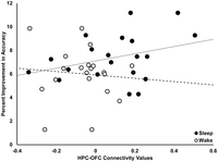
Interaction between sleep and wake groups on the relationship between percent improvement in accuracy on the Tower of Hanoi and the change in hippocampal–orbitofrontal cortex functional connectivity from rest 2 to rest 3. (HPC) Hippocampus, (OFC) orbitofrontal cortex.
Discussion
Previous studies have revealed that the hippocampal–striatal–cortical network of brain areas is involved in the consolidation of procedural memory for motor skills (Albouy et al. 2013b,c). This consolidation process preferentially benefits from a period of sleep (Debas et al. 2010; Boutin et al. 2018; Fang et al. 2021). In the present study, we observed (1) improved performance on the ToH when sleep (either nocturnal sleep or a daytime nap) occurred during the consolidation interval as compared with a period of wake, (2) strengthened functional connectivity from rest 2 to rest 3 in key brain areas associated with the acquisition of novel cognitive strategies and procedural problem-solving skills, (3) that these changes in functional connectivity differed following a consolidation period of sleep as compared with either a daytime nap or a period of wakefulness, and (4) that the extent of the strengthened functional connectivity between hippocampal–orbitofrontal areas was more positively correlated with the offline behavioral improvements for problem-solving skills in the sleep versus the wake group.
The role of subcortical–cortical communication in problem-solving skills
It has been said that “neurons wire together if they fire together” (Hebb 1949). At the system level, for most types of memory, this process initially involves a dialog between the hippocampus and neocortical areas. Sleep is thought to be an opportune time for this transformation of the memory trace to take place, which is apparent through sleep-dependent interactions between hippocampal–putamen areas (Debas et al. 2010; Barakat et al. 2013; Albouy et al. 2015), hippocampal–caudate areas (Cousins et al. 2016; van den Berg et al. 2021), striatal–cortical areas (Debas et al. 2014; Vahdat et al. 2017; Fang et al. 2021), and hippocampal–prefrontal areas (Samanta et al. 2021). During the offline consolidation period of sleep, cortical functional connectivity is decreased, whereas subcortical functional connectivity is increased (Vahdat et al. 2017). Notably, the putamen is a central hub of connectivity, and the strength of the within-network connectivity of the putamen is associated with offline improvements in motor sequence learning (Vahdat et al. 2017). Remarkably, following the offline consolidation period (i.e., upon awakening), subcortical–cortical resting-state functional connectivity is reinstated (Vahdat et al. 2017). In addition, functional communication between the putamen and motor cortex is increased following a consolidation interval of daytime sleep, as compared with an equivalent period of wake (Fang et al. 2021).
The results of the present study complement these recent findings and extend knowledge of how sleep is correlated with sleep-related offline gains in performance for novel cognitive strategies needed to solve problems. Interestingly, unlike motor skill memory consolidation, the strengthening of subcortical–cortical communication following a period of nocturnal sleep versus daytime wakefulness appears to involve the hippocampus and OFC but not the putamen. This was observed in both a hypothesis-driven ROI to ROI analysis and a complementary but less constrained seed to whole-brain approach. Follow-up analyses revealed that the extent of the change in functional connectivity over the consolidation interval was differentially correlated with performance improvements, depending on whether this interval contained either nocturnal sleep or daytime wakefulness. These results suggest that sleep-related changes in hippocampal–OFC functional connectivity are associated with offline gains in problem-solving skills.
The role of the striatum in problem-solving skills
The involvement of the striatum evolves as consolidation of simple motor procedural skills progresses over time (Lehéricy et al. 2005). Changes in functional connectivity in the striatum have been shown during (Vahdat et al. 2017) and following (Fang et al. 2021) a consolidation period of sleep. Communication between the putamen and other task-relevant brain areas is critical for simple motor sequence memory consolidation. Interestingly, unlike previous studies focused on the consolidation of simple motor skills, we observed enhanced connectivity with the caudate nucleus (to the somatosensory cortex), instead of the putamen when comparing sleep versus wake. Moreover, the putamen showed changes in functional connectivity only when comparing sleep versus nap. Thus, a full night of sleep might be involved in enhancing communication in regions involved in planning and problem-solving (e.g., the caudate), whereas a daytime nap might be involved in enhancing communication in brain areas involved in motor skills and motor execution (e.g., the putamen).
The role of the hippocampus in problem-solving skills
Both strengthened functional connectivity between the hippocampus and the OFC and improved problem-solving skills were observed following a full night of sleep compared with a day of wakefulness. This hippocampal–OFC strengthening across sessions was specific to nocturnal sleep relative to daytime wake, as demonstrated by both ROI to ROI and also the less constrained and more exploratory seed to whole-brain analyses. Importantly, the magnitude of this strengthened hippocampal–OFC functional connectivity was more positively correlated with performance improvements for the sleep versus the wake group. Prior research showed that the hippocampus and OFC work together during trial and error performance (Hornak et al. 2002) and goal-directed strategy switching (Young and Shapiro 2011). Consistent with these previous studies, our results suggest that hippocampal–OFC communication is associated with sleep-related offline gains in procedural skills involving the acquisition of novel cognitive strategies. This further suggests that enhanced functional communication at the system level is associated with improved performance at the behavioral level, which occurs preferentially during sleep compared with wake.
Differential effect on functional connectivity between sleep, nap, and wake
Previous studies have shown that for certain types of procedural memory consolidation (for example, perceptual memory [Mednick et al. 2003] or memory for motor sequences [Doyon et al. 2009]), a nap is just as beneficial as a night of sleep. However, at the neuronal system level, for changes that take place in brain areas that support higher-order problem-solving skills, an additional benefit comes from a longer period of sleep compared with either a day of wake or a daytime nap (van den Berg et al. 2021). Here, we show that a full night of sleep but not a daytime nap strengthens striatal–neocortical and hippocampal–neocortical communication in areas involved in memory for cognitive strategies and problem-solving skills.
Specifically, seed to whole-brain analyses (Table 1) revealed that the sleep group showed relatively strengthened functional connectivity between the caudate and somatosensory areas and between the hippocampus and the OFC as compared with the wake group. Meanwhile, the sleep versus nap comparison showed increased functional connectivity for the nap group in areas associated with simple motor skills; for example, between the putamen and cerebellum. These findings suggest that the changes following a nap involve brain areas that support the execution of the task but not the cognitive strategy per se. In contrast, sleep-related increased functional communication was observed for hippocampal and OFC seeds with areas involved in mental imagery for motor movements (i.e., the SMA). No significant ROI to ROI differences were observed when comparing nap versus wake groups for the hippocampus or OFC as seed regions; however, the less constrained seed to whole-brain approach revealed that the nap versus wake comparison showed increased negative functional connectivity between the motor cortex and cuneus and between the motor cortex and supramarginal gyrus. Thus, we speculate that while behavioral performance improvements can benefit from even a short period of daytime sleep (involving primarily motor cortical and related areas), a full night of sleep is optimal for more complete system-level consolidation (involving hippocampal and related areas).
Importantly, this does not lead to the conclusion that fewer or less important changes in functional communication occur from a daytime nap (as compared with wake), only that any changes in functional communication between brain areas during the resting state are not as robust or complete after only a nap. Moreover, we have previously shown that measures other than resting-state functional connectivity (e.g., enhanced brain activation) unfold following a nap. Indeed, in a related study (van den Berg et al. 2021), we showed that a nap strengthens activation of the parietal, motor, and prefrontal cortices, along with reduced activation of the hippocampus, but sleep does afford an additional boost to this memory trace strengthening over and above a nap or a period of wake.
The differential pattern of behavioral improvements and functional connectivity in the nap group might also indicate that consolidation is sensitive to time of day effects. At the neural system level, functional networks become less segregated following sleep deprivation (Chee and Zhou 2019) or when vigilance is impaired (Thompson et al. 2013). In other words, functional networks are generally more segregated when the brain is well rested. In the present study, it is possible that testing at different times of day between the sleep and nap groups, and in turn differences in these network segregations, might explain the differences observed between groups. However, it is unlikely that this confounds our results for two reasons. First, rather than identifying greater segregation between brain areas following sleep (i.e., decreases in functional connectivity), our findings are in the opposite direction and show increased functional connectivity between ROIs in sleep versus wake, with minimal changes in the nap versus wake comparisons (Table 1). Second, the brain–behavior analyses showed that the changes in functional connectivity were correlated with the extent of performance improvements. This correlation was more positive in the sleep group as compared with the wake group. This pattern of correlations cannot be easily explained by time of day alone.
Nonetheless, it is important to note that the time interval between rest 2 and rest 3 was shorter in the nap group (i.e., ∼5 h) compared with the ∼10-h intervals in the sleep and wake groups. The improved task performance in the nap group might be attributable to the effects of a short daytime sleep period or, alternatively, might be due to a shorter time window between test and retest. A separate, 90-min daytime wake group could clarify whether the behavioral effects are due specifically to a nap or to the time interval.
Another alternative interpretation is that sleep affords protection from interference, rather than enhanced performance (Rickard et al. 2008). Offline periods of quiet rest have shown evidence for “wake-dependent stabilization” (Craig et al. 2018; Humiston and Wamsley 2018). However, this does not appear to be a plausible explanation, given that previous work on this same memory task (i.e., ToH) showed that improvement in performance is observed after sleep and only after sleep (e.g., van den Berg et al. 2019). This alternative explanation is also not easily explained by the wake group in the current study, as a period of quiet wake has been shown to afford a protective effect similar to that of a sleep period (Craig et al. 2018; Humiston and Wamsley 2018). Thus, the improved behavioral performance in our study following a nap cannot be explained by wake-dependent stabilization or the simple passage of time.
Finally, it is possible that dissimilar effects of sleep and nap on functional connectivity may depend on nap architecture. In particular, REM sleep that occurs during a nap is shown to protect perceptual memories from interference (McDevitt et al. 2015). In addition, the presence of REM during a daytime nap can enhance perceptual memory consolidation to the same degree as a night of sleep. Specifically, it has been shown that naps that contained REM sleep enhanced subsequent performance, as compared with either naps without REM sleep or an equivalent period of wakefulness (Mednick et al. 2003). In the current study, 75% (n = 15) of participants in the nap group reached a single period of REM sleep, which may be insufficient to achieve the same extent of consolidation as a night of sleep. However, supplemental correlation analyses (Supplemental Table S1) did not reveal any significant relationships between REM sleep duration and either ToH performance improvements or hippocampal–orbitofrontal connectivity values in either the nap or sleep group.
Limitations and future directions
We selected a parsimonious number of ROIs in a hypothesis-driven approach to assess how functional connectivity changes within the hippocampal–striatal–cortical network of brain areas following a consolidation period of sleep, nap, or wake. These ROIs were selected based on peak coordinates in task-related activation areas of interest most consistently associated with sleep-related consolidation of procedural memories that comprise both strategy learning and problem-solving skills. However, the trade-off of this focused approach comes at the expense of limited exploration of whole-brain connectivity. We mitigated this limitation by including a complementary seed to whole-brain analysis, thus permitting us to examine how our ROIs functionally communicate with the rest of the brain.
In addition, it is worth noting that our ROIs were coincidentally all in the right hemisphere. ROIs were determined by task-related activations that demonstrated either unilateral activation exclusively in the right hemisphere or, in the case of regions with bilateral task-related activation, the right hemisphere consistently had stronger peak activations (van den Berg et al. 2021). Furthermore, these activations were ipsilateral to performance with right-handed task performance, which is consistent with meta-analysis findings of primarily right (ipsilateral) response activation in subcortical areas, which support spatial behaviors (Cieslik et al. 2015).
As discussed above (see “Differential Effect on Functional Connectivity between Sleep, Nap, and Wake”), time of day is an important consideration when comparing resting-state functional connectivity between intervals containing sleep or wake. Future neuroimaging studies could directly investigate this by including fMRI acquisitions using an a.m.–p.m.–a.m. versus p.m.–a.m.–p.m. group testing paradigm (van den Berg et al. 2019). Our group has previously used this paradigm in a behavior-only study for the ToH. This study revealed that improvement on the ToH occurs once given the opportunity to sleep (i.e., p.m. to a.m.), regardless of whether initial learning occurs in the a.m. or p.m. Inclusion of prepost resting-state acquisitions at each time point, as well as the inclusion of a daytime nap group, would allow for changes in brain activation and resting-state functional connectivity while examining time of day effects to be fully explored.
In addition, it is important to note that the observed changes in functional connectivity and their relationship to memory consolidation are entirely correlational. Including a condition using the same fMRI protocol, but where a control task is used, would allow for stronger evidence that changes in functional connectivity are indicative of memory consolidation per se, rather than a generalized benefit of sleep. Alternatively, simultaneous EEG–fMRI acquisitions during sleep would enable the investigation of memory trace reactivation in different sleep stages and time-locked to microarchitecture of sleep (e.g., spindles, slow waves, and eye movements), which would provide additional insight into how sleep actively participates in the memory consolidation process. Similarly, nocturnal sleep studies with more extensive investigation of the features of sleep that might change in response to this type of new learning would reveal the electrophysiological markers of sleep-related memory consolidation.
Finally, future studies could use a similar approach to investigate the relationship between sleep and other forms of memory (e.g., motor sequence learning and declarative memory). Specifically, ROI coordinates obtained from memory domain-specific neural activity can be examined during resting-state periods across the consolidation interval. This would also help to elucidate the specificity of the results of the current study.
Conclusions
These findings are consistent with recent evidence that the hippocampus is involved in sleep-related procedural memory consolidation not only for simple procedural motor sequences (Albouy et al. 2013a,b; Fogel et al. 2017; Boutin et al. 2018) but also for motor sequences that involve the acquisition of novel cognitive strategies required to solve problems (Cousins et al. 2016; van den Berg et al. 2021). Overall, our results suggest that a full night of sleep (vs. wake) strengthens functional communication between the hippocampal–striatal–cortical network of brain areas and that this enhanced functional communication is distinct from a daytime nap. Finally, the changes in hippocampal–orbitofrontal functional connectivity following nocturnal sleep versus daytime wake were associated with improved memory for cognitive strategies and problem-solving skills.
Materials and Methods
Participants
The data analyzed in this study were part of a larger polysomnographic, behavioral, and MRI protocol using various techniques intended to address a variety of research questions. The resting-state fMRI data have not been reported elsewhere and are unique to this study; see van den Berg et al. (2021) for additional details about the experimental protocol, specific to that study. All methods relevant to the current investigation are reported here. Participants were between 20 and 35 yr of age, were right-handed with no hand/finger mobility issues, were nonshift workers, took no medications known to interfere with sleep, and had consistent sleep schedules (i.e., 7–10 h of sleep, a bedtime between 2200 and 0100, and wake time before 0900). Participants had a body mass index (BMI) <30, had normal or corrected to normal vision, considered themselves a “nonsmoker,” consumed fewer than two caffeinated drinks per day, and consumed limited alcohol (i.e., fewer than seven drinks per week). Participants were excluded if they considered themselves a poor sleeper; had a history of chronic pain, seizures, or head injury; or had familiarity with the Tower of Hanoi (ToH) task. Furthermore, participants scored <10 on the Beck Anxiety (Beck et al. 1988) and Beck Depression (Beck and Beamesderfer 1974) Inventories and had no signs of sleep disorders as determined by the Sleep Disorders Questionnaire (Douglass et al. 1994).
Participants who met the above criteria underwent an overnight polysomnography (PSG) night to screen for sleep disorders, which took place 1 wk before the experimental sessions. Participants were excluded if their screening night sleep efficiency was <80%, if they had more than five respiratory events per hour (indicating signs of sleep apnea), or if they showed indications of parasomnias. This PSG night also served to acclimatize participants to the sleeping environment. Finally, all participants wore a Motionlogger actigraph (a wrist-worn accelerometer; Ambulatory Monitoring, Inc.) and were asked to complete a log of their sleep/wake habits for the duration of the study to verify compliance with the study protocol.
Power analyses determined the required sample size for observing both behavioral effects and MRI effects. Using α set to 0.05 to obtain 80% power, based on an effect size of f = 0.9 for behavioral effects, and a more conservative f = 0.7 for MRI effects, we would require a minimum of n = 15 per group. To ensure adequate power for all analyses, we aimed to have final sample sizes of n = 20 per group. Four participants were excluded from the study for noncompliance with the experimental protocol based on the results of their actigraph and sleep/wake log. One participant voluntarily dropped out of the study following the PSG screening night. The final sample of 60 healthy young adults (34 females, aged 20–35 yr, Mage = 24.38, SD = 4.44) were randomly assigned to either the sleep (n = 20), nap (n = 20), or wake (n = 20) group.
Ethics statement
All study procedures were approved by the Research Ethics Boards at the University of Ottawa and The Royal's Institute of Mental Health Research (IMHR). All participants provided written informed consent prior to participation and were financially compensated for their participation.
Experimental protocol
Following the PSG screening night, eligible participants returned to the laboratory for the experimental session 1 wk later (see Fig. 6 for the study design). Upon arrival at the laboratory, participants completed the training session by performing eight trials of the ToH task. The training session was flanked by 8-min resting-state scans (“rest 1” and “rest 2”) immediately before and after the training session. This was followed by either (1) an overnight PSG sleep recording (sleep group), (2) a 90-min daytime PSG recording (nap group), or (3) a daytime wake period (wake group). Following these intervals (and after a >30-min wake period to allow the effects of sleep inertia to dissipate in the sleep and nap groups), all participants completed the retest session. The retest session involved performing four trials of the ToH and was immediately preceded by an 8-min resting-state scan (“rest 3”). For all resting-state scans, participants were instructed to stay relaxed, keep their eyes open, and keep their gaze fixed on an on-screen plus sign (+).
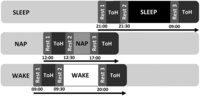
Study design. Following a screening and acclimatization night of PSG, participants returned 1 wk later to the laboratory for the experimental session. N = 20/group were assigned to either the sleep, nap, or wake group and underwent MRI imaging sessions to obtain resting-state fMRI BOLD images before training (rest 1), after training (rest 2), and after the retention interval but prior to retest (rest 3). In the sleep group, rest 1 began at ∼2100 h, and rest 2 began 20–30 min later (at ∼2130 h). The 8-h sleep opportunity began at ∼2200 h, and rest 3 began 10–11 h later (∼0900 h). In the nap group, rest 1 began at ∼1200 h, and rest 2 began 20–30 min later (at ∼1230 h). The 90-min sleep opportunity began at ∼1400 h, and rest 3 began 3 h later (∼1700 h). In the wake group, rest 1 began at ∼0900 h, rest 2 began 20–30 min later (at ∼0930 h), and rest 3 began 10–11 h later (∼2000 h). It is important to note that the intervals between sleep and wake were held constant. Participants in the wake group remained awake in between the training and retest sessions (confirmed via actigraphy).
Behavioral task and analysis
Tower of Hanoi
The ToH task was coded in Matlab R2016a (Mathworks, Inc.) using the Psychophysics Toolbox extension v3.0.12 (Brainard 1997; Kleiner et al. 2007) for Windows (Microsoft). The start configuration of the task included three identical, equally spaced pegs, with five disks stacked on the far-left peg in ascending order of size (i.e., largest disk on the bottom and the smallest disk on the top).
The ToH requires the participant to acquire a novel cognitive strategy (i.e., the use of recursive logic) to solve the problem in the minimum number of moves. There is only one optimal solution to the puzzle, which can be learned through trial and error.
Participants were instructed that, for each trial, the objective of the task was to transfer all disks from the initial position on the far-left peg to the final position on the far-right peg in the same ascending order. They were instructed that this was to be accomplished under the following constraints: (1) only one disk could be moved at a time, (2) only the uppermost disk from any one of the three pegs could be selected, and (3) a disk could only be placed on an empty peg or on another disk that is larger in size. Participants could control the on-screen movement of the disks with an MR-compatible fiber optic button pad (model HHSC-2X4-C, Current Designs, Inc.) using one of three push buttons equally spaced apart in a horizontal row. Participants were instructed to perform the task as quickly and accurately as possible with their right hand, where their index, middle, and ring fingers corresponded to the first (i.e., far-left peg), second (i.e., middle peg), and third (i.e., far-right peg) buttons, respectively. The buttons controlled the movement of the disks “from” and “to” one of the three pegs. A trial ended when all five disks were in ascending order on the far-right peg or if the maximum number of moves was reached before successful completion of the task. The maximum number of moves for the five-disk task was set at 93 (i.e., three times the optimal number of moves 2N − 1, where N is the number of disks; 25 − 1 = 31). Every trial of the ToH task was followed by a 20-sec rest period, during which the three ToH pegs were still visible, but with no disks. Participants were instructed to abstain from pressing any buttons during this time.
The variables of interest for the ToH task were speed (i.e., time to complete each ToH trial), accuracy (mean absolute percentage
error [MAPE]), and speed–accuracy trade-off (SATO). The MAPE can be interpreted as a percentage of perfect performance (e.g.,
100% reflects perfect performance, whereas 50% reflects making twice as many errors as perfect performance, and 0% represents
an infinite number of moves). The percent improvement for all variables was calculated by taking the performance in the retest
trials minus performance in the training trials, divided by performance in the training trials, multiplied by 100:

Behavioral analysis
All behavioral statistical analyses were carried out using SPSS Statistics version 25 (IBM). To ensure that task acquisition (i.e., learning) occurred during the training session, a block by trial ANOVA was performed across all participants. Next, ToH performance improvement across the interval of sleep, nap, or wake was measured using three separate one-way ANOVAs for measures of speed, accuracy, and SATO. Significant effects were followed up with planned comparison t-tests.
Polysomnographic recording and analysis
Polysomnographic recording parameters and analysis
Polysomnography (PSG) was recorded with the Embla N7000 32-channel amplifier system (Natus) sampled at 500 Hz. Scalp-recorded EEG from 13 channels (Fp1, Fp2, Fpz, F3, F4, Fz, C3, C4, Cz, P3, P4, Pz, and Oz), mastoid derivations (M1 and M2), and left and right electrooculogram (EOG) were referenced to Fpz online, all placed according to the international 10–20 system (Jasper 1958). In addition, a submental electromyogram (EMG) channel was recorded as a bipolar derivation.
EEG and EOG were rereferenced offline to average mastoid derivations (M1 and M2) and filtered from 0.3 to 35 Hz for EEG, 0.3 to 10 Hz for EOG, and 10 to 50 Hz for EMG. Manual sleep stage scoring was completed using RemLogic analysis software (Natus) by a single expert scorer according to standard criteria (Rechtschaffen and Kales 1968; Iber et al. 2007). The sleep variables of interest obtained from the polysomnographic recordings included total sleep time (TST; calculated as the total time spent asleep starting at sleep onset between “lights off” and “lights on”) and the time in minutes spent in stage 1 (NREM1) sleep, stage 2 (NREM2) sleep, stage 3 (NREM3) sleep, and rapid eye movement (REM) sleep. The percent duration of each stage from TST was also calculated. Sleep architecture results for both the sleep and nap groups are reported in Supplemental Table S2.
MRI acquisition and analysis
fMRI recording parameters
Functional magnetic resonance imaging (fMRI) data were collected on a Siemens Biograph mMR 3.0 Tesla whole-body MRI scanner (Siemens) using a 12-channel head coil. Anatomical images were acquired using a standard 3D multislice MPRAGE sequence (TR = 2300 msec, TE = 2.98 msec, TI = 900 msec, FA = 9°, 176 slices, FoV = 256 × 256 mm2, matrix size = 256 × 256 × 176, voxel size = 1 mm3). In addition, multislice T2-weighted fMRI images were acquired during the resting-state acquisition (TR = 2.16 sec, TE = 30 msec, FA = 90°, 40 transverse slices, 3-mm slice thickness, 10% interslice gap, FoV = 220 × 220 mm2, matrix size = 64 × 64 × 40, voxel size = 3.44 mm × 3.44 mm × 3 mm).
Preprocessing
Resting-state images were preprocessed using the Matlab-based Conn toolbox v20.b (Whitfield-Gabrieli and Nieto-Castanon 2012). Preprocessing was performed using the standard pipeline in Conn, comprising functional realignment, ascending-order slice-timing correction, coregistration to the MPRAGE structural scan, spatial normalization, and spatial smoothing using an 8-mm full-width half-maximum (FWHM) isotropic Gaussian kernel filter. Segmentation was performed on the individual T1 images. Normalization to Montreal Neurological Institute (MNI) space was performed. The denoising pipeline was also used, regressing out, from the realigned data, five noise components (for white matter and cerebral spinal fluid) and 12 motion effects (for X, Y, and Z translations; for pitch, yaw, and roll rotations; and for their first-order derivatives). The data were then band-pass-filtered from 0.008 to 0.09 Hz. Functional outliers were thresholded to the 97th percentile. Scrubbing was performed using artifact detection tools with the threshold for global signal >5 (Z-value) and for subject motion >0.9 mm overall. Visual inspection ensured each participant did not show abnormalities. Averaged time series for each ROI from the preprocessed images were extracted using Conn.
Seed regions
ROIs were computed as 6-mm spheres around each coordinate using FSLeyes v0.34.2 (Wellcome Center for Integrative Neuroimaging, https://fsl.fmrib.ox.ac.uk/fsl/fslwiki/FSLeyes) and were subsequently exported to Conn. ROIs were defined a priori, obtained from the extant literature. Specifically, they were selected based on areas associated with sleep-related consolidation of procedural memory, including memory for cognitively complex skills and problem-solving (e.g., the caudate, OFC, and dlPFC) and for regions typically implicated in a related motor sequence learning task (e.g., the hippocampus, putamen, and motor cortex). Coordinates for each region were determined from a companion study's task-related peak activations (van den Berg et al. 2021). This yielded five anatomical seed regions (i.e., the hippocampus, caudate, motor cortex, OFC, and dlPFC). In addition, the putamen is critically important for sleep-related procedural motor sequence learning (Albouy et al. 2013c, 2015; Fogel et al. 2017; Vahdat et al. 2017; Fang et al. 2021) and was thus also included as an ROI (Vahdat et al. 2017). In total, six anatomical seed regions were included (Table 2; Fig. 7). Coordinates for these ROIs either had unilateral activation exclusively in the right hemisphere or, in the case of regions with bilateral task-related activations, the right hemisphere demonstrated stronger peak activation.
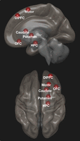
A priori ROIs included in resting-state functional connectivity analyses.
A priori ROIs, using 6-mm spheres drawn around task-related activation differences
ROI to ROI analyses
First, we examined changes in resting-state functional connectivity from before to after the training session (i.e., rest 2 > rest 1) across all participants. In addition, we tested for group differences at rest 2 (i.e., immediately following training) using a one-way ANOVA to determine whether groups differed in terms of their functional connectivity before the consolidation interval. No significant differences were observed from rest 1 to rest 2. These results are presented in the Supplemental Material.
To test our main hypotheses, we examined whether functional connectivity among our six ROIs changed from the resting-state scan immediately following the training session (rest 2) to the resting-state scan immediately prior to the retest session (rest 3) across an interval of sleep, nap, or wake. This was accomplished using a 3 × 2 group (sleep, nap, and wake) by session (rest 2 and rest 3) ANOVA. Planned post-hoc comparisons were conducted between each group at rest 2 and rest 3. All ROI to ROI F-tests and follow-up t-tests were thresholded at P < 0.001 uncorrected, with cluster-level correction for false discovery rate at PFDR < 0.05 to control for multiple comparisons.
Brain–behavior relationships
To further investigate whether there were any brain–behavior relationships from the ROI to ROI analyses described above, we entered performance improvements (speed and accuracy) as covariates of interest into the model to follow up significant ROI to ROI effects. We report the result from the group by session interaction effect with ToH performance improvements as covariates of interest. This allowed us to assess the relationship between behavioral performance and changes in functional connectivity from before to after the consolidation intervals in the sleep versus wake, nap versus wake, and sleep versus nap comparisons.
Seed to whole-brain analysis
To confirm the results of the hypothesis-driven, parsimonious a priori ROI to ROI approach, we used a less constrained seed to whole-brain analysis using the six ROIs as seed regions. A 3 × 2 group (sleep, nap, and wake) by session (rest 2 and rest 3) ANOVA was performed for each seed region. Planned comparison follow-up group by session ANOVAs tested for differences between (1) sleep versus wake, (2) nap versus wake, and (3) sleep versus nap groups. All tests were performed using statistical thresholds of uncorrected P < 0.001 at the whole-brain level and corrected for false discovery rate PFDR < 0.05 at the cluster level.
Acknowledgments
We are grateful to Katie Dinelle, Rahim Ismaili, Reggie Taylor, and the Brain Imaging Center at the Royal's Institute of Mental Health Research at the University of Ottawa for their support with magnetic resonance imaging. This work was supported by a Natural Sciences and Engineering Research Council (NSERC) Discovery grant (RGPIN/2017-04328 to S.M.F.), and by a Ministry of Research and Innovation Early Researcher Award (ERA; ER17-13-023 to S.M.F.).
Footnotes
-
[Supplemental material is available for this article.]
-
Article is online at http://www.learnmem.org/cgi/doi/10.1101/lm.053638.122.
- Received August 3, 2022.
- Accepted December 22, 2022.
This article is distributed exclusively by Cold Spring Harbor Laboratory Press for the first 12 months after the full-issue publication date (see http://learnmem.cshlp.org/site/misc/terms.xhtml). After 12 months, it is available under a Creative Commons License (Attribution-NonCommercial 4.0 International), as described at http://creativecommons.org/licenses/by-nc/4.0/.



