


Reconsolidation may incorporate state-dependency into previously consolidated memories
- Rodrigo O. Sierra1,2,
- Lindsey F. Cassini1,2,
- Fabiana Santana1,2,
- Ana P. Crestani1,2,
- Johanna M. Duran1,2,
- Josué Haubrich1,2,
- Lucas de Oliveira Alvares1,2,3,4 and
- Jorge A. Quillfeldt1,2,3,4
- 1Psychobiology and Neurocomputing Laboratory, Biophysics Department, Biosciences Institute, 91.501-970, Federal University of Rio Grande do Sul, Porto Alegre, Brazil
- 2Graduate Program in Neuroscience, Institute of Health Sciences, 90.046-900, Federal University of Rio Grande do Sul, Porto Alegre, Brazil
-
↵3 These authors contributed equally to this work.
Abstract
Some memories enter into a labile state after retrieval, requiring reconsolidation in order to persist. One functional role of memory reconsolidation is the updating of existing memories. There are reports suggesting that reconsolidation can be modulated by a particular endogenous process taking place concomitantly to its natural course, such as water or sleep deprivation. Here, we investigated whether an endogenous process activated during a natural/physiological experience, or a pharmacological intervention, can also contribute to memory content updating. Using the contextual fear conditioning paradigm in rats, we found that the endogenous content of an aversive memory can be updated during its reconsolidation incorporating consequences of natural events such as water deprivation, transforming a previously stored memory into a state-dependent one. This updating seems to be mediated by the activation of angiotensin AT1 receptors in the dorsal hippocampus and local infusion of human angiotensin II (ANGII) was shown to mimic the water deprivation effects on memory reconsolidation. Systemic morphine injection was also able to turn a previously acquired experience into a state-dependent memory, reproducing the very same effects obtained by water deprivation or local angiotensin II infusion, and suggesting that other state-dependent-inducing protocols would also be able to contribute to memory updating. These findings trigger new insights about the influence of ordinary daily life events upon memory in its continuing reconstruction, adding the realm of reconsolidation to the classical view of endogenous modulation of consolidation.
The classical idea that memories are immutable after consolidation has been recently challenged. A considerable amount of evidence has shown that consolidated memories can enter into a new label/unstable state after retrieval, during which they are again susceptible to pharmacological and behavioral disruption, requiring a later restabilization process known as reconsolidation (Duvarci and Nader 2004). It has been suggested that the biological function of memory reconsolidation may serve a wider purpose than simply restabilizing memories previously destabilized during reactivation (Lee 2010). Specifically, reconsolidation has been proposed to support processes such as memory strengthening (Lee 2008), updating (Hupbach et al. 2008), and precision keeping (de Oliveira Alvares et al. 2012).
Recent reports have shown that reconsolidation can be modulated by endogenous processes activated during natural events like water or sleep deprivation (Frenkel et al. 2005a; Shi et al. 2011). In the crab Chasmagnathus sp., concurrent water deprivation improves reconsolidation via endogenous angiotensin and strengthens memory expression (Frenkel et al. 2005a). In humans, a natural mild stressor like cold pressor stress, concomitant with memory reactivation, was able to improve long-term expression in a declarative memory paradigm (Coccoz et al. 2011). These observations indicate that new endogenous information may interact with a previously acquired memory during reconsolidation and open the debate about the influence of ordinary, natural, or real-life events in reconsolidation processes.
The concept of endogenous modulation of memory has had significant influence upon basic research on the neurobiological mechanisms of memory storage and expression (Cahill and McGaugh 1996). Particularly, the study of state-dependency allowed us to establish the role of the neurohumoral state upon different stages of memory (Izquierdo 1980). However, this type of endogenous modulation has not been previously studied in relation to reconsolidation, despite being already suggested elsewhere (Riccio et al. 2006). Our hypothesis is that reconsolidation can support updating of an established memory if some particular endogenous neurohumoral state is able to induce state-dependency, as it is known to influence consolidation.
Here we evaluated whether a particular endogenous state induced by a natural condition, or a pharmacological treatment, can update the content of a memory during reconsolidation, identifying its possible neurobiological mechanism. To address this question, we employed water deprivation (a natural condition) or morphine injection (a pharmacological treatment) in a reconsolidation protocol employing contextual fear conditioning (CFC) in rats.
Results
Reconsolidation under water deprivation can induce memory updating of its content
To study the updating of memory content by water deprivation, rats were submitted to the CFC and 48 h after training, reactivation took place in a water-deprived state. Twenty-four hours later animals were tested in a nonwater-deprived state (test 1), and 24 h after that they were retested again in a water-deprived state (test 2) (Fig. 1). ANOVA for repeated measures revealed significant effects of time (F(2,38) = 6.965, P = 0.002) and group × time interaction (F(2,38) = 5.017, P = 0.011), but not for group factor (F(1,19) = 1.182, P = 0.290). Post-hoc analysis showed that the water-deprived group expressed lower freezing levels if compared to the control group during test 1 (P < 0.05). Additionally, the water-deprived group expressed less freezing during test 1 when compared to the performance in the reactivation session (P < 0.001) and test 2 (P < 0.001). No significant differences were found between groups in the reactivation session and test 2 (P > 0.05) (Fig. 2A). This suggests that information about the water-deprivation experience during reactivation was somewhat incorporated into the memory content and, consequently, the presence of this endogenous information became necessary for an adequate memory retrieval. These results are consistent with the state-dependent memory phenomenon according to classical reports showing that the presence of neurohumoral and hormonal changes during or after training can also become necessary during retrieval (Izquierdo and Dias 1983b). A common, consistent endogenous state would thus connect memory acquisition/consolidation and retrieval sessions (Izquierdo and Dias 1983a).

Schematic design of the water-deprivation protocol. The experimental group was water deprived for 18 h prior to the memory reactivation session (the water remained available for >6 h after reactivation). Animals were not water deprived at test 1 (and for 6 h after it). However, test 2 was done in animals again under an 18-h water-deprived state. In these experiments, the control group was never water deprived.
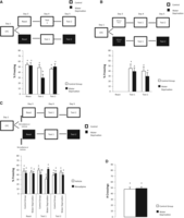
Endogenous memory content can be updated by natural events (such as water deprivation) during reconsolidation. Graphs show the mean ± SEM of percentage freezing time. The open field (OF) graph shows the mean ± SEM of the numbers crossing. Experimental design is depicted at the top of each panel. (A) The water-deprived group shows a significant decrease of memory expression during test 1, a phenomenon which is reverted by being water deprived again during test 2 (control group n = 10, water deprived n = 11). (B) When the reactivation session is omitted, water deprivation has no effect (control group n = 8, water deprivation n = 6). (C) Nimodipine injection before reactivation prevents the effect of water deprivation upon memory reconsolidation (control group/vehicle n = 7, control group/nimodipine n = 8, water deprivation/vehicle n = 10, water deprivation/nimodipine n = 11). (D) Water deprivation during 18 h has no effects upon motor performance (control group n = 7, water deprivation n = 8). Groups marked with the same letter (e.g., “a”) are statistically equal to each other and different from those marked with different letters (e.g., “a” groups differ from “b” groups), with significance level of P < 0.05.
We then asked whether the water-deprivation effect depends on memory reactivation. ANOVA for repeated measures revealed no significant effects either of group (F(1,12) = 0.721, P = 0.412) or time factors (F(1,12) = 1.358, P = 0.266), or group × time interaction (F(1,12) = 0.119, P = 0.735) when the reactivation session is omitted (Fig. 2B). These results suggest that the reactivation session is necessary for the incorporation of new endogenous information, modifying a previously acquired memory.
If it is true that the effect of water deprivation upon memory is mediated by a reconsolidation process, then the inhibition of the memory labilization mechanism should prevent it. To address this question, nimodipine was subcutaneously injected 30 min before memory reactivation. Nimodipine prevents labilization of a reactivated memory during retrieval, without affecting memory retrieval or storage (Suzuki et al. 2008). ANOVA for repeated measures revealed significant effects of the group × time × drug interaction (F(2,64) = 7.961, P = 0.000), but not of group (F(1,32) = 0.103, P = 0.749), drug (F(1,32) = 0.014, P = 0.940), or time factors (F(2,64) = 0.698, P = 0.501), or the interactions between group × drug (F(1,32) = 1.257, P = 0.270), group × time (F(2,64) = 0.468, P = 0.628), or drug × time (F(2,64) = 1.616, P = 0.206). Post-hoc analyses showed that the water-deprived, vehicle-injected group expressed lower freezing levels compared to the deprived group injected with nimodipine during test 1 (P < 0.05). Similarly, the water-deprived, vehicle-injected group expressed less freezing compared to the control group injected with vehicle (P < 0.05) in test 1. Additionally, the water-deprived group injected with vehicle expressed less freezing during test 1 compared to reactivation (P < 0.01) and test 2 session (P < 0.01). No significant differences were found among the groups in the reactivation session and test 2 (P > 0.05) (Fig. 2C). These results confirm that the endogenous memory update with water deprivation is mediated by the labilization/reconsolidation process.
No effect in exploratory performance was found between groups in animals with 18 h of water deprivation before the open field test (P = 0.697, independent t-test) (Fig. 2D), showing that our water-deprivation protocol has no apparent motor effects.
To our knowledge, this is the first evidence that an endogenous state can be incorporated into a previously acquired memory during retrieval through a reconsolidation mechanism. This memory is turned into a state-dependent trace with the endogenous state acting as an internal cue.
Angiotensin II and AT1 receptor in dorsal hippocampus mediates the effects of water deprivation upon memory reconsolidation
The central renin–angiotensin system (RAS) plays an important role in osmoregulatoy and dipsogenic systems (Hazon et al. 1989). The RAS is involved in learning and memory, but the actual role of angiotensin II (ANGII) in this process remains unclear. Dorsal hippocampus shows high levels of ANGII and different AT receptor subtypes (Haas et al. 1980). A recent study has shown that the ANGII plays an important role in the future memory expression by acting on consolidation and reconsolidation in crabs (Frenkel et al. 2005b). We hypothesized that the water-deprivation effects on memory reconsolidation could be associated with the increase of ANGII levels in dorsal hippocampus. Then, we would expect that the local infusion of ANGII could be sufficient to undergo a state-dependent memory. To address this question, rats were conditioned as previously described and injected with ANGII after memory reactivation. The treated group received vehicle 15 min before test 1 and ANGII 15 min before test 2. The control group received vehicle in all sessions of the experiment. ANOVA for repeated measures revealed significant effects of group (F(1,16) = 3.196, P = 0.092) and group × time interaction (F(2,32) = 3.567, P = 0.039), but not for time (F(2,32) = 1.969, P = 0.156) factor. Post-hoc analyses indicated that the ANGII group expressed lower freezing levels compared to the vehicle during test 1 (P < 0.05), as found before with a water-deprivation protocol. Additionally, the ANGII group expressed less freezing during test 1 compared to the reactivation session (P < 0.01) and test 2 (P < 0.05). No significant differences were found between the groups in the reactivation session and test 2 (P > 0.05) (Fig. 3A). These results confirm our hypothesis that the effect of water deprivation during memory reconsolidation is mediated by the increased ANGII levels in dorsal hippocampus.
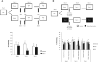
Angiotensin II and AT1 receptor in dorsal hippocampus mediates the endogenous updating. All graphs show percentage of freezing time, expressed as mean ± SEM. The experimental design is shown in the top of each panel. (A) ANGII mimics the water-deprivation effects on memory reconsolidation (vehicle n = 8, ANGII n = 10). (B) The antagonist of ANGII losartan reverts the effect of water deprivation on memory reconsolidation (control group/vehicle n = 10, control group/LOS n = 9, control group/PD n = 7, water deprivation/vehicle n = 8, water deprivation/LOS n = 7, water deprivation/PD n = 8). Groups marked with the same letter (e.g., “a”) are statistically equal to each other and different from those marked with different letters (e.g., “a” groups differ from “b” groups), with significance level of P < 0.05.
To test which receptor would be responsible for the water-deprivation effect on memory reconsolidation, we infused the antagonist of either AT1 or AT2 receptor in the dorsal hippocampus. Rats were submitted to CFC and deprivation protocol as described above. After memory reactivation, either the antagonist of AT1 receptors losartan, the antagonist of AT2 receptors PD 123319, or vehicle were infused into dorsal hippocampus. Test 1 was performed with nondeprived animals and test 2 again in a water-deprived state. The control group was not deprived in any session. ANOVA for repeated measures revealed significant effects of time (F(2,86) = 23.188, P = 0.000) and the interactions between group × drug (F(2,43) = 3.463, P = 0.040), group × time (F(2,86) = 8.067, P = 0.000), time × drug (F(4,86) = 3.123, P = 0.018), and among time × group × drug (F(4,86) = 2.753, P = 0.033), but not for group (F(1,43) = 3.547, P = 0.066) or drug (F(2,43) = 1.670, P = 0.200) factors. Post-hoc analyses showed that water-deprived animals infused with vehicle (P < 0.05) or PD 123319 (P < 0.01) expressed lower freezing levels compared to the deprived group infused with losartan during test 1. Similarly, the water-deprived group infused with vehicle expressed less freezing compared to the control group infused with vehicle (P < 0.01). The water-deprived group infused with PD 123319 expressed lower freezing levels compared to the control group infused with PD 123319 (P < 0.05). Additionally, the water-deprived group infused with vehicle expressed less freezing during test 1 compared to the test 2 session (P < 0.001) but not compared to the reactivation session (P = 0.465). The same pattern was shown by the group with water deprivation infused with PD 123319, expressing less freezing during test 1 compared to the test 2 session (P < 0.05) but not compared to the reactivation session (P = 0.946). The water-deprived group infused with losartan expressed less freezing during reactivation compared to test 1 (P < 0.001) and test 2 (P < 0.05). No significant differences were found between the groups in the reactivation session and test 2 (P > 0.05).
Taken together, these results show that (1) ANGII infusion in the hippocampus is sufficient to promote the water deprivation effect, inducing a state-dependent memory; and (2) such an effect is mediated by the AT1 receptors.
Water deprivation as internal cue strengthens the expression of a weak memory but is not sufficient to induce memory retrieval in a novel context by itself
Previous studies have postulated that interoceptive cues experienced under a given drug or hormonal state might act as a contextual cue for retrieval (Spear 1978; Riccio and Concannon 1981). This concept was consistently confirmed in classical experiments of state-dependent memory (Bormann and Overton 1993). If the water-deprivation event acts like an additional cue during memory retrieval, this information could enhance the expression of a weak memory and/or induce retrieval in a novel context after memory reconsolidation. In order to evaluate these possibilities, two experiments were performed. First, rats were submitted to a weak protocol of CFC (0.4 mA) and, 48 h later, reactivated under a water-deprivation condition and tested as described above. ANOVA for repeated measures revealed significant effects of group (F(1,16) = 4.540, P = 0.048) and group × time interaction (F(2,32) = 3.904, P = 0.030), but not for time (F(2,32) = 3.206, P = 0.053). Post-hoc analyses showed that the control group expressed lower freezing levels in test 2 compared to the water-deprived group (P < 0.05). Additionally, the water-deprived group expressed less freezing during test 1 compared to test 2 (P < 0.01). No significant differences were found among the groups in the reactivation session and test 1 (P > 0.05) (Fig. 4A). This result suggests that the water-deprivation state may act like an additional cue during memory retrieval, enhancing the expression of a state-dependent weak memory.
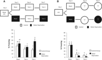
Water deprivation as internal cue strengthens the expression of a weak memory. All graphs show percent of freezing time, expressed as mean ± SEM. The experimental design is shown at the top of each panel. (A) Water-deprived group in a weak CFC training shows an increase of memory expression during test 2 (n = 9 per group). (B) No effect of water deprivation is verified when test sessions are performed in a novel context (control group n = 7, water deprivation n = 8). Groups marked with the same letter (e.g., “a”) are statistically equal to each other and different from those marked with different letters (e.g., “a” groups differ from “b” groups), with significance level of P < 0.05.
In the second experiment, rats were submitted to CFC (0.7 mA) in a water-deprivation protocol. Training and reactivation were performed in the same context, but the animals were tested in a novel context. The control group was not deprived in any sessions of the experiment. ANOVA for repeated measures revealed significant effects of time (F(2,24) = 37.555, P = 0.000) but not of group (F(1,12) = 0.262, P = 0.617) and group × time interaction (F(2,24) = 0.391, P = 0.680). Post-hoc analyses showed that the deprived group expressed lower freezing levels during test 1 (P < 0.001) and test 2 (P < 0.001) compared to the reactivation session. Additionally, the control group expressed lower freezing levels during test 1 (P < 0.001) and test 2 (P < 0.001) compared to the reactivation session. No significant differences were found between the groups in the reactivation session, test 1, and test 2 (P > 0.05) (Fig. 4B). This result shows that this procedure was not sufficient to induce memory retrieval in a novel context by itself.
Endogenous state induced by systemic injection of morphine can update the memory content during reconsolidation
To assess whether the memory update induced by water deprivation is a general memory property, we used a different approach to verify if it would render similar results on memory reconsolidation. Classically, state-dependent memory has been evaluated with the pre- or post-acquisition injection of different pharmacological agents and, posteriorly, with the pre-test administration of the same drug (Khavandgar et al. 2002; Homayoun et al. 2003; Rezayof et al. 2008; Jafari-Sabet and Jannat-Dastjerdi 2009; Sanday et al. 2012). It is well known that either post-training (Zarrindast et al. 2007) or pre-test (Colpaert et al. 2001) injection of morphine impairs memory. However, if morphine is injected both post-training and pre-test, there is no memory impairment (Rezayof et al. 2009). This phenomenon is known as morphine state-dependent learning (Nishimura et al. 1990).
The prediction here is that morphine administration after reactivation would make it state-dependent. To explore this hypothesis, rats were submitted to the same conditioning procedure but received a morphine (subcutaneously [s.c.]) injection immediately after reactivation. Animals were tested with vehicle injected 40 min pre-test 1 and morphine injected 40 min pre-test 2. Control groups received vehicle in all sessions of the experiment. ANOVA for repeated measures revealed significant effects of time factor (F(2,36) = 5.524, P = 0.008) and time × drug interaction (F(2,36) = 13.607, P = 0.000), but not of drug factor (F(1,18) = 0.400, P = 0.534). Post-hoc analyses showed that the morphine group expressed lower freezing levels compared to the vehicle group in test 1 (P < 0.05). Additionally, morphine-treated animals expressed less freezing compared to the reactivation session (P < 0.001) and test 2 session (P < 0.001). No significant differences were found among the groups in the reactivation session and test 2 (P > 0.05) (Fig. 5A). In accordance with our previous results on water deprivation or ANGII infusion, we found a similar state-dependent effect induced by memory reconsolidation using an exogenous procedure, suggesting that this endogenous updating is a general memory-related process.
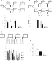
Systemic morphine injection can also induce endogenous updating. Graphs show percent of freezing time, expressed as mean ± SEM. The open field graph shows crossing number. The experimental design is shown in the top of each panel. (A) Morphine injection after memory reactivation impairs memory retrieval during test 1. This phenomenon is reverted with the injection of the same doses of morphine before test 2 (n = 10 per group). (B) Omitting the reactivation session, morphine impairs memory retrieval during test 2 (vehicle n = 7, morphine n = 8). (C) Nimodipine injection before memory reactivation prevents the impairment of morphine on memory reconsolidation and the state-dependent phenomenon (VVVV, n = 10, NMVV n = 8, VMVV n = 9, NMVM n = 9, VMVM n = 9). (D) Systemic morphine injection has no effects in motor performance (n = 7 per group). (V) Vehicle, (N) nimodipine, (M) morphine. Groups marked with the same letter (e.g., “a”) are statistically equal to each other and different from those marked with different letters (e.g., “a” groups differ from “b” groups), with significance level of P < 0.05.
ANOVA for repeated measures revealed significant effects of time (F(1,13) = 7.320, P = 0.017) and group × time interaction (F(1,13) = 7.639, P = 0.016), but not of group (F(1,13) = 1.004, P = 0.334) if the reactivation session is omitted. Post-hoc analyses showed that the morphine group expressed lower freezing levels compared to the vehicle group in test 2 (P < 0.05). Additionally, the morphine group expressed less freezing during test 2 compared to test 1 (P < 0.05). No significant differences were found between the groups in the reactivation session (P > 0.05) (Fig. 5B).
To confirm that the morphine state-dependent memory effect was mediated by the reconsolidation process, we used the same protocol described above, but injecting nimodipine (s.c.) 30 min before memory reactivation. Animals were divided into five groups: nimodipine pre-reactivation, morphine post-reactivation, and vehicle pre-test 1 and 2 (NMVV); vehicle pre-reactivation, morphine post-reactivation, and vehicle pre-test 1 and 2 (VMVV); nimodipine pre-reactivation, morphine post-reactivation, vehicle pre-test 1, and morphine pre-test 2 (NMVM); vehicle pre-reactivation, morphine post-reactivation, vehicle pre-test 1, and morphine pre-test 2 (VMVM); finally, a totally control group that received vehicle in all sessions of the experiment (VVVV). Repeated measures ANOVA revealed significant effects of treatment (F(4,40) = 11.535, P = 0.000), time (F(2,80) = 46.034, P = 0.000), and time × treatment (F(8,80) = 9.658, P = 0.000). Post-hoc test showed that VMVV and VMVM groups expressed less freezing levels compared to all other groups during test 1 (P < 0.05). Additionally, the NMVV group expressed lower freezing levels compared to the NMVM group (P < 0.05). During test 2, VMVM and NMVM groups expressed lower freezing levels compared to all other groups (P < 0.05). The VMVV group expressed lower freezing levels during test 1 and test 2 compared to the reactivation session (P < 0.001). The NMVM group expressed lower freezing levels during test 2 (P < 0.001) compared to test 1, but not compared with the reactivation session (P > 0.05). The group VMVM expressed lower freezing levels during test 1 compared to test 2 (P < 0.001) and the reactivation session (P < 0.001). No significant differences were found among the groups in the reactivation session (P > 0.05) (Fig. 5C). As described in our previous experiments, nimodipine prevented memory labilization and consequently the endogenous updating. In addition, we reproduced the state-dependent phenomenon and the amnestic pre-test morphine administration reported in other studies (Khajehpour et al. 2008). These results confirm that endogenous updating with morphine is dependent on memory reconsolidation.
No difference was found between morphine and control groups in motor performance (P = 0.088, independent t-test) (Fig. 5D), showing that our results cannot be attributed to any motor effect.
Discussion
Our data show that the endogenous memory content can be updated during reconsolidation by natural events, turning it into a state-dependent trace (Fig. 2). In a water-deprived state, this updating appears to be associated with the increase of ANGII levels and AT1 receptor signaling in dorsal hippocampus (Fig. 3). The internal state generated by water deprivation is incorporated as an endogenous cue that can modulate the memory expression in different ways. In the case of a weak memory, the endogenous updating strengthens memory expression in the presence of this internal state. However, the endogenous state alone is not sufficient to promote memory retrieval by itself (Fig. 4). Morphine injection after reactivation also turned memory dependent on a pre-test morphine treatment in order to express it properly (Fig. 5). Neurohumoral state is then changed either by an endogenous condition or the exogenous treatment. When such neurohumoral changes take place concomitantly with memory reactivation, they can be incorporated into the previously acquired memory, turning it into a requisite for further retrieval of the memory.
Water deprivation influences a whole plethora of physiological functions such as increase of corticoid secretion (Mornagui et al. 2010) and arginine-vasopressin levels (Knight et al. 2010), osmolality dysregulation (Stricker et al. 2002), increase of plasma renin activity (Nagai et al. 1993) and sodium concentration (De Luca et al. 2010), decrease of urinary volume (Jorgensen et al. 1993), and alterations in brain expression of angiotensin-converting enzyme (Bourassa and Speth 2010). However, we show that the induction of state-dependent memories may be supported, e.g., by the renin–angiotensin system (RAS), since the water deprivation effects were replicated by the infusion of ANGII into the hippocampus. Furthermore, that effect was blocked by the AT1 receptor antagonist losartan, strongly suggesting that water deprivation increased endogenous ANGII in the dorsal hippocampus and was able to update the memory trace through the activation of AT1 receptors. The discrepancy of this finding with a previous study where water restriction improved memory reconsolidation in crabs (Frenkel et al. 2005a) may be explained by several factors, including the difference between species, time of water restriction, and the memory paradigm employed (innate instead of acquired fear response). Although the ANGII system is well preserved across species and even phyla (in this case), it may not surprise us that osmoregulation mechanisms differ between mammals and crustaceans.
Interestingly, the water-deprivation protocol employed here did not affect memory retrieval when observed during the reactivation session. Actually, animals conditioned under water deprivation did not show any memory deficit when tested out of the deprived condition (data not shown). These results suggest that consolidation and reconsolidation seem to respond differently at least to a state-dependency inducing protocol involving water deprivation for the previous 18 h. This is also consistent with reports showing that, despite the fact that consolidation and reconsolidation share many similarities, there are several differences between them (Lee et al. 2004; Tronel et al. 2005; Bustos et al. 2006).
Our results may also have been influenced by other processes associated with the RAS system. Boccia et al. (2007) demonstrated that the inhibition of nuclear factor-κB (NF-κB) after memory reactivation impairs memory retention in mice. Interestingly, this nuclear factor is activated by ANGII in Chasmagnathus sp. (Frenkel et al. 2002). Considering that the RAS system has been highly preserved during evolution and the NF-κB transcription factor plays an important role in memory reconsolidation (Yang et al. 2011; Si et al. 2012), it is possible that NF-κB could be a process taking place downstream from the AT1 receptors activation in order to allow the establishment of a state-dependent memory through reconsolidation.
In the present study, we used nimodipine to ensure that the underlying mechanism for this plastic modification was a memory reconsolidation process. If an established memory becomes labile and requires reconsolidation in order to persist, then blocking destabilization would also prevent reconsolidation. One could argue that the effect found in this study was mediated by second order conditioning, creating a new independent association through a consolidation process (i.e., water deprivation → context → footshock), instead of simply updating an existing association through reconsolidation. Nimodipine prevents reconsolidation but does not interfere with the acquisition, consolidation, or retrieval processes (Suzuki et al. 2008) that would be affected if this was a second-order conditioning, i.e., an instance of first, new learning. In other words, if our results were supported by a mere memory consolidation process, no effects of nimodipine would be expected. In the present study, nimodipine injection prevented an already consolidated memory to become state-dependent.
This is the first evidence showing that reconsolidation can incorporate new information from neurohumoral states, and thus influence the future memory expression. Hence, the same internal state becomes necessary to warrant an adequate retrieval. For weak memories, it acts as an additional cue, complementing the contextual information in order to promote memory retrieval. However, in the absence of contextual information, the internal cue is not sufficient to retrieve fear memory by itself in a novel context. The convergent effects of both post-reactivation and pre-test water deprivation or morphine injection suggest that this endogenously driven updating mechanism may be a somewhat general memory property that enables reconsolidation-induced state-dependent memories.
Our data may claim for a careful reinterpretation of several previous studies that have shown reconsolidation deficits by treatments applied before or after reactivation; as previously suggested by Riccio et al. (2006), some of these results may be mediated by state-dependency, a possibility confirmed here.
These findings trigger new insights about the influence of ordinary daily life events upon memory in its continuing reconstruction, adding the realm of reconsolidation to the classical view of endogenous modulation of consolidation. A memory reactivated in a specific endogenous state may become state-dependent, and its posterior retrieval depends on the reinstallment of this state in order to take place properly. Thus, hormonal and neurohumoral modulation concomitant with a memory reconsolidation process can be a promising approach for future treatment of pathological memories.
Materials and Methods
Subjects
Male adult Wistar rats (270–320 g) from our breeding colony were used. Animals were housed in plastic cages, four to five per cage, under a 12-h light/dark cycle and at a constant temperature of 24°C, with water and food ad libitum before starting the experimental protocols. All experiments were conducted in accordance with local and national (Federal Law no 11.794/2008) guidelines for animal care and the project was approved by the University’s Ethics Committee.
Stereotaxic surgery and cannulae placement
Rats were deeply anesthetized by an i.p. injection of ketamine/xylazine (75 and 10 mg/kg, respectively) and bilaterally implanted with 27-gauge guide cannulae aimed at AP 24.2 mm (from bregma), LL ±3.0 mm, DV 1.8 mm, positioned just 1.0 mm above the CA1 area of the dorsal hippocampus (Paxinos and Watson 1998). After a 1-wk recovery from surgery, animals were submitted to the behavioral procedures. Following the behavioral experiments, subjects were sacrificed and their brains dissected and preserved in 10% formaldehyde to verify for cannula position. Only animals with correct cannula placements were included in the statistical analysis.
Drugs
L-Type voltage-gated calcium channels (LVGCCs) antagonist nimodipine and morphine sulfate (both from Sigma-Aldrich) were dissolved in sterile isotonic saline with 8% dimethylsulfoxide (DMSO) to a concentration of 16 mg/kg/mL and 7.5 mg/kg/mL, respectively, and injected subcutaneously (s.c.). Losartan (200 µM) PD123319 (200 µM) and human angiotensin II (0.5 nmol/side) (both from Sigma-Aldrich) were dissolved in 0.1% DMSO (pH 7.2) and bilaterally infused into the CA1 region of the dorsal hippocampus.
Intrahippocampal infusion
At the time of infusion, a 30-gauge infusion needle was fitted into guide cannulae, with its tip protruding 1.0 mm beyond the guide cannula end and aimed at the pyramidal cell layer of CA1 of the dorsal hippocampus. A volume of 0.5 μL was bilaterally infused at a slow rate (20 μL/h) and the needle was removed only after waiting an additional 30 sec.
Behavioral procedure
Each experiment consisted of four phases: conditioning, reactivation, test 1, and test 2, as described below.
Contextual fear conditioning (CFC)
The conditioning chamber consisted of an illuminated Plexiglas box, 25 × 25 cm, with a metallic grid floor (Context A). During training, rats were placed in the chamber for 3 min, received two footshocks (0.7 or 0.4 mA for 2 sec) separated by a 30-sec interval, and 60 sec after the last shock they were returned to their home cages.
Reactivation session
Subjects were reexposed to the Context A without footshocks during 180 sec for inducing memory retrieval.
For the experiments investigating the effects of natural events during memory reconsolidation, the experimental group was reactivated after a previous 18-h water deprivation. This deprivation was maintained for another 6 h after the memory reactivation session (Fig. 1A). Control groups were not deprived. Depending on the experiment performed, nimodipine was injected s.c. 30 min before the memory reactivation session.
For the experiments investigating the effects of intrahippocampal angiotensin II infusion upon memory reconsolidation, animals were infused with the hormone or its vehicle immediately after memory reactivation.
For the experiment investigating the effects of intrahippocampal infusion of angiotensin II antagonists in animals under a water deprivation condition, the same water-deprivation protocol was used but, immediately after memory reactivation, deprived groups were infused with losartan, PD 123319, or vehicle. Control groups infused with the same drugs were nonwater deprived.
For the experiment investigating the effects of morphine injection on memory reconsolidation, animals were injected s.c. with morphine or its vehicle immediately after memory reactivation.
Test 1
Animals were tested for 4 min either in Context A, or in a novel context on day 4 depending on the experiment performed. This session was performed in a nonwater-deprived state. Experiments with intrahippocampal human angiotensin II or systemic injection of morphine or their vehicle were injected 15- or 40-min pre-test, respectively.
Test 2
Animals were tested for 4 min in either Context A or a novel context on day 5, depending on the experiment performed. Contrary to test 1, this session was performed in a water-deprived state identical to the reactivation session. The time of deprivation was similar to the reactivation session (18-h pre). Control groups were tested under the same conditions as test 1 (without deprivation). Animals with intrahippocampal human angiotensin II or systemic injection of morphine during reactivation received the same drug 15- or 40-min pre-test, respectively. Control groups were tested with pre-test injection of vehicle.
Open field test
The OF chamber consisted of a 50-cm high, 60- × 40-cm plywood box with a frontal glass wall and a linoleum floor divided into 12 equal rectangles or “sectors.” Animals were exposed for 3 min and the number of crossings between sectors was registered. The number of crossings was considered a measure of motor performance.
Statistical analysis
Between- or within-group comparisons factorial ANOVA (here termed ANOVA with repeated measures) were employed, followed by the Fisher post-hoc test. In OF experiments, independent t-test was used for between-groups comparisons. Significance was set at P < 0.05.
Acknowledgments
This research was supported by fellowships and grants from the CAPES (MEC) (including a PNPD fellowship for L.O.A.), CNPq (MCT), PROPESQ (UFRGS), and FINEP (“Rede Instituto Brasileiro de Neurociências,” IBN-Net, No. 01.06.0842-00). We acknowledge Zelma Regina V. de Almeida for her kind technical assistance, as well as Flávia Zacouteguy, Fabrício Dutra, and Querusche Zanona. We are especially grateful to Dr. Janete Anselmo Franci (USP-RP) for providing us with some of the drugs necessary to complete this work.
Footnotes
-
↵4 Corresponding authors
E-mail quillfe{at}ufrgs.br
E-mail lucas_alvares{at}yahoo.com
- Received December 14, 2012.
- Accepted April 18, 2013.
- © 2013, Published by Cold Spring Harbor Laboratory Press
This article is distributed exclusively by Cold Spring Harbor Laboratory Press for the first 12 months after the full-issue publication date (see http://learnmem.cshlp.org/site/misc/terms.xhtml). After 12 months, it is available under a Creative Commons License (Attribution-NonCommercial 3.0 Unported), as described at http://creativecommons.org/licenses/by-nc/3.0/.



