


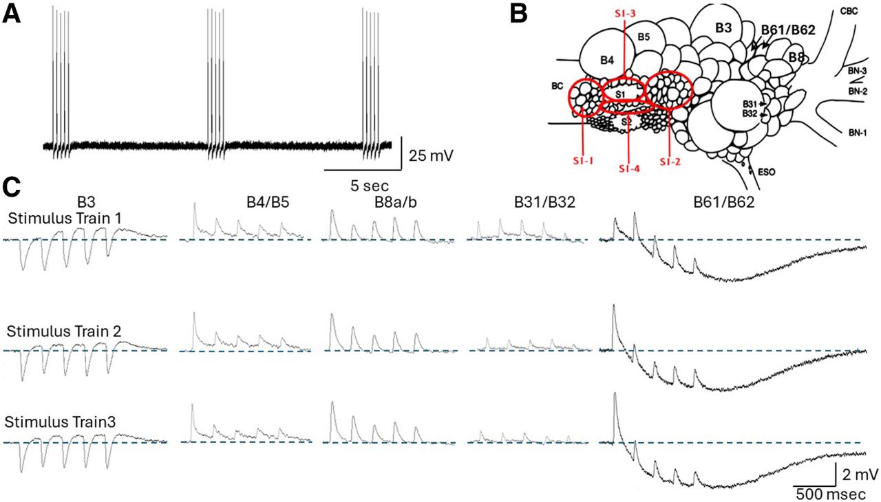
(A) Pattern of stimulation. Each presynaptic S1 neuron was stimulated with three trains of brief intracellular pulses, with each eliciting a single action potential. (B) Locations of the four subareas of the S1 cluster that were separately mapped for connectivity to the five follower neurons, as well as the locations of the follower cells investigated as targets of the S1 cells. (BN-1) Buccal nerve 1, (BN-2) buccal nerve 2, (BN-3) buccal nerve 3, (CBC) cerebral–buccal connective, (ESO) esophageal nerve, (BC) buccal commissure. The buccal ganglion is viewed from the caudal surface. (C) Examples of the response of the five follower neurons to three stimulus trains of five spikes each to an S1 neuron. Note the fast and slow responses, which may be of different signs in the various followers. Also note the changes in PSP amplitude within a stimulus train or between stimulus trains. The large slow IPSP in B61/B62 is particularly striking. Blue dashed lines denote the baseline membrane potentials.



