


Emotional memory consolidation during sleep is associated with slow oscillation–spindle coupling strength in young and older adults
- 1Neuroscience and Behavior Program, University of Massachusetts Amherst, Amherst, Massachusetts 01003, USA
- 2Institute for Applied Life Sciences, University of Massachusetts Amherst, Amherst, Massachusetts 01003, USA
- 3Developmental Sciences Program, University of Massachusetts Amherst, Amherst, Massachusetts 01003, USA
- Corresponding author: rspencer{at}umass.edu
Abstract
Emotional memories are processed during sleep; however, the specific mechanisms are unclear. Understanding such mechanisms may provide critical insight into preventing and treating mood disorders. Consolidation of neutral memories is associated with the coupling of NREM sleep slow oscillations (SOs) and sleep spindles (SPs). Whether SO–SP coupling is likewise involved in emotional memory processing is unknown. Furthermore, there is an age-related emotional valence bias such that sleep consolidates and preserves reactivity to negative but not positive emotional memories in young adults and positive but not negative emotional memories in older adults. If SO–SP coupling contributes to the effect of sleep on emotional memory, then it may selectively support negative memory in young adults and positive memory in older adults. To address these questions, we examined whether emotional memory recognition and overnight change in emotional reactivity were associated with the strength of SO–SP coupling in young (n = 22) and older (n = 32) adults. In younger adults, coupling strength predicted negative but not positive emotional memory performance after sleep. In contrast, coupling strength predicted positive but not negative emotional memory performance after sleep in older adults. Coupling strength was not associated with emotional reactivity in either age group. Our findings suggest that SO–SP coupling may play a mechanistic role in sleep-dependent consolidation of emotional memories.
Emotion and mood disorders have a prevalence of 6%–38% in older adults (Strauss et al. 2020). Although less prevalent than in adolescents and young adults, the lasting emotional impacts of the COVID-19 pandemic (Morrow-Howell et al. 2020) and the increasing size of the older adult population (He et al. 2016) add urgency to understanding mechanisms underlying mood and emotion processing in older adult populations. It has been suggested that age-related changes in sleep may contribute to deficits in emotion processing. For example, the presence of insomnia in older adults predicts the onset of major depressive disorders 1 yr later (Perlis et al. 2006) and depressive symptoms 4 yr later (Jaussent et al. 2011). Sleep-dependent consolidation of negative memories is reduced in older adults (Jones et al. 2016). However, unlike young adults, older adults consolidate positive memories during sleep, an emotional bias that may be protective of mood in aging (Jones et al. 2019). Thus, understanding the relation between sleep and emotion in older adults is a promising avenue toward understanding and treating emotion and mood disorders in aging.
Emotion has a strong effect on declarative memory; noradrenergic signaling by the amygdala enhances memory processing, leading to more vivid and longer-lasting memories (McGaugh 2004; LaBar and Cabeza 2006). Whether sleep-dependent consolidation mechanisms contribute to the enhancing effect of emotion on memory is unclear. Sleep has been proposed to have a larger effect on emotional relative to neutral memories, and some evidence supports this claim (Payne et al. 2008, 2015; Bennion et al. 2015). However, more recent review and meta-analytical work call this view into question, suggesting instead that emotional and neutral memories benefit equivalently from sleep (Lipinska et al. 2019; Schäfer et al. 2020; Berres and Erdfelder 2021; Davidson et al. 2021). Many factors, including the type of retrieval test and the placement of sleep relative to encoding and retrieval, may be relevant when considering whether sleep preferentially benefits emotional memory (Lipinska et al. 2019; Cunningham et al. 2022). Furthermore, potential carryover effects between emotional and neutral stimuli should be considered when stimuli are intermixed, as emotional brain states persist and enhance memory for unrelated neutral information (Tambini et al. 2017). In line with a carryover effect of negative emotion, we observed a sleep benefit in young adults on memory of neutral pictures when they were learned together with negative pictures but not when they were learned together with positive pictures (Jones et al. 2016). Thus, emotion carryover effects may mask a preferential sleep benefit for emotional memories in some widely used paradigms.
The majority of research on sleep and emotional memory has focused on negative memory in young adults. Many aspects of sleep decline with aging, leading to reduced memory consolidation in some cases (Harand et al. 2012; Scullin and Bliwise 2015). Thus, sleep-dependent consolidation of emotional memories may be impaired in aging. Indeed, we saw no benefit of sleep on negative memory in older adults, in contrast to our observation in young adults (Jones et al. 2016). However, sleep did confer a benefit on positive memory in older adults (despite no benefit in young adults), suggesting that sleep-based memory mechanisms remain at least partially intact and functional with advancing age and may require emotional salience (positive emotion) to be engaged. Recent work supports that sleep selectively consolidates negative emotional aspects of memory across young adulthood and into middle age (Denis et al. 2022b) and that this bias favors positive memory in older age (Huan et al. 2020).
The sleep-based mechanisms underlying emotional memory consolidation are unclear, with some evidence pointing to REM sleep (Nishida et al. 2009; Walker 2009; Popa et al. 2010) and other evidence pointing to NREM sleep (Groch et al. 2011; Hauner et al. 2013; Payne et al. 2015; Girardeau et al. 2017). It may be that NREM sleep strengthens the episodic contents of emotional memory and REM sleep confers additional, emotion-related processing (Cairney et al. 2015; Genzel et al. 2015). In young adults, negative emotional memory consolidation during sleep is associated with slow-wave sleep (SWS), suggesting a shared mechanism with neutral declarative memory consolidation, which is also associated with SWS (Groch et al. 2011; Hauner et al. 2013; Payne et al. 2015). Likewise, positive memory consolidation is associated with SWS in older adults (Jones et al. 2016). Sleep continuity or less awakening at night may further moderate this relationship between positive memory and sleep in older adults (Xie et al. 2022).
Active systems consolidation theory proposes that the temporal coupling of the up state of the slow oscillations, thalamocortical sleep spindles, and hippocampal sharp-wave ripples during NREM sleep supports transfer of the memory from short-term hippocampal storage to more efficient stable storage in the cortex (Rasch and Born 2013). Supporting this, tightly timed coordination of slow oscillations and sleep spindles (SO–SP coupling) is associated with memory consolidation (Niknazar et al. 2015; Mikutta et al. 2019; Zhang et al. 2020). This coupling is reduced and less precise in older adults; however, the functional association between coupling characteristics and memory retention is similar across young and older ages (Helfrich et al. 2018; Muehlroth et al. 2019). Muehlroth et al. (2020) emphasize the need to study sleep oscillatory dynamics such as SO–SP coupling in aging in order to help explain the inconsistent findings regarding sleep-dependent memory benefits in older adulthood.
Whether SO–SP coupling is also essential for emotional memory consolidation is poorly understood. The only study to date found a negative relationship with emotional memory and SO–SP coupling in young adults who were exposed to a stress test, warranting further exploration into this relationship (Denis et al. 2022a). We hypothesize that associations between SWS and emotional memory consolidation during sleep reflect an underlying SO–SP coupling mechanism. Given the positive shift in the emotional bias of sleep-dependent consolidation with aging (Jones et al. 2016), we hypothesized that SO–SP coupling would support negative memory in young adults and positive memory in older adults. Older adults have reduced SO–SP coupling, which corresponds to diminished neutral declarative memory consolidation (Helfrich et al. 2018; Muehlroth et al. 2019). Although reduced, SO–SP coupling during sleep could still yield a sleep benefit (relative to wakefulness) for positive memory in older adults if it is coordinated with hippocampal reactivation of positive memories. Alternatively, amygdala modulation during encoding of positive memories may lead to enhanced SO–SP coupling in older adults. Finally, it is also possible that SO–SP coupling does not support memory consolidation in older adults regardless of emotional valence and that the positive memory benefit from sleep does not reflect hippocampal–neocortical interactions (e.g., perhaps instead relying on the hippocampus and/or amygdala).
Reactivity is the emotional response that accompanies the content of the emotional memory being formed. Therefore, sleep may preserve and strengthen emotional reactivity (Ashton et al. 2019; Jones and Spencer 2019). The parallel protection of emotional reactivity by sleep for negative memories in young adults and positive memories in older adults is thought to be a distinct process from consolidation of the emotional episodic contents. Specifically, changes in emotional reactivity were not associated with SWS in prior reports; rather, changes in emotional reactivity following an interval of sleep were associated with REM sleep (Lara-Carrasco et al. 2009; Werner et al. 2015; Jones et al. 2016). In the present analyses, we also explored whether SO–SP coupling is related to sleep-related protection of emotional reactivity. On the one hand, if change in reactivity is a REM-dependent process, a significant correlation between SO–SP coupling and change in emotional reactivity over sleep would seem unexpected. On the other hand, REM-dependent processing of emotional reactivity may be tied to the success of earlier NREM consolidation and thus likewise reliant on the coupling of SO–SP.
Results
Results are from a secondary analysis of the data from the study by Jones et al. (2016). In short, participants viewed images that were positive or negative in valence and rated them for valence and arousal (Fig. 1A). Those images intermixed with foils were presented again during the recognition test, and participants again rated the images for valence and arousal as well as indicated whether they had seen the image before. Encoding took place in the evening and the recognition test took place 12 h later, following an interval containing overnight sleep (Fig. 1B).
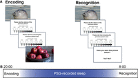
(A) Participants viewed 60 pictures (targets) and rated the valence and arousal for each on a nine-point Likert-type scale during encoding. Participants viewed 180 pictures (targets and foils) and rated the valence and arousal during recognition. During recognition, participants indicated whether they recognized the picture by responding yes or no. (B) Encoding took place in the evening followed by recognition 12 h later. Polysomnography (PSG) was administered overnight.
Sleep physiology characteristics
A summary of sleep macrostructure across age groups and conditions is in Table 1; two (young vs. older) × two (negative vs. positive) ANOVAs were conducted on sleep stage variables. There were no significant main effects or interactions between age and condition for total sleep time, sleep efficiency, or percent of sleep time spent in NREM2 and REM (all Ps > 0.05). However, the interaction between age and condition was significant for percent of sleep time spent in NREM stage 1 (F(1,48) = 8.25, P = 0.006). Specifically, younger adults in the negative condition had 4.92 min more time in NREM stage 1 on average than older adults in the negative condition (P = 0.02, 95% CI of the difference = 0.78–9.06), although after corrections this was not a significant difference. In contrast, percent time in NREM stage 1 between younger and older adults in the positive condition did not significantly differ (P = 0.11). Additionally, older adults in the negative condition had 8.78 min more time in NREM stage 2 on average than younger adults in the negative condition (P = 0.004, 95% CI of the difference = 2.97–14.59). In contrast, percent in NREM stage 2 between younger and older adults in the positive condition did not significantly differ (P = 0.65). Finally, there was a significant main effect of age (P = 0.01) but not condition (P = 0.52) on percent of sleep time in NREM3. For additional descriptions of sleep characteristics for the full sample, see Jones et al. (2016).
Sleep characteristics (mean [SEM])
A summary of SO and SP characteristics from N2 (measured at central electrodes) and SWS (measured at frontal electrodes) across age groups and conditions is in Table 2; two (young vs. older) × two (negative vs. positive) ANOVAs were conducted on SO and SP variables. No main effects or interactions were observed in N2 (all Ps > 0.05). In SWS, there was a main effect of condition for coupling strength (F(1,47) = 19.1, P < 0.001; higher in the negative condition). There were significant interactions between age and condition for spindle count, spindle density, slow oscillation count, and SO density (F(1,47) = 7.08, P = 0.01; F(1,47) = 6.81, P = 0.01; F(1,47) = 14.47, P = <0.001; F(1,47) = 4.72, P = 0.04). As can be seen in Table 2, young adults had a significantly higher spindle count, spindle density, and SO count than older adults in the positive memory condition, but there was no age difference in any of these variables in the negative memory condition. There were no other significant effects (all Ps > 0.05).
SO and SP characteristics (mean [SD])
Associations between emotional memory and SO–SP coupling
We first considered relations between emotional memory following sleep (corrected recognition) and SO–SP coupling strength. As predicted, in young adults there was a positive association between SO–SP coupling strength and memory performance for negative emotional stimuli following sleep (Fig. 2A). This relationship was present for coupling strength during N2 (R = 0.78, P = 0.01; assessed at central electrodes) but not SWS (R = −0.09, P = 0.81; assessed at frontal electrodes). There was no association between coupling strength and positive emotional memory after sleep in young adults in either N2 (R = −0.25, P = 0.47; assessed at central electrodes) or SWS (R = −0.51, P = 0.11; assessed at frontal electrodes). In older adults, there was a positive association between SO–SP coupling strength and memory for positive stimuli after sleep (Fig. 2B). This relationship was apparent for coupling strength during SWS (R = 0.64, P = 0.01; assessed at frontal electrodes) and N2 (R = 0.55, P = 0.03; assessed at central electrodes), although the significance did not survive correction for multiple comparisons in the latter case. The association between coupling strength and negative memory performance after sleep was not significant in older adults in either SWS (R = −0.18, P = 0.55; assessed at frontal electrodes) or N2 (R = 0.15, P = 0.62; assessed at central electrodes). There was no significant association between corrected recognition of neutral stimuli and coupling strength in either the positive or negative condition for both younger and older adults (all Ps > 0.0125) (Supplemental Fig. S1A,B).
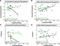
Memory performance and change in reactivity and SO–spindle coupling. Green circles denote experiment 1 (negative), and black triangles denote experiment 2 (positive). (A) Correlation between coupling strength in N2 (central electrodes) and corrected recognition in younger adults. (B) Correlation between coupling strength in SWS (frontal electrodes) and corrected recognition in older adults. (C) Correlation between coupling strength in N2 (central electrodes) and change in valence in younger adults. (D) Correlation between coupling strength in SWS (frontal electrodes) and change in valence in older adults. (*) P < 0.0125.
Next, we also explored relations between coupling strength and d′. While we observed a similar pattern of results (positive relationship between coupling and negative memory in young adults and positive relationship between coupling and positive memory in older adults), after correcting for multiple comparisons these would be significant and trend-level associations did not hold. Additionally, there was no significant association between coupling strength and d′ of neutral stimuli in either condition (positive or negative) in younger or older adults (all Ps > 0.0125) (Supplemental Fig. S2A–D).
Associations between change in emotional reactivity and SO–spindle coupling
We next explored relations between change in emotional reactivity (valence ratings) and SO–SP coupling strength. Change in reactivity was not associated with coupling strength in either N2 or SWS for either positive or negative memory in either age group (all Ps > 0.0125) (Fig. 2C,D). Likewise, there was no association between change in reactivity for neutral stimuli and coupling strength in either sleep stage in either the positive or negative condition in either age group (all Ps > 0.0125).
Discussion
Previous literature indicates an association between SO–SP coupling and sleep-dependent memory consolidation for nonemotional declarative memories, but whether the same is true for emotional memories is unknown. This study aimed to examine sleep-dependent emotional memory processing and SO–SP coupling in younger and older adults. We found that, as with declarative memories (Muehlroth et al. 2019; Hahn et al. 2022), SO–SP coupling strength is associated with greater emotional memory consolidation. Specifically, coupling strength predicted better negative emotional memory recognition following sleep for younger but not older adults and better positive emotional memory recognition following sleep for older but not younger adults. Furthermore, we did not find SO–SP coupling strength to be associated with a change in the reactivity of emotional memories in either younger or older adults.
Prior studies have linked memory consolidation to NREM macrostructure and microstructure. Markers of NREM stage 2, such as spindles and sigma activity, have been associated with declarative and emotional memory processing (Cairney et al. 2018; Jones et al. 2019). Time spent in SWS has been linked to not only neutral declarative memory but also emotional memory (Payne et al. 2015; Jones et al. 2016; Alger et al. 2018). To date, there has been only one other study examining emotional memory and SO–SP coupling (Denis et al. 2022a). Denis et al. (2022a) found that time spent in SWS was positively associated with memory for emotional and neutral items when a stressor was present but not in the control group. When they examined SO–spindle coupling specifically, they found a negative association between emotional memory and SO–SP coupling in the stress group and no relationship was found in their control group. However, their study only examined SWS, whereas our finding in young adults was in N2. Furthermore, this relationship in the stress group was strengthened in participants with higher cortisol, in line with a deleterious effect of high stress on hippocampal memory (Kim et al. 2015). Although stress was not manipulated in our study, these prior results are overall at odds with our current findings, perhaps pointing to the importance of specific sleep stage, electrode site, and methodologies used in examining SO–spindle coupling and emotional memory.
The relationship between coupling strength and negative memory consolidation in younger adults was specifically found in NREM stage 2 rather than SWS, while the relationship between coupling strength and positive memory consolidation in older adults was most apparent in SWS. This finding points to the importance of examining the functional relevance of SO–SP coupling events in specific sleep stages. Recent evidence supports the possibility that SO–SP coupling may have various subtypes (McConnell et al. 2021, 2022). Specifically, SO–SP coupling can be subdivided into two subtypes based on spindle timing relative to the peak of the slow oscillation up state: Early fast spindle coupling occurs primarily in NREM2 across central regions, and late fast spindle coupling occurs predominately in SWS across frontal regions. Furthermore, these studies provide support for our results, as they found that older adults shift toward increases in the late fast, frontal coupling subtype in SWS. Thus, it is possible that different subtypes of coupling primarily supported memory consolidation in young and older adults in our study. Alternatively, the N2 versus SWS difference that we observed may be due to younger adults being faster at consolidating during sleep; specifically, consolidation may start in NREM stage 2 and be finished by the time young adults reach SWS, unlike in older adults, where consolidation is still ongoing into SWS. The observation that positive memory was associated with coupling at frontal electrodes and negative memory was associated with coupling at central electrodes is also broadly aligned with evidence of greater prefrontal involvement in positive memory and posterior sensory involvement in negative memory (Bowen et al. 2018).
Unlike young adults, older adults do not show over-sleep changes in negative memory that differ from over-wake changes in negative memory, suggesting that negative memories are not consolidated preferentially during sleep (relative to waking) with aging (Jones et al. 2016).
Consistent with this interpretation, we did not observe any relationship between SO–SP coupling and negative memory in older adults here. However, in line with the sleep (relative to waking) benefit on positive memory in older adults, the present analysis suggests that sleep mechanisms, specifically SO–SP coupling, still support positive emotional memory consolidation with aging. Evidence indicates that older adults exhibit greater amygdala activation while viewing positive stimuli, while young adults exhibit greater activation while viewing negative stimuli (Leclerc and Kensinger 2010). Thus, amygdala modulation of hippocampal encoding processes (LTP) may lead to preferential replay of positive memory traces in older adults and negative memory traces in young adults. When coordinated with SO–SP coupling, this valence-based difference in replay may underlie the opposing valence biases in emotional memory consolidation in young and older adults.
Some prior reports point to a role of REM sleep in emotional memory consolidation (Goldstein and Walker 2014; Carr and Nielsen 2015). However, like neutral memories, emotional memories include episodic content. We propose that the episodic content is consolidated over NREM sleep, while the emotional tone is processed during REM sleep. Consistent with this, we and others found a link between emotional memory performance and SWS (Groch et al. 2011; Hauner et al. 2013; Payne et al. 2015), while changes in emotional reactivity are associated with REM (Baran et al. 2012). Our current findings further support a role of NREM sleep in consolidating emotional episodic content, specifically implicating SO–SP coupling, consistent with prior studies of declarative memories (Niknazar et al. 2015; Mikutta et al. 2019; Zhang et al. 2020; Denis et al. 2021). The lack of association between coupling strength and changes in emotional reactivity is in line with the processing of emotional reactivity occurring separately from the strengthening of episodic content, perhaps during REM sleep. More research is needed to better understand the roles of different sleep stages in emotional memory processing.
Typically, aging is associated with disrupted sleep. Here, sleep stage composition was overall similar between the young and older adult groups; the only difference was that a smaller percent of sleep was spent in SWS for older compared with younger adults, consistent with meta-analyses of life span changes in sleep (Ohayon et al. 2004). Contrary to previous reports (Mander et al. 2017), we did not observe widespread reductions in SO and SP characteristics. We found no age-related decline in the number or density of spindles or slow oscillations or their coupling strength in N2 sleep. In SWS, the number and density of spindles and slow oscillations (but not their coupling strength) were indeed lower in older adults than in young adults but puzzlingly only in the positive memory condition. Our sample included healthy older adults, and study requirements may have further biased the sample to healthy sleepers. At any rate, despite well-documented reductions in many aspects of sleep quantity and quality with aging, our current results add to the evidence that SO–SP coupling still supports memory consolidation in older adults (Ladenbauer et al. 2017; Helfrich et al. 2018; Muehlroth et al. 2019). Furthermore, as we found no effect of age on coupling strength, reduced SO–spindle coupling cannot explain the age-related reduction in the effect of sleep on negative memory in our data.
There are several limitations of this study to consider. First, we used a between-groups design for emotion (negative vs. positive). A within-subject design would increase sensitivity and may further elucidate the difference of positive versus negative sleep-dependent memory consolidation. Second, we did not administer an immediate recognition memory test. Without an assessment of memory prior to sleep, we cannot directly assess the change in memory performance after sleep. Third, as emphasized by Muehlroth and Werkle-Bergner (2020), it is important that future research consider algorithm adjustments for the amplitude of SOs across the life span for more accurate comparisons across age groups. However, by including both N2 and SWS in our analysis, we potentially captured most occurrences of SO–SP coupling. Last, our sample size was limited relative to Jones et al. (2016). We note that other studies have reported correlational analysis examining SO–SP coupling using similar sample sizes (Wilhelm et al. 2011; Hauner et al. 2013; Denis et al. 2022a). Nonetheless, future studies with a larger sample size are warranted to further investigate the role of SO–SP coupling in emotional memory with aging.
In conclusion, we found a relation between emotional memory consolidation and slow oscillation–spindle coupling, suggesting a sleep mechanism similar to that indicated for nonemotional memory consolidation. This mechanism is biased toward negative memory for young adults and positive memory for older adults, suggesting that it is still functional but differentially engaged with aging. Importantly, these results may point to potential mechanisms that could eventually be targeted to ameliorate deficits in mood and emotion processing that sometimes occur in older adults. Potential therapeutics such as noninvasive transcranial direct current stimulation (Ladenbauer et al. 2017) have shown promise in increasing slow oscillation activity. Further research into therapeutics that target SO–SP coupling increases could prove beneficial for improvements in positive memory and potentially mood and emotion disorders.
Materials and Methods
Participants
We used data from a previously published study of emotional memory consolidation in young and older adults (Jones et al. 2016), specifically data from the sleep condition. Polysomnography (PSG) was available in a subset of participants. Comparisons with negative and neutral stimuli (experiment 1 in Jones et al. 2016) included 10 younger adults (M = 19.55 yr, SEM = 1.04) and 16 older adults (M = 61.56 yr, SEM = 10.98). Comparisons of positive and neutral stimuli (experiment 2 in Jones et al. 2016) included 12 younger adults (M = 20.50 yr, SEM = 1.35) and 16 older adults (M = 63.13, SEM = 7.16). Fourteen of the 36 younger adults and six of the 38 older adult participants were excluded from this secondary analysis due to preprocessing issues with the PSG recording. Eligibility criteria in the study included normal or corrected-to-normal vision, no history of neurological or sleep disorder, no prior head injury, and no use of medications that affect sleep or cognitive function.
Emotional memory task
Stimuli were 90 negative (experiment 1), 90 positive (experiment 2), and 90 neutral (experiments 1 and 2) images. Images were obtained from the International Affective Picture System (IAPS) (Bradley and Lang 2017) and an in-house set that matched the content and emotionality of the IAPS images (Baran et al. 2012). Emotional pictures were moderate to high arousal, and neutral pictures were low arousal. The mean arousal and valence ratings for the images used in this study are detailed in the previously published study (Jones et al. 2016).
The task consisted of an encoding phase and a recognition phase (Fig. 1A). During the encoding phase, participants viewed 60 images—30 negative (experiment 1) or positive (experiment 2) images interleaved with 30 neutral images. After viewing the image for 1 sec, participants were cued to rate the picture's valence on a nine-item valence scale (1 = negative, 5 = neutral, and 9 = positive), followed by another prompt asking them to rate the picture's arousability on a nine-item arousal scale (1 = no arousal, 5 = moderate arousal, and 9 = highly arousing). A 1.5-sec interval separated trials. Participants were not aware that their memory for the stimuli would be tested afterward. During the recognition phase, participants were shown 180 images that included the 60 images from encoding (targets) mixed with 120 novel stimuli (foils; 60 negative [experiment 1] or 60 positive [experiment 2] and 60 neutral). Stimuli were presented for 1 sec, and participants were again asked to rate each picture on the valence and arousability scales. After each scale was presented, an additional question prompted participants to indicate whether they had seen the stimuli previously by pressing either a “y” for yes or an “n” for no. For more information on other metrics collected during the procedure, see Jones et al. (2016).
Procedure
Informed consent was obtained prior to the experiment, and all procedures were approved by the University of Massachusetts Amherst Institutional Review Board. Participants completed two sessions separated by 12 h (Fig. 1B). They were assigned to either a sleep condition or a wake condition. Data described here are from the sleep condition. Participants in that condition completed the first session in the evening and the second session in the morning (e.g., 8:00 p.m. and 8:00 a.m., respectively).
In the first session, participants completed the Pittsburgh Sleep Quality Index (PSQI), the Mini-Mental State Examination (MMSE; older adult participants only), and a sleep–wake diary. Participants completed the positive and negative affect schedule (PANAS) at each session. Subsequently, the participants completed the encoding phase of the emotional memory task and then were equipped with PSG and encouraged to have a normal night's sleep. In the morning, PSG was removed. Twelve hours after encoding, participants completed the recognition phase of the emotional memory task.
Polysomnography
PSG was recorded using the Aura PSG ambulatory system (Grass Technologies). Electrode sites included two EOG, two EMG (chin), and six EEG leads at O1, O2, C3, C4, F3, and F4. All channels were referenced to the contralateral mastoid. Each sleep record was scored in accordance with the American Academy of Sleep Medicine (Berry et al. 2015). Prior to preprocessing, PSG data analysis was performed by using Matlab R2018a (Mathworks, Inc.); functions from the Fieldtrip toolbox (Oostenveld et al. 2011), EEGLab toolbox (Delorme and Makeig 2004), and an in-house toolbox (https://github.com/afitzroy/psgpower); and in-house scripts.
EEG preprocessing
Preprocessing was conducted by following the description from Muehlroth et al. (2019) using customized open source scripts (Muehlroth and Werkle-Bergner 2020). Specifically, EEG data were segmented to 1-sec segments for artifact detection. The segmented data regarded as body movements or artifacts (segments that had amplitude difference >500 µV) were excluded for further analyses. The data in each of the segments were z-normalized and removed if the mean value of each segment had a z-score >5. Additionally, any bad EEG channels were visually removed by experts in the laboratory.
Event detection
The algorithm for event detection was conducted by following the descriptions from Helfrich et al. (2018) and Staresina et al. (2015). The open source scripts shared by Muehlroth and Werkle-Bergner (2020) were used in event detection as well. Slow oscillations were detected by filtering the artifact-free data in the frequency of 0.16–1.25 Hz by using a two-pass FIR bandpass filter (order = three cycles of the low-frequency cutoff). After filtering was applied, data that had zero crossings and met criteria of duration (0.8–2 sec) and amplitude (>75th percentile of SO candidates) were detected as final SOs. Extracted SOs included ±2.5 sec around the nadir of the SO trough from the raw signal. Spindles were extracted by filtering the data in 12–15 Hz with two-pass FIR bandpass filter (order = three cycles of the low-frequency cutoff). The amplitude criteria of detecting SP were determined by the 75th percentile of the root mean square, which was calculated by using the moving average of 0.2 sec. If SPs had a larger amplitude than the threshold for 0.5–3 sec, they were extracted as final SPs from the raw signal. We detected SOs (0.16–1.25 Hz) and SPs (12–15 Hz) in NREM stage 2 (N2) from central electrode sites (C3 and C4) and NREM stage 3 (SWS) from frontal electrode sites (F3 and F4) separately from each of the participants. We focused on these electrode sites in these stages based on recent evidence of SO–SP coupling subtypes (McConnell et al. 2021, 2022).
Slow oscillation–spindle (SO–SP) coupling
SO–SP coupling was analyzed following the methods described by Ladenbauer et al. (2021). Only −2 to +2 sec of SO segments (0.16–1.25 Hz) was used for coupling measurement to avoid edge artifacts. We detected instantaneous phases from SO segments by using the Hilbert transform. Spindles were detected from the same segments in the SP frequency band (12–15 Hz). The amplitude of SPs was extracted using the Hilbert transform. Coupling strength was first measured for each participant by averaging across frontal and central electrode sites in N2 and SWS using the Circstat toolbox (Berens 2009; Hahn et al. 2022). Reported coupling strength was the mean resultant vector length for the circular data, quantifying how precisely SPs were embedded in the phase of the SOs (Hülsemann et al. 2019).
Behavioral performance
Participants’ valence ratings were used to categorize the stimuli; targets were categorized based on encoding phase ratings, and foils were categorized based on the recognition phase ratings. To quantify memory performance, we examined corrected recognition, which was calculated by subtracting the false alarm rate from the hit rate. Hit rate is defined as the percent correct of the emotional target stimuli. False alarm rate is defined as the percent of foil stimuli incorrectly identified as seen previously. These measures were computed separately for emotional (negative or positive) and neutral stimuli. While the results from the previous study used hit rate, here we focused on corrected recognition to increase sensitivity (Brady et al. 2022). Furthermore, the only other study that examined SO–SP coupling and emotional memory also examined corrected recognition, thus allowing for comparable interpretations (Denis et al. 2022a). The change in reactivity or change in valence ratings was calculated for target pictures at both the encoding and recognition phase (Δvalence = recognition valence rating − encoding valence rating). A positive Δvalence score for negative stimuli indicates a decrease of the initial negative rating (toward neutral). A positive Δvalence score for positive stimuli indicates an increase of the initial positive rating. To minimize the number of comparisons, we focused on valence. In our prior study (Jones et al. 2016), significant outcomes were found for valence and not arousal. Details on the behavioral results from the full sample are reported in Jones et al. (2016) and this subset in Supplemental Table S1.
Statistical analysis
To assess differences in sleep and SO–SP coupling metrics between experimental conditions (negative vs. positive) and age (young vs. old), a series of two × two ANOVAs was conducted. Student's t-tests were used to compare age groups within each experiment. For the coupling analysis, we conducted a test for outliers (more than three SD from the mean) based on other reports (e.g., Denis et al. 2022a). No data points were identified as outliers. Correlations between memory performance (corrected recognition) and reactivity (Δvalence) as well as SO–SP coupling strength were computed using Spearman correlations. To control for the family-wise error rate, we used the Holm–Bonferroni method to adjust for multiple comparisons (Holm 1979). Specifically, due to the large number of tests, ANOVAs on sleep microstructure, correlations, and t-tests between age groups were corrected using the Holm–Bonferroni method. T-tests on sleep macrostructure (Table 1) were considered in groups of six (six tests per condition), and only P-values <0.01 survived the correction. ANOVAs on sleep microstructure were considered in groups of five (five tests per sleep stage), and P-values <0.05 survived the correction. T-tests on sleep microstructure (Table 2) and correlations were considered in groups of four (each metric or relationship assessed in two conditions × two sleep stages), and only P-values <0.0125 survived the correction in each case.
Acknowledgments
We extend our gratitude to Julia Ladenbauer and Bernhard Staresina, who assisted as we developed the coupling analyses. This work was supported by National Insitutes of Health grants R01 AG040133 and R56 AG058685 to R.M.C.S.
Footnotes
-
[Supplemental material is available for this article.]
-
Article is online at http://www.learnmem.org/cgi/doi/10.1101/lm.053685.122.
- Received April 13, 2023.
- Accepted August 29, 2023.
This article is distributed exclusively by Cold Spring Harbor Laboratory Press for the first 12 months after the full-issue publication date (see http://learnmem.cshlp.org/site/misc/terms.xhtml). After 12 months, it is available under a Creative Commons License (Attribution-NonCommercial 4.0 International), as described at http://creativecommons.org/licenses/by-nc/4.0/.



