


Neuronal and astrocytic protein degradation are critical for fear memory formation
- 1School of Animal Sciences, Virginia Polytechnic Institute and State University, Blacksburg, Virginia 24061, USA
- 2School of Neuroscience, Virginia Polytechnic Institute and State University, Blacksburg, Virginia 24061, USA
- Corresponding author: tjjarome{at}vt.edu
-
↵3 These authors contributed equally to this work.
Abstract
Strong evidence has implicated proteasome-mediated protein degradation in the memory consolidation process. However, due to the use of pharmacological approaches, the cell type specificity of this remains unknown. Here, we used neuron-specific and novel astrocyte-specific CRISPR–dCas9–KRAB–MECP2 plasmids to inhibit protein degradation in a cell type-specific manner in the amygdala of male rats. We found that while inhibition of neuronal, but not astrocytic, protein degradation impaired performance during the training session, both resulted in impaired contextual fear memory retention. Together, these data provide the first evidence of a cell type-specific role for protein degradation in the memory consolidation process.
It is widely accepted that the process of long-term memory formation requires increased transcriptional regulation and de novo protein synthesis (Bailey et al. 1999; Schafe et al. 1999; Schafe and LeDoux 2000; Kwapis and Wood 2014). In addition to this, strong evidence now supports a role for ubiquitin–proteasome-mediated protein degradation in the memory consolidation process (Jarome and Helmstetter 2013). To date, protein degradation is increased in several brain regions following behavioral training, including the amygdala, hippocampus, prefrontal cortex, and insular cortex (Lopez-Salon et al. 2001; Jarome et al. 2011; Reis et al. 2013; Orsi et al. 2019; Beamish et al. 2022). Additionally, inhibition of functional proteasome activity has been shown to impair the consolidation of memories for a wide variety of aversive and nonaversive tasks, including auditory and contextual fear conditioning, trace fear conditioning, inhibitory avoidance, Morris water maze, conditioned taste aversion, object recognition, and object location tasks (Lopez-Salon et al. 2001; Artinian et al. 2008; Lee et al. 2008; Choi et al. 2010; Jarome et al. 2011; Rodriguez-Ortiz et al. 2011; Reis et al. 2013; Figueiredo et al. 2015; Furini et al. 2015; Cullen et al. 2017; Devulapalli et al. 2019). Furthermore, this need for proteasome-dependent protein degradation is shared across sexes (Devulapalli et al. 2021), although this does vary based on the brain region examined (Martin et al. 2021). Regardless, it is now accepted that protein degradation via the ubiquitin–proteasome system is an important regulator of long-term memory formation in the brain.
The ubiquitin–proteasome machinery is present in every subcellular compartment and common to neuronal and nonneuronal cell types (Tai and Schuman 2008; Jansen et al. 2014). However, although studies investigating the role of protein degradation in memory formation have been conducted for nearly 20 yr, this work has focused on pharmacological manipulations that lack cell type specificity and quantification methods that are unable to distinguish neuronal from nonneuronal cell populations. Thus, the cell type specificity of this process remains unknown. Using an unbiased proteomic approach, we recently found that GFAP, a critical component of the astrocyte structure (Hol and Pekny 2015), was targeted for degradation by the proteasome in the amygdala of both male and female rats following contextual fear conditioning (Farrell et al. 2021). This suggests that protein degradation could be occurring in both neuronal and nonneuronal cell populations during memory consolidation. However, due to the aforementioned technical limitations, this has never been directly tested.
Here, we used a novel CRISPR–dCas9 approach to test the necessity of neuronal and astrocytic protein degradation in the consolidation of a contextual fear memory. Expanded details on methodology can be found in the Supplemental Material. To examine neuron-specific protein degradation, we took advantage of a recently developed neuron-specific dCas9–KRAB–MECP2 transcriptional repressor plasmid under the control of the human synapsin (SYN) promoter (Duke et al. 2020). In order to examine astrocyte-specific protein degradation, we took the aforementioned plasmid and substituted the SYN promoter with the condensed GFAABC1D promoter, allowing targeting of astrocytes (Heffernan et al. 2022). Importantly, this condensed promoter is similar in size to SYN, allowing the two dCas9 plasmids to be nearly identical in every aspect except for the promoter used and thus cell type-targeted (Fig. 1A).
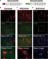
Characterization of cell type-specific CRISPR–dCas9 plasmids. (A) Schematics of the CRISPR–dCas9–KRAB–MECP2 transcriptional plasmids under the human synapsin (SYN) and GFAABC1D (GFAP) promoters that were used to target neurons and astrocytes, respectively. The GFAP–dCas9 plasmid was developed by substituting the SYN promoter. (B) Immunofluorescence of hippocampus tissue transfected with SYN–dCas9 plasmid expression in neurons. (C,D) Immunofluorescence of hippocampus tissue transfected with GFAP–dCas9 plasmid expression in astrocytes (C) but not neurons (D). NeuN stains neurons, GFAP stains astrocytes, FLAG stains for the dCas9 plasmid, and DAPI stains DNA. The dotted white box indicates the region focused on in the zoomed images.
We first confirmed the specificity of these dCas9 plasmids via transfection into the rat hippocampus and immunofluorescence against the 3xFLAG label carried on each. The hippocampus was chosen due to lower background fluorescence and because slice quality was consistently higher in this region compared with the amygdala, which made accurate visualization of the FLAG label more reliable. Based on FLAG expression 4 wk after transfection, the SYN–dCas9 plasmid was expressed only in NeuN-positive cells (Fig. 1B), consistent with the extensive validation that was previously done during its development (Duke et al. 2020). For the GFAP–dCas9 plasmid, we confirmed FLAG expression in GFAP-positive cells (Fig. 1C) that was absent from NeuN-positive cells (Fig. 1D). Thus, our plasmids appear to express predominantly in neurons (SYN–dCas9) or astrocytes (GFAP–dCas9).
Next, we combined these cell type-specific dCas9 plasmids with our previously developed and validated Uba52 and Psmd14 guide RNAs (gRNAs), which target critical ubiquitin and proteasome subunit coding genes (Devulapalli et al. 2021), allowing cell type-specific inhibition of protein degradation in the brain. We infused these plasmids into the amygdala of male rats 4 wk prior to contextual fear conditioning and tested memory retention the following day (Fig. 2A). During training (Fig. 2B), we found a significant effect of treatment (F(2,11) = 4.428, P = 0.0388), time (F(4,44) = 32.34, P < 0.0001), and treatment × time interaction (F(8,44) = 2.295, P = 0.0378). Interestingly, the SYN–dCas9, but not GFAP–dCas9, impaired performance during training relative to control injected animals, an effect not previously seen when broad CMV promoters were used (Devulapalli et al. 2021). Furthermore, the SYN–dCas9 group did not show an effect for time (F(4,12) = 2.473, P = 0.1923), indicating they did not properly acquire the fear conditioning task. Despite this, during testing we observed a significant effect for treatment, as both SYN–dCas9 and GFAP–dCas9 plasmids resulted in similar impairments in long-term memory relative to controls (F(2,11) = 5.017, P = 0.0283) (Fig. 2C). These data indicate that both neuronal protein degradation and astrocytic protein degradation are critical for contextual fear memory consolidation in the amygdala of male rats.
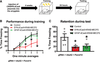
Inhibition of neuronal and astrocytic protein degradation in the amygdala impairs contextual fear memory in male rats. (A) Schematic of the experimental design. CRISPR–dCas9 plasmids (from Fig. 1) were injected into the amygdala of male rats (N = 4–5 per group). Four weeks after transfection, animals were trained to contextual fear conditioning and tested 24 h later. In all cases, guide RNAs (gRNAs) targeted the ubiquitin gene Uba52 and proteasome subunit Psmd14. Control animals received only the gRNAs. (B) Performance during the training session. Inhibition of neuronal (SYN–dCas9), but not astrocytic (GFAP–dCas9), protein degradation reduced freezing behavior during the training session relative to controls. (C) Memory retention during the testing session. Inhibition of neuronal (SYN–dCas9) and astrocytic (GFAP–dCas9) protein degradation impaired long-term memory during the testing session. (*) P < 0.05 from control.
Prior work has consistently shown a need for proteasome-mediated protein degradation in the process of long-term memory formation (for review, see Jarome and Helmstetter 2013). However, the cell type specificity of the protein degradation process to memory consolidation has yet to be explored. Using a cell type-specific CRISPR–dCas9 approach, we report the first evidence that neuronal and astrocytic protein degradation is critical for the consolidation of a contextual fear memory in the amygdala of male rats. Collectively, these data open a new avenue of research in examining the role of protein degradation in neuronal and nonneuronal cell populations during activity and learning-dependent synaptic plasticity.
In our prior work, we found that GFAP was targeted for degradation in the amygdala following contextual fear conditioning (Farrell et al. 2021), suggesting a role for astrocytic protein degradation in the memory consolidation process. Consistent with this, in our present study we found that inhibiting astrocytic protein degradation impaired contextual fear memory consolidation. Furthermore, inhibition of neuronal protein degradation impaired contextual fear memory consolidation to a similar degree, though it also impaired performance during the training session. While it is unclear why the neuronal and astrocytic manipulations differ in these training deficits following manipulation of protein degradation, it does suggest that more work is needed in this area, as no training effects were observed with prior cell type-independent pharmacological or genetic approaches. In terms of the genetic approaches, we previously found that cell type-independent CRISPR–dCas9-mediated inhibition of protein degradation in the amygdala impaired contextual fear memory consolidation without altering performance during training (Devulapalli et al. 2021), the latter result of which is in direct contrast to what we observed with neuron-specific protein degradation blockade in the present study. While it is unclear what led to these contrasting results, it is possible that the cell type-independent manipulation did not result in impaired training performance because the effects of neuronal protein degradation inhibition could have been masked from loss of proteasome function in other cell types. Importantly, we did not observe training effects from our astrocyte manipulation of protein degradation, and the CMV-driven broader manipulation used in the prior study would also target other cell types, including microglia and oligodendrocytes. Thus, future studies should use the approach described here to better elucidate the cell type-specific role of protein degradation in the memory acquisition and consolidation processes while also developing approaches that can target other nonneuronal cell types.
Related, another important consideration is that as neuronal inhibition of protein degradation significantly impaired performance during the training session, it is unclear whether the memory deficits observed during test were from impaired memory consolidation or from the animals never correctly acquiring the task to begin with. Importantly here, animals receiving the SYN–dCas9 plasmid did not show a significant learning curve across time during the training session, indicating that they never properly acquired the fear conditioning task. This makes it difficult to fully separate the effects of neuronal protein degradation inhibition on the memory acquisition versus consolidation processes. Unfortunately, the persistent genetic manipulation that also takes several weeks to express makes it impossible to separate these stages of memory formation and storage. Future studies will want to improve the temporal control of the tools described here, perhaps with the use of light-activated CRISPR–dCas9 constructs (Polstein and Gersbach 2015), to better answer the importance of neuronal protein degradation to the memory acquisition and consolidation processes.
Our data strongly suggest that both neuronal protein degradation and astrocytic protein degradation are critical for fear memory formation in the amygdala; however, it is unclear how protein degradation in both these cell types contributes to this process. While much of the focus has remained on the importance of learning-induced molecular changes in neurons, recent evidence has provided strong support for an involvement of astrocytes in memory formation (Adamsky and Goshen 2018). Interestingly, astrocytes have been reported to have relatively unique functions during learning, such as modulating communication between brain regions to support remote memory formation (Adamsky et al. 2018; Kol et al. 2020). Furthermore, other evidence suggests transport of substances between astrocytes and neurons during memory formation (Suzuki et al. 2011). It is possible, then, that simultaneous increases in protein degradation in neurons and astrocytes help support the communication and interaction between these two cell types during the memory consolidation process. Of note is our recent finding that GFAP was a target of the proteasome following learning (Farrell et al. 2021), which would suggest changes to the astrocyte structure. Such activity-driven changes to the astrocyte structure could help support communication between other cell types, including neurons (Zhou et al. 2019). Consequently, protein degradation in neurons and astrocytes might help support communication between these cell types during the memory consolidation process. Future studies should aim to use the cell type-specific approaches described here to better understand whether and how neuronal and astrocytic protein degradation changes interact following learning.
While our data provide the first evidence of a role for neuronal and astrocytic protein degradation in fear memory consolidation, it should be noted that there are several limitations that must be considered. Most notable is that due to technical limitations, we could not directly quantify cell type-specific inhibition of the protein degradation process. For example, while recent approaches using magnetic-activated cell sorting (MACS) could provide an opportunity to quantify neuronal and astrocytic protein degradation in brain tissue (Holt et al. 2019), such methods have only recently been described and have primarily been used in mice. As a result, we were unable to confirm cell type-specific inhibition of protein degradation in the present study. However, we did previously confirm that our gRNA, when combined with a similar cell type-independent (CMV promoter) dCas9–KRAB–MECP2 plasmid, could inhibit protein degradation in the amygdala (Devulapalli et al. 2021). As the current plasmids were derived from this original dCas9 construct, we are confident that our manipulation was able to sufficiently inhibit the protein degradation process. Furthermore, the promoters used, hSYN and GFAABC1D, have been extensively characterized by prior studies to have selectively toward neurons or astrocytes, respectively. Thus, we are confident that we were able to selectively inhibit protein degradation in specific cell types. Regardless, our study still provides the first attempt, to date, at testing the need for cell type-specific protein degradation in memory formation, which opens a new avenue of research in this rapidly expanding field.
As noted above, another important limitation is that our CRISPR plasmids are persistently active, resulting in constant inhibition of protein degradation throughout the entire training and testing procedure. This makes it difficult to separate training and retrieval effects from those on the consolidation process itself. Unfortunately, temporally controlled cell type-specific CRISPR–dCas9 manipulations do not currently exist, limiting our ability to selectively inhibit protein degradation in neurons or astrocytes during only the posttraining consolidation window. Despite this, our approach still provides significant advantages over the cell type-independent pharmacological approaches that have been used by prior work. Finally, as both males and females need protein degradation in the amygdala to form a fear memory, it would be interesting to test whether these cell type-specific effects observed here vary between sexes. However, our data do provide a starting point for future studies to better understand how sex differences in protein degradation vary by sex during the memory consolidation process.
In conclusion, we provide the first evidence that protein degradation in neurons and astrocytes is critical for fear memory consolidation in the amygdala. These data add to our rapidly expanding knowledge of the role of protein degradation in long-term memory formation and extend it by providing the first evidence that this could occur in a cell type-specific manner.
Acknowledgments
This work was supported by National Institutes of Health grants MH122414, MH120498, MH120569, MH123742, AG071523, and AG079292 to T.J.J.
Footnotes
-
[Supplemental material is available for this article.]
-
Article is online at http://www.learnmem.org/cgi/doi/10.1101/lm.053716.122.
- Received November 22, 2022.
- Accepted February 21, 2023.
This article is distributed exclusively by Cold Spring Harbor Laboratory Press for the first 12 months after the full-issue publication date (see http://learnmem.cshlp.org/site/misc/terms.xhtml). After 12 months, it is available under a Creative Commons License (Attribution-NonCommercial 4.0 International), as described at http://creativecommons.org/licenses/by-nc/4.0/.



