


A recollection of mTOR signaling in learning and memory
- Department of Neurology and Neurosurgery, Montreal Neurological Institute, McGill University, Montreal, Quebec H3A-2B4, Canada
Abstract
Mechanistic target of rapamcyin (mTOR) is a central player in cell growth throughout the organism. However, mTOR takes on an additional, more specialized role in the developed neuron, where it regulates the protein synthesis-dependent, plastic changes underlying learning and memory. mTOR is sequestered in two multiprotein complexes (mTORC1 and mTORC2) that have different substrate specificities, thus allowing for distinct functions at synapses. We will examine how learning activates the mTOR complexes, survey the critical effectors of this pathway in the context of synaptic plasticity, and assess whether mTOR plays an instructive or permissive role in generating molecular memory traces.
mTOR in the developing, growing, and dying brain
mTOR is a multifunctional and highly conserved serine/threonine kinase that, in recent years, has proven to be a critical integrator of cell signaling; so much so that the original “mammalian” TOR nomenclature has yielded to the more apt “mechanistic” TOR. An anabolic autocrat of sorts, mTOR directs the overall growth of all cells in the organism primarily through protein homeostasis, controlling both the production of proteins through the regulation of mRNA translation, and the degradation of proteins through the regulation of autophagy. Whether it is oxygen, genotoxic stress, energy status, or amino acid availability, mTOR senses them all (Laplante and Sabatini 2012).
In the developing brain, mTOR not only dictates the overall growth of differentiating neuronal stem cells (Magri et al. 2011) and post-mitotic neurons (Kwon et al. 2003), it is critical in defining neuronal polarity (Li et al. 2008), axon guidance (Jaworski and Sheng 2006), and dendritic arborization (Urbanska et al. 2012). In the adult brain, mTOR acts in the hypothalamus to integrate the actions of nutrients (e.g., amino acids) and both chronic anorexigenic (e.g., leptin) and acute orexigenic (e.g., ghrelin) hormones to direct appetite (Cota et al. 2006; Martins et al. 2012).
Pathological hyperactivation of mTORC1-dependent protein synthesis in the aging brain may lead to the accumulation of inclusion bodies that are associated with neurodegenerative diseases such as Alzheimer’s, Huntington’s, and Parkinson’s; in these contexts, inhibiting mTORC1 may be restorative by blocking translation of toxic proteins and by promoting autophagy (Bove et al. 2011; Laplante and Sabatini 2012). However, as we will later see in this review, severe inhibition of mTOR-dependent protein synthesis can impair learning and memory. Therefore, precise regulation of mTOR activity to prevent hypo- or hyperactivation is necessary for optimal cognitive function throughout life. This equilibrium is also necessary during neuronal development, as pathological activation of mTORC1 deregulates excitatory/inhibitory balance at synapses (Gkogkas et al. 2013; Santini et al. 2013) and has been implicated in autism spectrum disorder and schizophrenia, where inhibitors of mTOR are also being considered for treatment (Sahin 2012). Therefore, balanced mTOR activity is necessary for many aspects of neuronal biology and consideration for the former is an important part of any rational design of psychoactive therapeutics involving mTOR signaling pathways.
It is generally accepted that activity-dependent plasticity (i.e., strengthening and weakening) of neuronal connections represents the basis of learning and memory—the synaptic plasticity and memory (SPM) hypothesis (Martin et al. 2000). Changes in synaptic strength require stimuli that activate discrete biochemical pathways (learning) to invoke a specific and temporally correlated modification of the synaptic proteome (a memory). As we will see in this review, mTOR serves a critical role in this process. To begin to decipher how mTOR might act to modulate learning and memory, we must first understand the interplay of complex biochemistry and signaling pathways that comprise the mTOR regulatory nexus.
What is the target of rapamycin?
Rapamycin, a macrolide first isolated from Streptomyces hygroscopicus on the Polynesian island of Rapa Nui (Easter Island), was gaining attention throughout the 1980s with its demonstrated immunosuppressive, antiproliferative, and antifungal properties (Vezina et al. 1975). Although the mechanism by which it functioned was known to require binding to the prolyl isomerase FK506-binding protein 12 (FKBP12), it was unclear how this led to its toxic effects on yeast cells. Two loci in Saccharomyces cerevisiae, TOR1 and TOR2, were originally identified in spontaneous mutant strains that confer resistance to rapamycin (Heitman et al. 1991). A subsequent study independently isolated and mapped these loci and defined the “target of rapamycin” protein products as paralogous serine–threonine kinases that interact with the rapamycin–FKBP12 complex (Cafferkey et al. 1993). The conserved function of rapamycin in mammals suggested the existence of TOR homologs in higher eukaryotes, and several groups subsequently isolated a single mammalian homolog of TOR (mTOR) (Brown et al. 1994; Sabatini et al. 1994; Sabers et al. 1995).
mTOR possesses a domain architecture with a high degree of similarity to that of its yeast paralogs. The kinase domain together with the FKBP12–rapamycin binding domain (FRB) reside near the C-terminus, and are flanked by FRAP–ATM–TTRAP (FAT) and FAT C-terminal (FATC) domains that are important for the regulation of kinase activity through protein–protein and intermolecular interactions (Su and Jacinto 2011). The N-terminal of the protein consists of several HEAT (huntingtin, elongation factor 3, protein phosphatase 2A [PP2A], TOR1) repeats. These repeats consist of pairs of antiparallel α helices and are thought to facilitate protein–protein interactions.
The multifaceted topology of mTOR allows it to form at least two functionally distinct core complexes in metazoans. mTOR complex 1 (mTORC1) brings together mTOR and the regulatory associated protein of mTOR (Raptor), while mTOR complex 2 (mTORC2) consists of mTOR and a complex of mSIN1 (SAPK interacting protein 1) and the rapamycin-insensitive companion of mTOR (Rictor). In addition, both mTOR complexes contain mLST8, although this, while necessary for modulating the kinase activity of mTORC2, is dispensable for normal mTORC1 signaling (Guertin et al. 2006; Wang et al. 2012). These complexes are mutually exclusive; mTOR is bound to either Raptor or Rictor/mSIN1 and never to both at the same time (Sarbassov et al. 2004). It remains possible that additional mTOR complexes exist, but as yet there is no strong evidence for this.
One major consequence of the differential binding to Raptor or Rictor/mSIN1 to mTOR is to affect substrate specificity. The two major downstream targets of mTORC1 are the eukaryotic initiation factor (eIF) 4E binding proteins (4EBPs) and ribosomal protein S6 kinases (S6Ks), and their specificities for mTORC1 lie in their interactions with Raptor (Hay and Sonenberg 2004). In contrast, the major downstream targets of mTORC2, through binding with Rictor/mSIN1, include AKT (also known as protein kinase B), serum/glucocorticoid regulated kinase 1 (SGK1, indirectly via the SGK1-binding protein PROTOR), and some isoforms of protein kinase C (PKC; the classic PKCs α, β, and γ as well as the novel PKCε) (Su and Jacinto 2011).
General mechanisms of activation
Research over the past 5 yr has given us a much deeper understanding of how and where mTORC1 and mTORC2 are regulated in the cell. Although these models are based on data derived from nonneuronal cells, they are likely applicable to neuronal and synaptic contexts as well, although much of this regulation remains to be formally documented in neurons.
mTORC1 is turned on (typically measured by the phosphorylation of its substrates, 4EBP1 and S6K1) by binding of the small GTP-binding protein Ras homolog enriched in brain (RHEB) when it is in the activated GTP-binding state. Indeed, RHEB overexpression is probably the easiest mechanism to directly activate mTORC1 independently of growth factors. The GTP state of RHEB is mainly regulated by tuberous sclerosis complex (TSC), a RHEB GTPase-activating protein (GAP) heterodimer consisting of TSC1 and TSC2. In turn, many of the upstream regulators of mTORC1, such as oxygen (mediated through regulated in development and DNA damage responses 1 [REDD1]), energy status (mediated through AMP kinase [AMPK]), or growth factors (mediated through AKT or extracellular signal-regulated kinase [ERK]) regulate the GAP activity of TSC1/2 (Fig. 1).
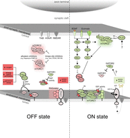
A model of mTORC1 and mTORC2 activity at the synapse. mTOR interacts with distinct adaptor proteins forming mTORC1 and mTORC2 in the postsynaptic density. mTORC1 can be allosterically inhibited by the small molecule rapamycin bound to FKBP12. Chronic inhibition with rapamycin can indirectly lead to a down-regulation in mTORC2 activity. Both mTORC1 and mTORC2 can be acutely inhibited with kinase site inhibitors. In physiological contexts that are not conducive to growth (e.g., hypoxia, low energy, DNA damage, amino acid starvation), mTORC1 is kept in the “OFF-state” through the enhanced GAP activity of the TSC1/2 heterodimer. During amino acid starvation, the GATOR1 complex acts as a GAP for the Rag proteins. Under normal conditions and, indeed, during behavioral learning, mTORC1 and mTORC2 become active (“ON-state”). In this context, release of neurotransmitters from presynaptic neurons activates postsynaptic receptors (tyrosine-related kinase B [TrkB] in the case of homeostatic plasticity, mGluR and NMDAR during Hebbian stimulation), which leads to signal transduction through RAS and PI3K-dependent pathways. These signals converge at the surface of late endosomes where the resulting inhibition of TSC1/2 GAP activity allows charged RHEB to associate with PA-producing PLD1. The increased local concentration of PA leads to direct activation of mTORC1 activity. mTORC1 itself is localized to endosomal membranes via a Rag protein shuttle that is maintained through activation of the RAGulator, a protein complex whose activity is unlocked through structural rearrangement of a vacuolor–ATPase complex that is sensitive to amino acids. In contrast, mTORC2 is anchored to postsynaptic lipid rafts via Syndecan-4 (S4) and can be activated either directly through PI3K-dependent signaling or PLD1-synthesized PA. It is important to note that most of this model has been developed in nonneuronal cells, and therefore the relevance of mTOR localization in neurons has not been formally demonstrated. Moreover, although a postsynaptic density is pictured, this signaling can also occur presynaptically or in the cell soma. Green and red colors indicate active and inactive components, respectively.
Although some evidence suggests that RHEB activates mTORC1 through direct binding (Long et al. 2005), RHEB may also activate mTORC1 through its ability to bind and promote phospholipase D1 (PLD1) activity in a GTP-dependent manner (Sun et al. 2008). PLD1 hydrolyzes phosphatidyl choline (PC) to phosphatidic acid (PA), an important second messenger lipid in neurons that modulates synaptic signaling (Holler et al. 1993; Attucci et al. 2001). PA is required to activate mTORC1 (Fang et al. 2001), perhaps through direct binding (Yoon et al. 2011b). Indeed, it has been proposed that rapamycin inhibits mTORC1 through direct competition with PA (Veverka et al. 2006).
Interestingly, activation of mTORC1 is highly localized to late endosomes/lysosomes (expressing RAB7), since overexpressed RHEB is concentrated at these compartments (Saito et al. 2005; Sancak et al. 2008). Localizing mTORC1 to these endosomes appears to be mediated by a Raptor-dependent interaction with the Rag proteins, a family of Ras-related small GTP-binding proteins. Amino acid entry into the lumen of the lysosome triggers a conformational shift in the ATP-dependent vacuolar proton pump (v-ATPase) (Zoncu et al. 2011). This activates Rag guanine-nucleotide exchange factor (GEF) activity within a membrane-anchored pentameric complex of proteins termed the RAGulator (Sancak et al. 2010; Bar-Peled et al. 2012). The GEF activity of the RAGulator, in turn, charges the obligate heterodimer RagA/B, leading to the recruitment and subsequent RHEB-dependent activation of mTORC1 (Sancak et al. 2010; Bar-Peled et al. 2012). In contrast, the novel trimeric GATOR1 complex acts as a GAP for the Rag proteins and thus down-regulates mTORC1 activation during amino acid deprivation (Bar-Peled et al. 2013). During times of amino acid sufficiency, a second complex, GATOR2, inhibits GATOR1 activity and is required for amino acid-dependent activation of mTORC1 but, unlike the RAGulator, is not involved in localizing Rag proteins to lysosomal membranes (Bar-Peled et al. 2013). Interestingly, PLD1 has been found to also translocate to lysosomal membranes in the presence of amino acids (Yoon et al. 2011a). Therefore, a model can be suggested in which sufficient amino acid levels lead to translocation of mTORC1 to lysosomal membranes, where associated RHEB induces PC hydrolysis yielding PA that can activate mTORC1 (Fig. 1).
Clearly, much more work needs to be done to clarify the nature of the sensor that detects luminal amino acids and determine how it alters the association of the ATPase with the RAGulator. The effect of such a sensor in the neuronal context cannot go unnoticed. Endosomes are abundant in spines (Kennedy and Ehlers 2006) and play major roles in regulating plasticity through trafficking of AMPA receptors and other proteins involved in spine shape and size (Park et al. 2004, 2006). However, it is not known which types of endosomes (recycling or late endosomal) RHEB is associated with at synapses. In axons, RAB7-positive endosomes are strongly implicated in retrograde trafficking (Ng and Tang 2008), and they play a role in sequestering AMPA receptors from membranes during long-term depression (LTD, a form of synaptic plasticity) (Fernandez-Monreal et al. 2012). Moreover, it is not clear how amino acid sensing would work on these nonlysosomal structures. It would be informative to determine if the Rag proteins (which shuttle mTOR to endosomal surfaces) are, indeed, required for mTOR-dependent synaptic plasticity. Alternatively, neurons may have specific mechanisms to bypass the normal requirement for activation by amino acids since, at least in mature neurons, there is no obvious need to limit the induction of neuronal plasticity to periods of nutrient excess.
In contrast to mTORC1, the mechanisms for activation of mTORC2 are just beginning to be elucidated. It has recently been shown that mTORC2 can be directly activated (typically measured by phosphorylation of its best known substrate AKT at S473) in vitro by phosphatidylinositol (3,4,5)-trisphosphate (PIP3)—the product of phosphatidylinositol 3-kinase (PI3K) activity (Gan et al. 2011). Like mTORC1, mTORC2 activity also has a requirement for PA (Toschi et al. 2009). Therefore, these data suggest the possibility that PA can alter kinase activity within either mTORC1 or mTORC2 (Foster 2009). This scenario posits that differential activation of mTORC1 and mTORC2 at the synapse could be the result of varying subcellular localization and concentrations of PA (Fig. 1).
The mTORC2 substrates AKT and the PKCs primarily act in transducing receptor-mediated signaling close to the plasma membrane. Interestingly, mTORC2 has been localized to lipid rafts via the transmembrane protein Syndecan-4 (Partovian et al. 2008). In addition, a pleckstrin homology-like (PH) domain in AVO1 targets this yeast ortholog of mSIN1 to the plasma membrane and this localization is essential for cell viability (Berchtold and Walther 2009). It remains to be seen whether the PH domain of mSIN1 serves a similar purpose. From these data, it seems likely that localization to the plasma membrane facilitates the activation of mTORC2 at the synapse (Fig. 1). In contrast, as discussed above, activation of mTORC1 at the synapse may be restricted to intracellular endosomes.
Pharmacological inhibition
As we found out at the beginning of this review, rapamycin was the first “potent” and specific small molecule inhibitor of mTOR. It has been almost exclusively used in the past to attach function to mTORC1 at both the level of the cell and the animal (especially for assessing the function of mTOR on learning and behavior). Surprisingly, the detailed molecular mechanisms of rapamycin action are still unclear. The rapamycin–FKBP12 complex has been proposed to either directly inhibit binding of RHEB, PA (a product of RHEB activity, as we saw in the last section), or Raptor, which would explain its specificity for mTORC1 (Jacinto 2008). In addition, a recent crystal structure of mTOR/mLST8 shows that the FRB domain (the domain that binds rapamycin–FKBP12) restricts access to the catalytic site and is important for directing substrates into this site (Yang et al. 2013). Moreover, rapamycin–FKBP12 binding is predicted to directly block substrate access to the catalytic site. The structure was deduced from cocrystals of full-length mLST8 and a fragment of mTOR lacking the N-terminal HEAT repeats. Therefore, it is possible that in the cellular context, conformational changes occur after binding of Rictor and/or Raptor (which interact with the HEAT repeats) that alter this role of the FRB domain in mTORC1 and mTORC2. It should be noted that prolonged incubation with rapamycin sequesters mTOR in unproductive complexes, preventing efficient mTORC2 complex formation (Sarbassov et al. 2006). Thus, rapamycin–FKBP12 can bind to and disrupt mTORC1 function, but it cannot disrupt preformed mTORC2 complexes. The lower sensitivity of mTORC2 to rapamycin has also been suggested to be due to its inability to displace PA in mTORC2 as opposed to mTORC1 (Foster 2009). As of now, there are no specific pharmacological inhibitors of mTORC2. Presumably, these would have to target the specific binding of Rictor/mSIN1 to mTOR.
In the cancer clinic, rapamycin and its analogs are subject to resistance, largely due to its inability to quench hyperactivation of mTORC2 under these conditions, leading to feedback activation of PI3K-AKT-dependent growth (O’Reilly et al. 2006). Recently, mTOR kinase inhibitors such as Torin, PP242, and INK128 have been developed to get around this issue (Feldman et al. 2009; Thoreen et al. 2009; Hsieh et al. 2012). Directed against the mTOR ATP site, these are potent inhibitors of both mTORC1 and mTORC2. Unfortunately, feedback is still an issue with these inhibitors, as the mTOR inhibitory activity of PP242 has been found to generate apoptotic resistance through the potent activation of the RAF–MEK–ERK signaling pathway (Hoang et al. 2012). Interestingly, phosphorylation of some targets of mTORC1, such as 4EBP, is more strongly inhibited by ATP-based inhibitors than by rapamycin, suggesting that rapamycin does not completely block all targets of mTORC1 (Thoreen et al. 2012). The increased inhibition of 4EBP can also be linked to the activation of the RAF–MEK–ERK pathway in myeloma cells (Hoang et al. 2012). Although this information is of obvious importance to oncobiologists, the possibility of feedback-mediated changes in other signaling pathways induced by mTOR inhibition must also be heeded by neurobiologists wishing to employ them in experiments that assess the role of mTOR in learning and memory.
Translational control at the synapse
As we have seen, mTOR is a central regulator of mRNA translation. In neurons, translational control can be localized to specific cellular compartments, including in dendritic spines (Sutton and Schuman 2005). This decentralized translation, where messages can be translated independently of the somatic translation machinery, gives synapses the power to shape their own proteome subject to the local environment. This model necessitates active somato-dendritic transport of specific mRNAs repressed at the level of translation. Indeed, a unique feature of neuronal cells is large RNA transport complexes (neuronal RNA granules) that are regulated in this manner (Sossin and DesGroseillers 2006). A number of recent reviews have examined the importance of local translation for synaptic plasticity and learning (Sutton and Schuman 2005; Costa-Mattioli et al. 2009; Richter and Klann 2009; Liu-Yesucevitz et al. 2011) and there is abundant evidence that mTORC1 is active at synapses (Costa-Mattioli et al. 2009), while synaptic mTORC2 activity is also evident (Huang et al. 2013). An outstanding question is whether synaptic mTORC1 participates in removing the block in translation on these mRNAs, or instead stimulates their translation only after they have been released from their transport complex through other means.
mTORC1, synaptic plasticity, and memory
It is well known that the formation of long-term memory requires protein synthesis (Squire and Davis 1981). Thus, given the important role of mTORC1 in controlling protein synthesis, it is not surprising that inhibiting mTORC1 with rapamycin blocks long-term memory formation in a number of paradigms (Tischmeyer et al. 2003; Parsons et al. 2006; Bekinschtein et al. 2007; Blundell et al. 2008; Belelovsky et al. 2009; Glover et al. 2010; Gafford et al. 2011; Deli et al. 2012; Halloran et al. 2012; Jobim et al. 2012). Probably the most convincing evidence uses a mTOR heterozygotic mouse to show that both consolidation and reconsolidation of memories are blocked at lower concentrations of rapamycin, directly tying the actions of rapamycin on memory to mTOR (Stoica et al. 2011).
Most models of memory depend on modifications in synaptic strength. Therefore, if mTORC1 is involved in memory formation, it should also be implicated in the synaptic changes that underlie memory. Many of these paradigms, including late-phase long-term potentiation (L-LTP) in CA1 neurons of the rodent hippocampus, and long-term facilitation (LTF) in Aplysia sensory-motor neuron synapses, demonstrate sensitivity to rapamycin (Casadio et al. 1999; Tang et al. 2002). There are, however, exceptions; protein synthesis-dependent LTP at the connection between the entorhinal cortex and the dentate gyrus (the circuit where LTP was first observed) is insensitive to rapamycin (Panja et al. 2009). Metabotropic receptor-mediated long-term depression (mGluR-LTD), another model of synaptic plasticity in CA1 rodent neurons, has also been reported to be sensitive to rapamycin by several groups (Hou and Klann 2004; Sharma et al. 2010; Lebeau et al. 2011; Bozdagi et al. 2012). However, despite similar protocols used, not all laboratories have reported rapamycin sensitivity in mGluR-LTD (Auerbach et al. 2011). This suggests that under some conditions, mTORC1 activation is dispensable for mGluR-LTD, presumably because the specific products of rapamycin-sensitive mRNA translation are already present in sufficient quantity prior to stimulation. The role of mTORC1 in mGluR-LTD is discussed in more detail in an upcoming section describing the downstream targets of mTORC1, which paradoxically appears to have both positive and negative roles in this form of plasticity.
A major question in the literature that cannot be answered simply by blocking mTORC1 activity is whether this complex plays an instructive role or merely acts a gatekeeper in memory consolidation. Does mTORC1 merely lead to a nonspecific increase in overall protein synthesis that facilitates synaptic plasticity and memory? This seems exceedingly unlikely given the current data. Rapamycin only blocks a small component of overall protein synthesis (Choo et al. 2008), including in the nervous system (Yanow et al. 1998) and in some cases the mTORC1-dependent increase in overall translation has been dissociated from the requirement of mTORC1 for plasticity (Weatherill et al. 2010). In most cases, however, the protein synthesis under control of mTORC1 is not sufficient to induce plasticity. Instead, additional mTORC1-independent regulatory signals are required to induce plasticity, or even determine the type of plasticity that occurs (i.e., increases vs. decreases in synaptic strength), as in the concept of cross-tagging, where all necessary proteins required for L-LTP and L-LTD are synthesized following a stimulus and then interact with a protein synthesis-independent synaptic “tag” to generate plasticity (Frey and Frey 2008). Thus, stimulation of mTORC1 probably generates a set of proteins important for plasticity, but not necessarily sufficient for plasticity.
One exception to this rule is a form of homeostatic plasticity observed in both Drosophila and mammals, where a decrease in basal synaptic strength via a blockade of postsynaptic AMPA receptors activates mTORC1. This leads to a mTORC1-dependent increase in the synthesis of a retrograde messenger that then acts presynaptically to increase neurotransmitter release, thus restoring basal synaptic strength (Henry et al. 2012; Penney et al. 2012). In these cases mTORC1 activation appears to be sufficient, as either overexpression of RHEB (the mTORC1-specific activator), or overexpression of a constitutively active downstream target of mTORC1 in Drosophila, induces the increase in presynaptic neurotransmitter release (Henry et al. 2012; Penney et al. 2012). Thus, a major physiological effect of activating mTORC1 at the postsynaptic density is to increase neurotransmitter release from the presynaptic neurons. Consistent with this, a recent study examining the effects of mTORC1 hyperactivation through loss of the phosphatase and tensin inhibitor (PTEN) that suppresses PI3K signaling found a general increase in presynaptic release of both the excitatory and inhibitory neurotransmitters glutamate and γ-aminobutyric acid (GABA), respectively (Weston et al. 2012). In vertebrates, this retrograde messenger may be brain-derived neurotrophic factor (BDNF), which is encoded by a dendritically localized mRNA under tight translational control by a number of pathways (Zheng et al. 2012). In Drosophila, however, no ortholog of BDNF, or its receptor, tyrosine-related kinase B (TrkB) exists and the retrograde messenger has not been identified (Penney et al. 2012).
Notably, none of these experiments revealed either a LTP- or LTD-like change in the levels of postsynaptic receptors following mTORC1 hyperactivation, consistent with a gatekeeping role for mTORC1 in these forms of synaptic plasticity.
Functions of mTORC1 targets in learning and memory
Given the importance of mTORC1 activation for synaptic plasticity and memory, it becomes essential to understand what downstream targets of mTORC1 are involved in learning and memory. Below, we review what is known about these targets.
4EBP
The functional role of 4EBP is very well defined. It competes with the scaffold protein eIF4G for binding to the 7-methyl cap binding protein eIF4E, preventing formation of the pre-initiation complex that catalyzes the recruitment of mRNAs to the ribosome (Fig. 1). There are four conserved phosphorylation sites in all mammalian 4EBP isoforms (T37, T46, S65, T70). Sequential phosphorylation of all four sites is required to block eIF4E binding and activate translation (Gingras et al. 2001). Although only two of these sites are rapamycin-insensitive, using the more potent mTOR kinase domain inhibitor Torin, Thoreen et al. (2009) were able to show a profound block in the phosphorylation of T37/46 and S65 (compared to rapamycin) and, most importantly, substantially increased association with eIF4E.
Three paralogs of 4EBP are encoded in the vertebrate genome, 4EBP1, 4EBP2, and 4EBP3. In the adult brain, 4EBP2 is by far the most abundant isoform, with very little 4EBP1 and no detectable 4EBP3 (Tsukiyama-Kohara et al. 2001; Banko et al. 2005). Therefore, although all 4EBPs have similar function (mTOR-dependent repressors of translation), we will restrict our discussion here to the brain-specific isoform 4EBP2. Curiously, 4EBP2 is post-translationally regulated not only by phosphorylation, but also by spontaneous deamidation of asparagine residues. These residues are conserved in mammalian 4EBP2 but not in the other two isoforms or, indeed, in any organisms that possess a single copy of the Eif4ebp gene, suggesting a novel role in chordate brain function (Bidinosti et al. 2010). Deamidation is a nonenzymatic process resulting in conversion of asparagine to aspartic acid and has been proposed to serve as a molecular timer regulating protein turnover (Robinson and Robinson 2001). In the case of 4EBP2, deamidation in proximity to the TOR signaling motif (TOS, present in all mTOR substrates and required for binding to Raptor and thus mTOR-dependent regulation [Schalm and Blenis 2002]) enhances its association with Raptor. The increased binding to Raptor prevents deamidated 4EBP2 from efficiently interacting with eIF4E and down-regulating translation, as a deamidated 4EBP2 mimic (asparagines replaced with aspartic acids) lacking the TOS motif (preventing Raptor binding) is still able to interact with eIF4E. Critically, the long half-life of 4EBP2 in the brain allows for a considerable fraction of the protein to become deamidated during neural development, a time when mTORC1 signaling wanes as evidenced by decreased 4EBP2 phosphorylation (Bidinosti et al. 2010). Therefore, despite a reduction in upstream mTORC1 signaling in the mature brain that results in enhanced affinity of hypophosphorylated 4EBP2 for eIF4E, increased deamidation over the same time period short-circuits this pathway, allowing for continued translation. It should be noted that there is no evidence that decreased 4EBP2 phosphorylation in the mature brain is due to deamidation. In the context of active mTORC1, enhanced association of Raptor and deamidated 4EBP2 would, instead, be expected to increase 4EBP2 phosphorylation.
Most studies have examined the role of the 4EBPs by studying genetic knockouts. Removing the brain-specific isoform 4EBP2 increases both excitatory and inhibitory synaptic transmission in the hippocampus, but increases the excitatory/inhibitory ratio, thus favoring excitatory neurotransmission and resulting in autistic-like phenotypes in mice (Gkogkas et al. 2013). At the level of synaptic plasticity, induction of Late-LTP is impaired in eif4ebp2−/− hippocampal neurons, presumably due to excessive translation of an inhibitor of this process (Banko et al. 2005). In contrast, Early-LTP stimulation (a phenomenon that is independent of protein synthesis) leads to Late-LTP in these mice, suggesting that some plasticity-related proteins (PRPs) may already be translated prior to stimulation (Banko et al. 2006). However, this LTP still requires protein synthesis, suggesting that not all proteins required for L-LTP are regulated by 4EBP2. LTP of inhibitory neurons induced by mGluR stimulation was also enhanced in the absence of 4EBP2 (Ran et al. 2009). mGluR-LTD in excitatory neurons lacking 4EBP2 is similarly enhanced, and activation of 4EBP2 explains the sensitivity of mGluR-LTD to rapamycin in wild-type neurons observed in this study (Banko et al. 2006).
Since 4EBP is a repressor whose activity is removed by mTORC1 signaling, knocking out 4EBP2 is equivalent to strong constitutive activation of this branch of mTORC1 signaling in the neuron. However, this type of experiment does not address whether, under physiological conditions, removal of 4EBP repression is important for inducing plasticity. One attempt to address this is to use 4EBP mutants that still bind to eIF4E, but are not regulated by mTOR. In Aplysia, overexpressing 4EBP lacking the TOS site, thus rendering it insensitive to mTORC1, repressed overall translation, but did not block rapamycin-sensitive LTF suggesting that in this case the target of mTORC1 important for plasticity was not 4EBP (Weatherill et al. 2010).
4EBP targets
Although the target of 4EBP is clearly inhibition of eIF4E binding to eIF4G, the mRNAs regulated by 4EBP are not yet well defined. Initiation of translation on cellular mRNAs necessitates a methylguanosine “cap” structure located at their 5′ termini. Although some viral and cellular mRNAs circumvent this requirement by recruiting the ribosome to an internal ribosome entry site (IRES) independently of the 5′ cap, most (>90%) cellular mRNAs undergo cap-dependent translation (Graber and Holcik 2007). Thus, one could conclude that the vast majority of mRNAs would be under control of 4EBP.
It has been suggested that mRNAs with long and structured 5′ untranslated regions (complex 5′ UTRs) have a higher dependence on eIF4E, therefore rendering them acutely sensitive to regulation by 4EBP (Koromilas et al. 1992). Neuroligin is one such mRNA; its up-regulation in eif4ebp2−/− mice contributes to the autistic phenotypes exhibited by this mutant, as down-regulation of neuroligin can rescue many of the phenotypic abnormalities (deficits in social interaction, altered communication, and repetitive behaviors) observed in these animals (Gkogkas et al. 2013). In another study using the same mice, a selective increase in GluA1 and GluA2 (AMPA receptor subunits) mRNA translation was observed (Ran et al. 2013). Although neuroligin, GluA1, and GluA2 mRNAs have complicated 5′ UTRs, there is no direct evidence that they contribute to the 4EBP2-mediated translational control of their messages.
In contrast, Thoreen et al. (2012) used ribosome profiling (a recent technique that quantifies translational efficiency of the entire transcriptome at codon resolution) in mouse embryonic fibroblast (MEF) to determine that acute (2 h) inhibition of mTORC1 by Torin, which leads to maximal activation of 4EBPs, has no discernible effect on the translational efficiency of mRNAs harboring complex 5′ UTRs, but reduces translation of 99.8% of the transcriptome by an average of 60%. Thoreen et al. (2012) did find, however, that Torin preferentially represses translation of terminal oligopyrimidine (TOP) mRNAs and that this was through 4EBP1/2, as Torin-treated double-knockout MEFs or HeLa cells expressing 4EBP1/2 RNA interference caused little decrease in TOP mRNA translation relative to that of non-TOP mRNAs. TOP mRNAs mainly constitute proteins involved in translation, including all mRNAs encoding ribosomal proteins and elongation factors. This study also shows that eIF4E binding to TOP mRNAs is more sensitive to 4EBP activity than for other capped messages. Using a comparable methodology but in a cancer cell line, Hsieh et al. (2012) found a similar sensitivity of TOP mRNAs using the mTOR active site inhibitors PP242 and INK128.
Opposing observations from the Meyuhas laboratory have shown that the critical mTORC1 component Raptor is not required for TOR-dependent activation of TOP mRNAs (Patursky-Polischuk et al. 2009). One distinction between these studies is that the Sabatini group examined basal repression of TOP mRNAs by mTORC1, while the Meyuhas group examined stimulation of TOP mRNAs by growth factor activation of mTORC1. This suggests multiple mechanisms by which mTORC1 can regulate translation of TOP mRNAs. The role of TOP mRNAs in plasticity will be discussed separately below. Overall, although progress is clearly being made in determining which mRNAs are 4EBP targets, the question of defining the specific mechanism that makes an mRNA sensitive to 4EBP is still an open question.
S6 kinase
Another major target of mTORC1 is S6 kinase. Learning leads to increases in S6 kinase activity in most systems, and this is mainly mediated by mTORC1 as it is strongly blocked by rapamycin. In vertebrates there are two isoforms of S6 kinase, S6K1 and S6K2, and both are expressed in the brain. Although most targets can be phosphorylated by either isoform, some are more affected by S6K1, and others by S6K2. Consistent with separate but overlapping functions, behavioral and electrophysiological effects of the individual knockouts are distinct. Mutant mice lacking S6K1 show normal translation-dependent L-LTP, but impaired translation-independent Early-LTP (Antion et al. 2008). Despite having intact L-LTP, the S6K1 knockout mouse exhibits a variety of behavioral learning deficits, including the impairment of spatial learning, contextual fear memory, and conditioned taste aversion. S6K2 knockout mice similarly showed intact L-LTP in conjunction with impaired contextual fear memory and conditioned taste aversion, but unlike S6K1 knockouts, had normal spatial learning (Antion et al. 2008).
Unfortunately, S6K1 and S6K2 can compensate for one another, complicating the interpretation of these results. Also problematic is that most double knockout mice die in utero or shortly after birth (Pende et al. 2004), making assessment of the role of S6K in the brain more difficult. Several specific inhibitors of S6 kinase have been recently developed that may help address functional questions (Pearce et al. 2010). Interestingly, in Aplysia, the overexpression of a dominant negative form of S6K with a mutated TOS site, so that it did not interfere with other targets of mTORC1, blocked the induction of LTF (Weatherill et al. 2010), suggesting this kinase is important for some forms of plasticity. Overexpression of a constitutively active S6K was sufficient to induce the synthesis of a retrograde messenger to increase presynaptic strength in Drosophila, suggesting that S6K is also an important downstream target of mTORC1 in this pathway (Penney et al. 2012). In general, the importance of S6Ks for most forms of synaptic plasticity is still an open question.
S6 kinase targets
S6K targets several players that regulate translation including ribosomal protein S6, eIF4B, eukaryotic elongation factor 2 kinase (eEF2K), and fragile mental retardation protein (FMRP). Surprisingly, removing S6K has no effect on either general translation or translation of specific messages in the liver. In contrast, these experiments showed that the major effect of S6K was to stimulate the transcription of mRNAs encoding proteins important in ribosome biogenesis (Chauvin et al. 2013). These proteins regulate pre-rRNA synthesis, cleavage, post-transcriptional modifications, ribosome assembly, and export (Chauvin et al. 2013). This was largely mediated through phosphorylation of S6, as mice expressing a nonphosphorylatable S6 had a deficit in the up-regulation of these proteins (Chauvin et al. 2013). This transcriptional pathway is likely to be important for the role that S6K-mediated S6 phosphorylation plays in regulating cell size (Meyuhas and Dreazen 2009; Ruvinsky et al. 2009). However, the detailed pathway leading from phosphorylation of S6 to transcriptional control of ribosome biogenesis-encoding mRNAs is not known. In the neuronal context, there is a correlative increase in the phosphorylation of S6 during LTP and mGluR-dependent LTD (Tsokas et al. 2007; Antion et al. 2008), as well as during LTF in Aplysia (Khan et al. 2001). In many cases, this increase can be seen locally at the synapse, suggesting roles independent of transcriptional effects on ribosome biogenesis. However, the consequence of local S6 phosphorylation on translation in this context remains a mystery.
S6K also phosphorylates the eIF4A regulator, eIF4B in most cell-types, although its regulation in neurons has not been studied as yet (Shahbazian et al. 2006). This S6K pathway stimulates translation initiation and, like that of 4EBP, is thought to target mRNAs with complicated 5′ UTRs (Shahbazian et al. 2010).
Although there is a paucity of data linking S6 and eIF4B to plasticity, there have been numerous reports that S6K-directed effects on translation elongation are critical factors in learning and memory. S6K indirectly stimulates elongation by phosphorylating and inhibiting eEF2K, itself an inhibitor of eEF2 (Wang et al. 2001). In turn, eEF2 binds tightly to the ribosome and catalyzes its GTP-dependent translocation. When phosphorylated (e.g., by eEF2K), eEF2 loses affinity for the ribosome causing a decrease in elongation rate (Dever and Green 2012). Critically, eEF2K is activated by calcium/calmodulin and consequently regulated by neural activity (Nairn et al. 2001). Indeed, eEF2K has been implicated as a control point in the synthesis of proteins important for memory since either increasing or decreasing eEF2K activity affects memory formation in distinct plasticity paradigms (Im et al. 2009; Gildish et al. 2012). eEF2K has been shown to be particularly important in conveying information mediated by spontaneous release (Sutton et al. 2007) and signaling though mGluR receptors (Park et al. 2008).
Decreasing eEF2 phosphorylation has been proposed to specifically activate translation of mRNAs associated with plasticity. Specifically, Late-LTP and hippocampus-dependent long-term memory are blocked in mice by forcing eEF2 phosphorylation through overexpression of eEF2K (Im et al. 2009). S6K has also been linked to decreases in eEF2 phosphorylation after persistent activation in Aplysia neurites (Carroll et al. 2004; Weatherill et al. 2010) and after BDNF application in hippocampal cultures (Inamura et al. 2005). However, the situation is probably more complicated. In a number of cases, mTORC1 activation is correlated with increased eEF2 phosphorylation, such as during taste conditioning (Belelovsky et al. 2005) and mGluR-LTD (Banko et al. 2006; Park et al. 2008). Indeed, during LTP in the dentate gyrus, the increase in eEF2 phosphorylation was also mTORC1-dependent (Panja et al. 2009).
In principle, slowing elongation via phosphorylation of eEF2 will affect all mRNAs. Paradoxically however, translation of several mRNAs is actually increased under conditions where elongation should be repressed (Sossin and Lacaille 2010). These transcripts include those that encode CAMK2, ARC, and MAP1B—important regulators of synaptic architecture (Scheetz et al. 2000; Chotiner et al. 2003; Belelovsky et al. 2005; Davidkova and Carroll 2007; Park et al. 2008). Thus, both increases and decreases in eEF2 phosphorylation have been linked to synaptic plasticity. The mechanisms behind this phenomenon remain unknown. Any depression in elongation brought on by increased eEF2 phosphorylation would necessarily reduce the number of initiation events on the majority of mRNAs. Thus, it has been hypothesized that this frees critical initiation factors that, in the context of fully active eEF2, are limiting to the translation of specific mRNAs involved in plasticity (Sossin and Lacaille 2010).
FMRP is another target of S6K that plays a critical role in synaptic plasticity. Mutations in Fmr1, the X-linked gene that encodes FMRP, result in fragile X syndrome (FXS), a disease that is typified by impaired cognition and is the leading monogenic cause of autism (Martin and Huntsman 2012). This pathology is phenocopied in Fmr−/y mice, and is due to the loss of the translation repression function of FMRP (Brown et al. 2001). The obvious clinical relevance of this protein has resulted in a flurry of research and controversy regarding the specific role of FMRP in translation that is beyond the scope of this review.
Here, we focus on the role of the mTORC1-S6K1 signaling arm that targets phosphorylation of FMRP. This phosphorylation, thought to be important in mediating constitutive repression of translation by FMRP (Ceman et al. 2003; Narayanan et al. 2008; Muddashetty et al. 2011), is sensitive to rapamycin treatment and abolished in S6K1−/− hippocampal neurons (Narayanan et al. 2008). However, mGluR-LTD (the form of plasticity most tightly associated with FMRP) depends on PP2A activity that targets removal of FMRP phosphorylation within seconds after stimulation, while mTOR-S6K-dependent rephosphorylation follows (Niere et al. 2012). These data fit a model in which mRNAs encoding proteins required for LTD are held in check by constitutive S6K1-mediated activation of FMRP. Upon mGluR stimulation, transient phosphatase activity releases the FMRP “brake,” and subsequent mTORC1 activation stimulates the translation of newly unlocked LTD target mRNAs, although, as described earlier, not all studies indicate a requirement for mTORC1 activation to support this subsequent translation (Auerbach et al. 2011). The accompanying mTORC1 activity also resets the system through S6K1-mediated rephosphorylation of FMRP that then associates with new LTD target mRNAs.
The requirement of S6K for FMRP-mediated repression is consistent with the enhanced mGluR-LTD that is seen in S6K1/2 double knockout mice similar to the Fmr−/y knockout animals (Antion et al. 2008). These results are complicated due to additional roles of S6K in mGluR-LTD. The mTORC1 pathway, including S6K activity, is up-regulated after loss of FMRP (Hoeffer et al. 2012). Indeed, synaptic and behavioral phenotypes of FMRP knockout mice were rescued by genetic removal of S6K1, including the loss of enhanced mGluR-LTD, and this was clearly independent of S6K1-mediated regulation of FMRP (Bhattacharya et al. 2012). Thus, S6K plays multiple roles in the signal transduction pathways involved in mGluR-LTD (Fig. 2). The situation is even more complex as hyperactivation of mTORC1 through removal of TSC1/2 components in mice impairs mGluR-LTD (Auerbach et al. 2011; Bateup et al. 2011), perhaps through constituitively active S6K-FMRP signaling in these mutant animals. Consistent with this model, rapamycin “rescues” mGluR-LTD in this context of hyperactivated mTORC1 (Auerbach et al. 2011). In conclusion, since mTORC1 is important in mGluR-LTD both for the repression of mGluR-LTD target mRNA translation through S6K phosphorylation of FMRP and the subsequent activation of the translation of these mRNAs through phosphorylation of 4EBP (Banko et al. 2006), it may not be surprising that manipulations of mTORC1 can give conflicting results in this paradigm of plasticity.
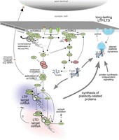
A model of signaling pathways downstream of mTORC1 and mTORC2 that regulate synaptic plasticity. In addition to canonical receptor-mediated signaling, the activation of mTORC1 following synaptic stimulation can be enhanced indirectly through mTORC2-mediated cotranslational stabilization of AKT. Translation of both LTP- and LTD-target mRNAs is targeted following mTORC1 activation. Release of 4EBP-mediated repression preferentially stimulates translation initiation of TOP mRNAs, while release of eEF2K-mediated repression through S6K stimulates translation elongation of LTP-target mRNAs. While S6K also targets ribosomal protein S6 and eIF4B (not shown), their roles in synaptic plasticity are unknown. Many mRNAs that are required for LTD have an additional level of translation repression in the form of FMRP. FMRP is thought to repress translation by promiscuously binding to the coding sequence of the mRNA, thus preventing ribosomal translocation, perhaps through blocking the activity of eEF2. By acting as a brake, this protein impedes translation of LTD-target mRNAs despite mTORC1-dependent stimulation of translation. On the other hand, stimulation of mGluR receptors leads to activation of the phosphatase PP2A, which transiently inactivates FMRP activity, releasing the brake. This allows subsequent mTOR-dependent stimulation of LTD target mRNA translation at the expense of LTP target mRNAs. Sustained stimulation resets FMRP activity through S6K-mediated phosphorylation, allowing for the replenishment of the LTD-target mRNA pool. mTORC1-dependent protein synthesis can translate mRNAs encoding retrograde signals to increase presynaptic strength. However, mTORC1 activation during LTP and LTD is not sufficient to mediate synaptic plasticity and requires additional signaling. One step required for LTP may be activation of the RAC1–PAK1 pathway by mTORC2 that leads to inhibition of the actin depolymerase, Cofilin. Alternatively, actin stabilization may be important upstream of mTORC1 for activation of the translational control pathway. The combined result of mTORC1- and mTORC2-signaling during LTP or LTD is mediated by altered trafficking of AMPA receptors and changes in the synaptic architecture. Green and red colors indicate active and inactive components, respectively.
An important point to remember about S6K target sites is that they can often be regulated by other parallel signaling pathways, particularly via MAP kinase-activated ribosomal S6 kinase 2 (RSK2). RSK2 has been shown to target S6, eEF2K, and eIF4B in mitotic cells, although its importance in modifying synaptic plasticity remains an open question (Wang et al. 2001; Shahbazian et al. 2006; Roux et al. 2007).
TOP mRNAs
As described above, a major class of mRNAs regulated downstream of mTORC1 are TOP mRNAs. Critically, all ribosomal proteins are encoded by TOP mRNAs, as are a number of other translation factors such as eEF1a and eEF2 (Meyuhas 2000). In mitotic cells, mTORC1 activation increases ribosomal protein synthesis and subsequent ribosome biogenesis in the nucleolus—a pathway that highlights the role that mTORC1 plays in controlling cell size and proliferation. However, TOP mRNA regulation in neurons may serve additional functions, since TOP mRNAs are transported into dendrites and axons where they are locally translated (Tsokas et al. 2007). Indeed, TOP mRNAs are one of the more abundant localized mRNAs (Moccia et al. 2003; Poon et al. 2006). Moreover, the local translation of TOP mRNAs depends on mTORC1 activity and the integrity of the TOP sequence (Gobert et al. 2008). What are TOP mRNA-encoded proteins doing in neurites?
It seems unlikely that new ribosomes are assembled in this compartment, as this process requires specialized activities only found within the nucleolus (Shaw and Jordan 1995). The simplest possibility is that the localization of these proteins reflects the passive transport of TOP mRNAs (an abundant class of mRNA) out of the soma and into dendrites. A second possibility is that translation is regulated in dendrites through selective removal and replacement of peripheral ribosomal proteins. A third and equally attractive possibility is that the proteins encoded by TOP mRNAs serve extraribosomal functions. Indeed, eEF1α, one of the TOP mRNAs translated during plasticity (Tsokas et al. 2005) may play important roles in regulating the actin-bundling program and surface expression of at least one muscarinic acetylcholine receptor subtype (McClatchy et al. 2006) in addition to its canonical activity on the ribosome (Murray et al. 1996). However, without a specific inhibitor of this target pathway, the importance of local TOP translation in neurons is still largely speculative.
A surprising effect of mTORC1 in suppressing translation of an ion channel
Although we have seen that, in most cases, mTORC1 activation leads to increased protein synthesis, there is an intriguing case in which basal mTORC1 activity suppresses translation of a target mRNA in neurons. Raab-Graham et al. (2006) reported that either rapamycin treatment or addition of the NMDA receptor antagonist AP5 leads to decreased mTOR activity yet triggers increased dendritic translation of Kv1.1 potassium channel mRNA, monitored using a photoconvertible translational reporter. This may provide a mechanism for mTORC1-dependent decreases in synaptic potassium currents that could result in increased excitability. How basal mTOR activity represses translation of Kv1.1 mRNA remains to be elucidated.
mTORC2, synaptic plasticity, and memory
The lack of mTORC2 inhibitors has made examination of the specific role of mTORC2 difficult. Moreover, as discussed below, mTORC2 is required cotranslationally for the stabilization of a number of AGC kinases, such as AKT, PKC, and SGK1, and thus removal of mTORC2 (accomplished with rictor knockout animals) is lethal (Shiota et al. 2006). Additionally, neuron-specific knockouts of rictor have many developmental deficits making it difficult to use these mice to examine learning and memory (Siuta et al. 2010; Carson et al. 2013).
To avoid these issues, Huang et al. (2013) have recently crossed these mice with ones expressing Cre recombinase driven by a Camk2a promoter that specifically removes mTORC2 activity from excitatory neurons in limbic and cortical areas after development. Functionally, this study showed that mTORC2 activity (measured by AKT phosphorylation at S473) is enhanced by stimuli associated with learning, and this stimulation-dependent increase in AKT phosphorylation was not seen in the absence of mTORC2. Critically, these mice are incapable of establishing L-LTP and consequently demonstrate an impairment in learning and retaining long-term fear memories, suggesting a necessary role for mTORC2 in synaptic plasticity and memory (Huang et al. 2013).
Functions of mTORC2 substrates and their targets in learning and memory
The elongation step of translation also appears to be an important target of mTORC2. mTORC2 associates with polysomes through direct binding of mSIN1 and Rictor to large ribosomal subunit proteins (Fig. 2), and protein synthesis in mSIN1−/− MEFs is markedly depressed (by ∼40%) (Oh et al. 2010). Although a role for negative feedback to mTORC1 could not be ruled out in that study, others have shown that mTORC1 activity is not affected in the absence of mTORC2, perhaps due to the fact that AKT activity is not completely missing in this context despite the lack of mTORC2 that is needed to phosphorylate it (Jacinto et al. 2006).
The interaction of mTORC2 with polysomes is required for the ability of mTORC2 to cotranslationally phosphorylate several AGC kinases (AKT, PKCα,β,γ,ε) at the turn motif (TM) as it emerges from the ribosomal exit tunnel (Ikenoue et al. 2008; Oh et al. 2010). This cotranslational and constitutive modification stabilizes and protects these kinases from degradation by the proteasome, as the absence of Rictor or mSIN1 in MEFs leads to their ubiquitin-dependent degradation (Ikenoue et al. 2008; Oh et al. 2010). This is contrary to the mTORC2-dependent phosphorylation in the hydrophobic motif (HM) of these kinases; a modification that is necessary for their activation but that occurs independently of mRNA translation (Zinzalla et al. 2011). Although there is some evidence that the HM site can also be autophosphorylated in the absence of mTORC2 activity, SGK, AKT, and some PKCs exhibit dramatically less HM phosphorylation in the absence of mTORC2, suggesting that mTORC2 is the major kinase regulating these sites (Ikenoue et al. 2008).
Interestingly, the sequence context of this mTORC2-specific HM site is equivalent to the mTORC1-specific phosphorylation site present on S6K. Thus, although the substrate specificity of the mTOR kinase proper is the same, presumably the adaptor proteins that are unique to mTORC1 and mTORC2 dictate a higher order of substrate specificity. Importantly, HM sites can be regulated by synaptic activity in a mTORC2-dependent manner. This is evident in Aplysia, where phosphorylation of the novel PKC Apl II at the hydrophobic site increases during serotonin-mediated plasticity at the sensory-motor neuron (Lim and Sossin 2006). This is mediated through mTORC2 as the effect is blocked either by Torin, or by decreasing levels of Rictor by RNAi (Labban et al. 2012).
In addition to its importance in plasticity of Aplysia neurons, we have seen earlier that mTORC2 is required for both L-LTP and long-term memory in mice. What is the mechanism? Evidence suggests that this effect is mediated by mTORC2-dependent regulation of the cytoskeleton (Fig. 2). Interestingly, mTORC2 enhances actin polymerization and cytoskeleton stability in nonneuronal cells through the activation of the RAC1–PAK–Cofilin pathway, and cells deficient in Rictor exhibit smaller, depolymerized filamentous actin (F-actin) presumably owing to enhanced activity of the actin depolymerase Cofilin (Jacinto et al. 2004; Sarbassov et al. 2004). Indeed, rearrangement of the postsynaptic actin cytoskeleton is a critical factor in neural plasticity (for review, see Fortin et al. 2012).
Linking these two facts, Huang et al. (2013) were able to show that actin polymerization in neurons is substantially impaired in the Rictor conditional knockout mouse. Although they could not rule out the participation of the other mTORC2 substrate PKCα in this process, they convincingly show that mTORC2 provides a scaffold for binding of RAC1 and its GEF Tiam1. Subsequent mTORC2-mediated RAC1 activation triggers the RAC1–PAK–Cofilin cascade that inhibits Cofilin depolymerase activity, thus stabilizing F-actin (Fig. 2). In some cases, this process may be rate-limiting, as pharmacological activation of mTORC2 was sufficient to convert weak neural stimulation into a long-lasting potentiation and to enhance the consolidation of memory (Huang et al. 2013). This is presumably through activation of actin polymerization as the inducer of actin polymerization, jasplakinolide, had similar effects (Huang et al. 2013). Although actin polymerization may be thought of as a downstream effector of changes in protein synthesis, this role of mTORC2 may also be upstream of activation of gene expression and protein synthesis as the Late-LTP induced by the combination of a weak stimuli and jasplakinolide still required protein synthesis.
Retrospective and future outlook
The past few years have yielded a wealth of information pertaining to the regulation of mTOR. Although the bulk of this data has been produced in mitotic cells, the tools that have resulted from these studies have allowed neurobiologists to begin to chip away at the gargantuan task of clarifying the role of this signaling nexus in synaptic plasticity, and specifically in learning and memory. It is clear that both mTORC1 and mTORC2 play vital roles in the signal transduction pathways leading to synaptic plasticity and memory. However, many of the details remain to be elucidated.
While activation of mTORC1 is sufficient in some paradigms, such as homeostatic plasticity, the proteins produced downstream of mTORC1 probably require interactions with other mTORC1-independent changes to mediate plasticity in other cases. This may explain why mTORC1 is required for both increases in synaptic strength, such as during Late-LTP, and decreases in synaptic strength, such as during mGluR-LTD.
Determining how mTORC1 specifies which mRNAs are to be translated after a given stimulus still remains a daunting task, especially given the impact of mTORC1 on a number of distinct pathways, such as FMRP and eEF2 signaling, as well as the more direct targets of 4EBP and S6K. In the same vein, it will be important to determine whether mTORC1 regulates translation locally by removing the repression exerted during mRNA transport, or independently regulates initiation or elongation of those mRNAs after repression has been removed.
A number of interesting findings concerning mTORC1 in nonneuronal cells (the regulation of TOP mRNAs by 4EBP, the localization of mTORC1 on endosomes, the role of the RAG proteins in determining amino acid sufficiency, the compensatory up-regulation of other pathways following mTORC1 inhibition) have not been tested in neurons, and certainly not at local synaptic sites. Therefore, it will be worth establishing if neurons use the same mechanisms, or whether they bypass some or all of these requirements by substituting their own regulatory mechanisms.
Acknowledgments
P.K.M. and T.E.G. are supported by a Fonds de la recherche en santé du Québec Studentship and post-doctoral fellowship, respectively. W.S.S. is a James McGill Professor. This work was supported by CIHR grant MOP 119286 to W.S.S.
- Received April 2, 2013.
- Accepted June 25, 2013.
- © 2013, Published by Cold Spring Harbor Laboratory Press
This article is distributed exclusively by Cold Spring Harbor Laboratory Press for the first 12 months after the full-issue publication date (see http://learnmem.cshlp.org/site/misc/terms.xhtml). After 12 months, it is available under a Creative Commons License (Attribution-NonCommercial 3.0 Unported), as described at http://creativecommons.org/licenses/by-nc/3.0/.
References
- ↵
- ↵
- ↵
- ↵
- ↵
- ↵
- ↵
- ↵
- ↵
- ↵
- ↵
- ↵
- ↵
- ↵
- ↵
- ↵
- ↵
- ↵
- ↵
- ↵
- ↵
- ↵
- ↵
- ↵
- ↵
- ↵
- ↵
- ↵
- ↵
- ↵
- ↵
- ↵
- ↵
- ↵
- ↵
- ↵
- ↵
- ↵
- ↵
- ↵
- ↵
- ↵
- ↵
- ↵
- ↵
- ↵
- ↵
- ↵
- ↵
- ↵
- ↵
- ↵
- ↵
- ↵
- ↵
- ↵
- ↵
- ↵
- ↵
- ↵
- ↵
- ↵
- ↵
- ↵
- ↵
- ↵
- ↵
- ↵
- ↵
- ↵
- ↵
- ↵
- ↵
- ↵
- ↵
- ↵
- ↵
- ↵
- ↵
- ↵
- ↵
- ↵
- ↵
- ↵
- ↵
- ↵
- ↵
- ↵
- ↵
- ↵
- ↵
- ↵
- ↵
- ↵
- ↵
- ↵
- ↵
- ↵
- ↵
- ↵
- ↵
- ↵
- ↵
- ↵
- ↵
- ↵
- ↵
- ↵
- ↵
- ↵
- ↵
- ↵
- ↵
- ↵
- ↵
- ↵
- ↵
- ↵
- ↵
- ↵
- ↵
- ↵
- ↵
- ↵
- ↵
- ↵
- ↵
- ↵
- ↵
- ↵
- ↵
- ↵
- ↵
- ↵
- ↵
- ↵
- ↵
- ↵
- ↵
- ↵
- ↵
- ↵
- ↵
- ↵
- ↵
- ↵
- ↵
- ↵
- ↵
- ↵
- ↵
- ↵
- ↵
- ↵
- ↵
- ↵
- ↵



