


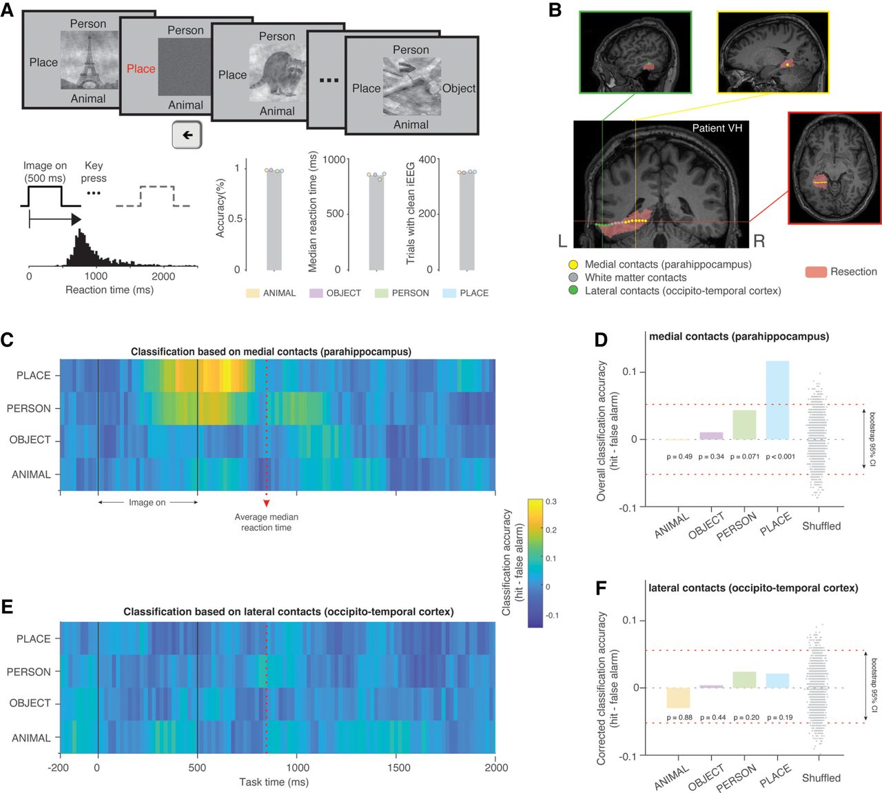
Sensitivity of resected brain tissues to scene images in Patient VH. (A) During epilepsy monitoring, Patient VH performed a visual categorization task, identifying images as ANIMAL, OBJECT, PERSON, or PLACE with high accuracy. (B) The electrode probe that later covered resected tissues (pink) had 16 effective contacts, with seven in the parahippocampus (yellow), four in white matter (gray), and six in occipital-temporal cortex (green). (C) Highest classification accuracy (hit—false alarm) occurred for scene images during image presentation until response (median reaction time: 848 msec). Classification for other image categories over time is much attenuated. (D) Classification accuracy for scene images significantly exceeded chance within a task-related time window (bootstrap P < 0.001), whereas other categories did not. No significant category-sensitive patterns were found in lateral recording sites (E and F). (CI) Confidence interval. Each dot in D and F represents decoding results based on shuffled data from a single iteration.



