


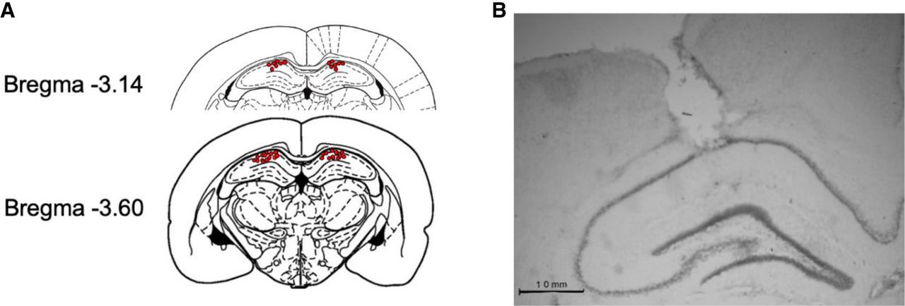
Figure 1.
Schematic representation of the cannula placements for intra-dHPC infusions. (A) Individual placements for each subject included in Experiment 5 (n = 21). (B) Micrograph of an example cannula tip placement terminating in the CA1 region of the dHPC.



