


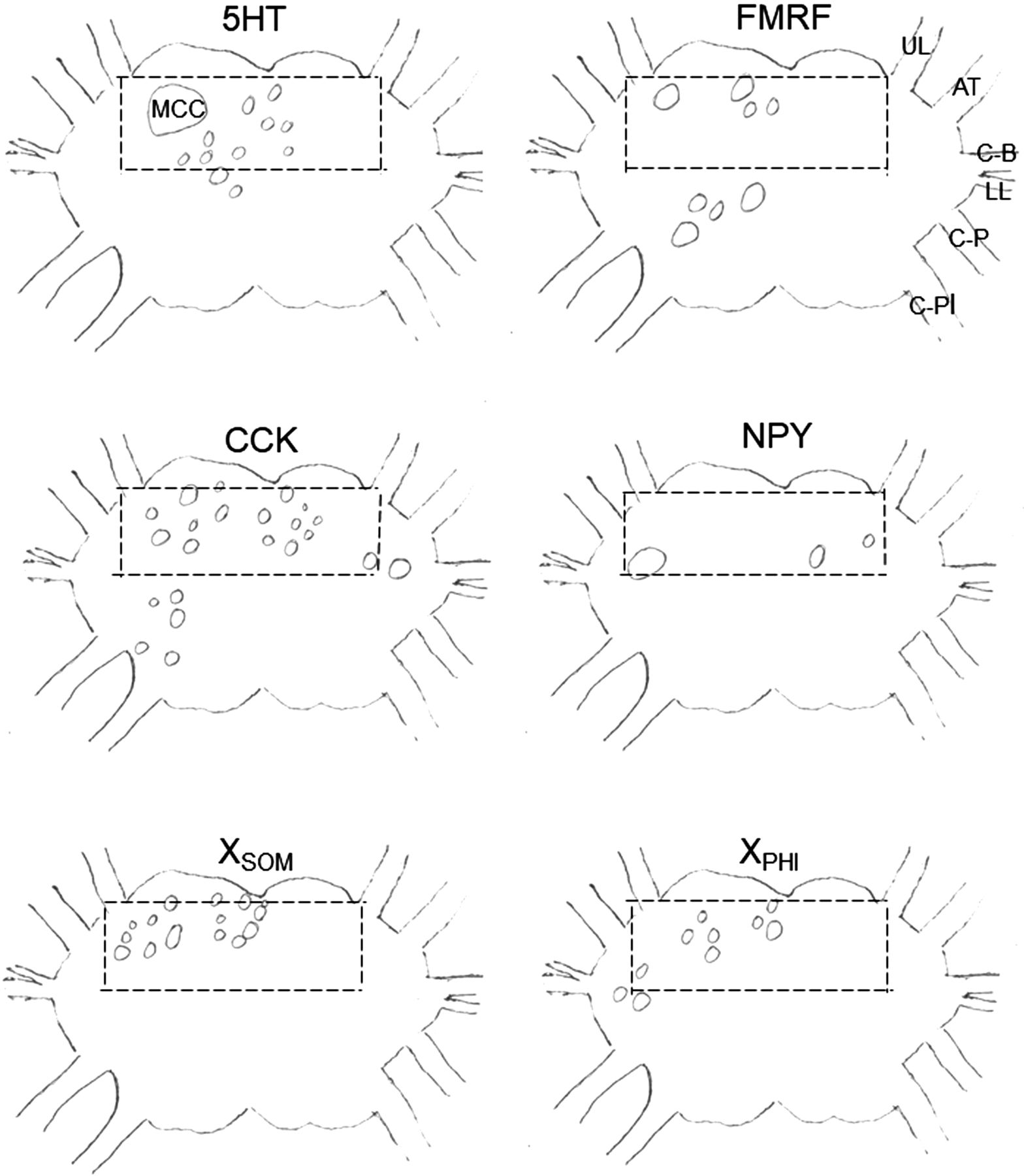
Schematic drawings illustrating the outline (dashed lines) of a region in the cerebral ganglion that has staining with antisera raised against many different transmitters, and the locations and sizes of cells that stained with antisera raised against different transmitters in that region from representative sections. The anti-SOM and ant-PHI antisera stain the unknown peptides XSOM and XPHI, respectively. (MCC) Metacerebral cell, (C-B) cerebral–buccal connective, (C-P) cerebral–pedal connective, (C-Pl) cerebral–pleural connective, (UL) upper labial nerve, (AT) anterior tentacular nerve, (LL) lower labial nerve. (Drawing of the ganglion modified from Rosen et al. 1991.)



