


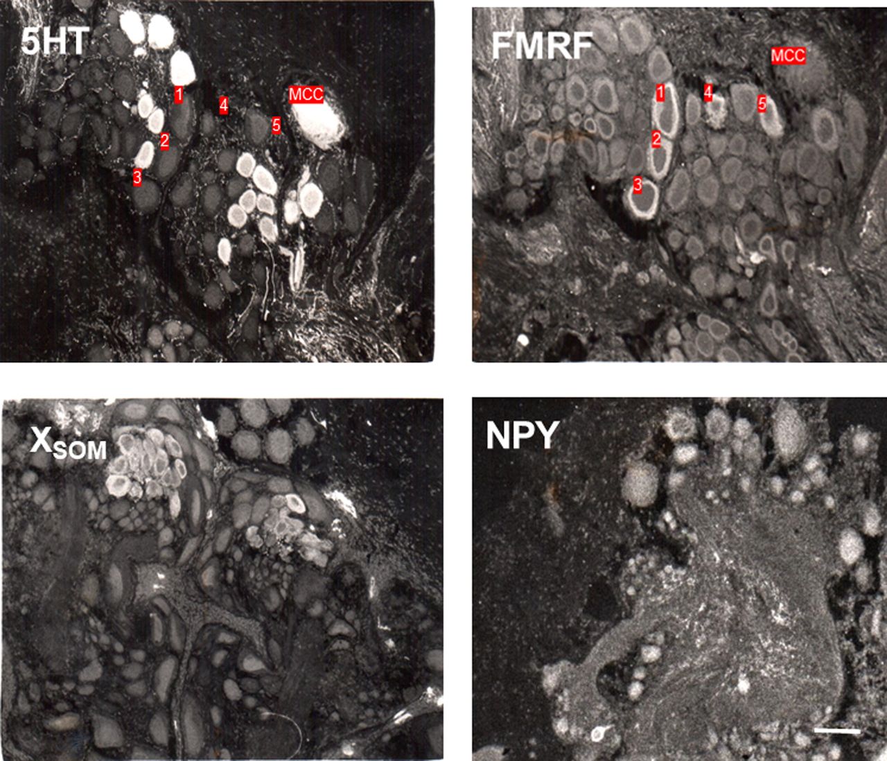
Figure 3.
Examples of staining of the cytoplasm with antisera raised against serotonin (5HT), Aplysia FMRFamide, and mammalian somatostatin (SOM) and neuropeptide Y (NPY) in a region at the anterior end of the cerebral ganglion that has staining with antisera raised against many different transmitters. The anti-SOM antiserum stains an unknown peptide, XSOM. (MCC) Metacerebral cell, which was in the plane of section of two of the four micrographs. The numbers indicate individual neurons, none of which were double-labeled with antisera raised against 5HT and FMRFamide in adjacent sections.



