


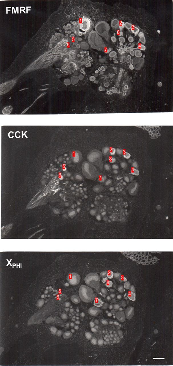
Figure 1.
Examples of staining with antisera raised against Aplysia Phe–Met–Arg–Phe–NH2 (FMRFamide [FMRF]) and mammalian cholecystokinin (CCK) and peptide histidine isoleucine (PHI) in the cytoplasm of some cell bodies and also fibers in adjacent cryostat sections of the abdominal ganglion of Aplysia. The anti-PHI antiserum stains an unknown peptide, XPHI. The numbers indicate individual neurons that stained with antisera raised against FMRFamide alone (1), CCK alone (5 and 6), FMRFamide and PHI (7), or all three (2, 3, 4, and 8). Scale bar, 200 µm.



