


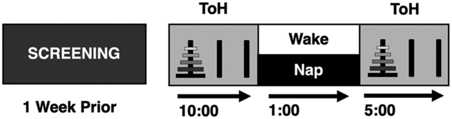
Study design. Overview of experimental protocol in the nap and wake conditions. After an initial screening and acclimatization polysomnography (PSG), participants were divided into young nap (YN), young no nap (YNN), older nap (ON), and older no nap (ONN) conditions. Nap condition: Participants arrived at the sleep laboratory at 9:00 a.m. and were trained and tested on the ToH at 10:00 a.m. They then had a nap opportunity from 1:00 p.m. to 2:30 p.m. while their sleep was recorded via PSG. Upon awakening, but after sleep inertia dissipation, participants were retested at 5:00 p.m. Wake condition: The same procedure was used in the eake condition, except that instead of a daytime nap opportunity, individuals remained awake and read in a supine position with polysomnographic recording.



