


Figure 1.
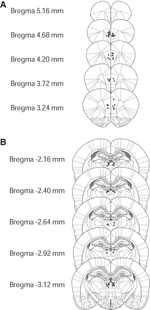
(A) Approximate locations of infusions into the mPFC. (B) Approximate locations of infusions into the mdThal. Illustrations were adapted from Paxinos and Watson (2006), with permission from Elsevier 2019.
(Downloading may take up to 30 seconds. If the slide opens in your browser, select File -> Save As to save it.)
Click on image to view larger version.
(A) Approximate locations of infusions into the mPFC. (B) Approximate locations of infusions into the mdThal. Illustrations were adapted from Paxinos and Watson (2006), with permission from Elsevier 2019.
 CiteULike
 Delicious
 Digg
Digg Facebook
 Reddit
 Twitter
