


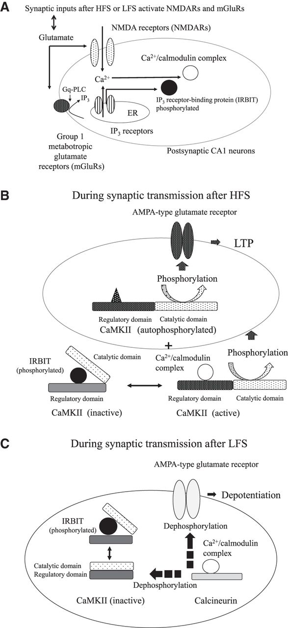
Schematic depiction of the mechanism of LFS-induced depotentiation in hippocampal CA1 neurons. (A) Synaptic inputs after HFS or LFS activate NMDARs and mGluRs on postsynaptic CA1 pyramidal neurons. The activation of IP3Rs on the membrane of the ER occurs downstream from group I mGluRs and phospholipase C in the signaling cascade. The increase in [Ca2+]i due to the coactivation of NMDARs and IP3Rs induces an increase in the free cytoplasmic levels of Ca2+/calmodulin complexes and IRBIT in the postsynaptic dendritic spines of CA1 neurons. (B) CaMKII autophosphorylated during and after priming HFS may continue to phosphorylate AMPA-type glutamate receptors on the dendritic spines of CA1 neurons to potentiate synaptic responses (to induce LTP), while IRBIT that is released from IP3Rs into the cytoplasm and is phosphorylated may block the binding of Ca2+/calmodulin complexes to the regulatory domain of CaMKII and inhibit further CaMKII activation in postsynaptic CA1 neurons. CaMKII is active when Ca2+/calmodulin complexes are bound to its regulatory domain. (C) During synaptic transmission after LFS, the levels of free Ca2+/calmodulin complexes, which are not sequestrated by CaMKII in the presence of IRBIT, are increased to a point at which calcineurin is activated in the dendritic spines to dephosphorylate CaMKII and AMPARs at CA1 synapses, leading to a reduction in synaptic potentiation (depotentiation) in CA1 neurons.



