


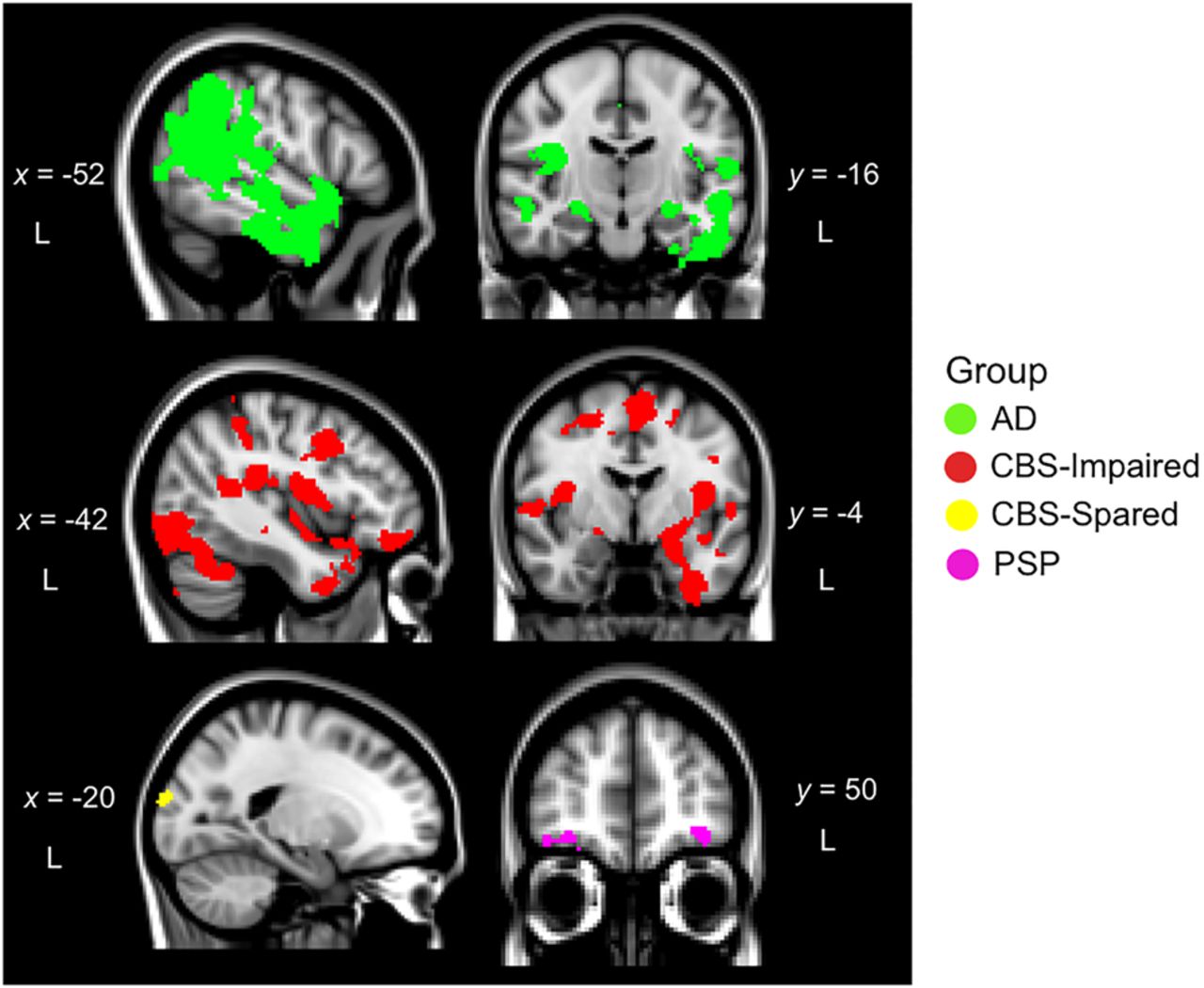
Regions of significant gray matter intensity decrease that correlate with delayed episodic recall performance. All x and y coordinates reported are Montreal Neurological Institute (MNI) coordinates. Colored voxels show regions that emerged as significant in the VBM analyses at a strict uncorrected threshold of P < 0.001 with a cluster threshold of 100 contiguous voxels. All clusters reported at t > 3.56. Age was included as a covariate in the analyses. Clusters are overlaid on the MNI standard brain; (L) Left, (AD) Alzheimer's Disease, (CBS-Impaired) Corticobasal Syndrome-Impaired memory (<−1.5 z-score on delayed recall), (CBS-Spared) Corticobasal Syndrome-Spared memory (≥−1.5 z-score on the delayed recall), (PSP) Progressive Supranuclear Palsy.



