


Cover image
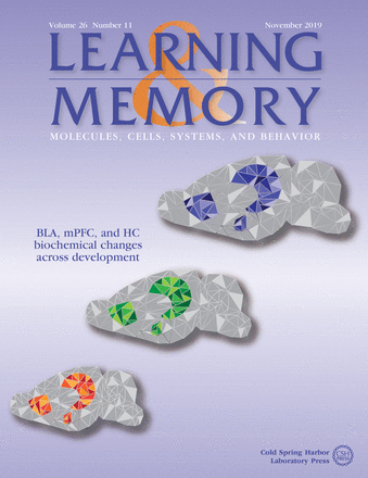
Graphical illustration of a sagittal view of the rat brain at different developmental ages. From bottom to top: infant, juvenile, and adult brain. The hippocampus (HC), the medial prefrontal cortex (mPFC), and the basolateral amygdala (BLA) are shown in color. See Bessières et al. (LearnMem 26: 436–448).

