


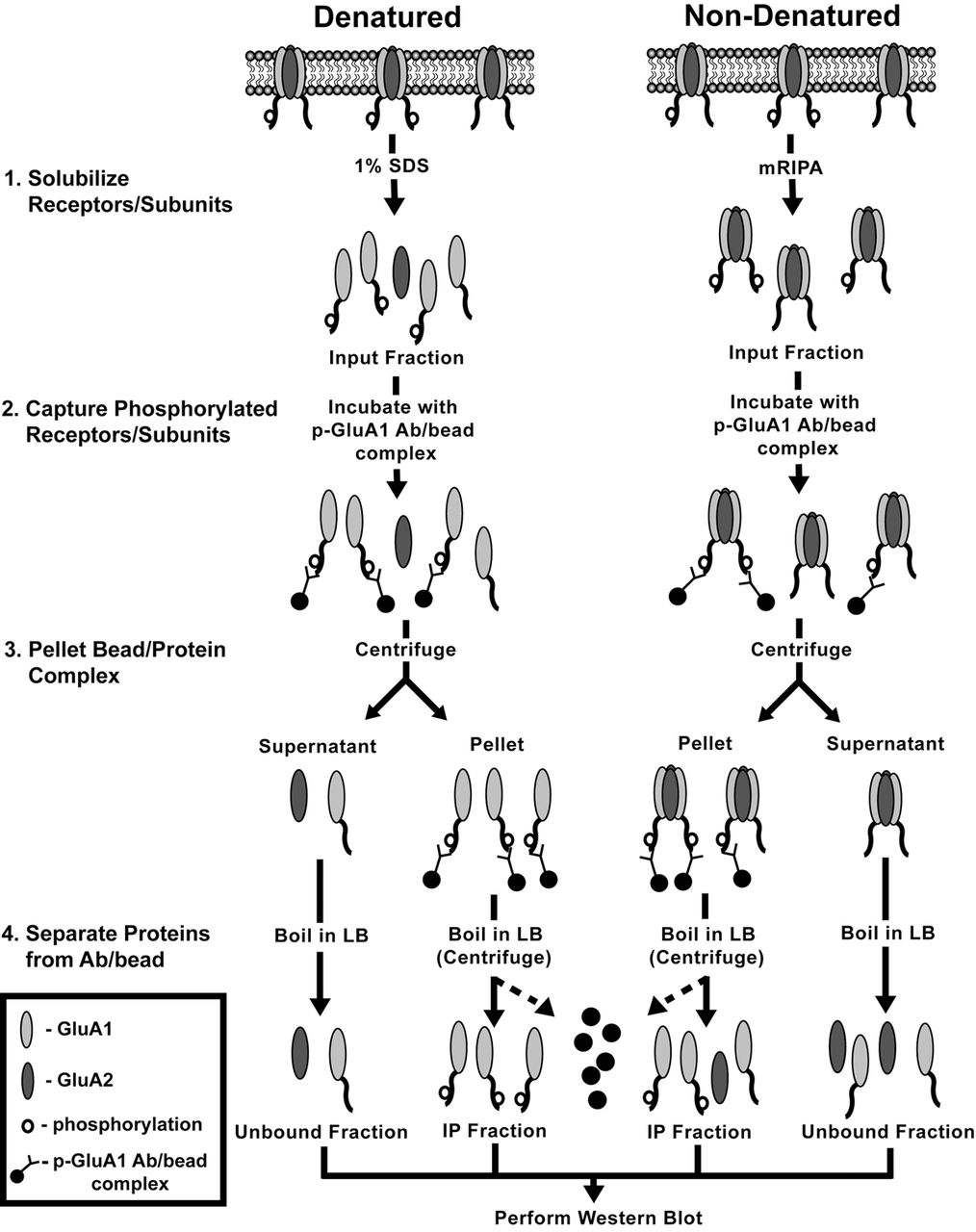
Schematic of immunoprecipitation/depletion assays using denatured (left) and nondenatured homogenization buffers (right). The starting material for immunoprecipitation (input fraction) consists of either individual subunits obtained from denatured homogenates prepared using 1% sodium dodecyl sulfate (SDS) buffer or intact receptors obtained from nondenatured homogenates prepared in modified radioimmunoprecipitation assay (mRIPA) buffer (Step 1). The input fraction (containing phosphorylated and nonphosphorylated subunits) is incubated with phospho-T840 or phospho-S845-specific antibodies (p-GluA1 Ab) conjugated to protein A coated agarose beads (Step 2). Beads (and attached proteins) are then pelleted by centrifugation, leaving nonphosphorylated subunits/receptors in the supernatant/unbound fraction (Step 3). The pelleted fraction is then resuspended in loading buffer (LB) and boiled to release proteins and antibodies (not shown) from the beads (Step 4). Phosphorylated subunits are collected by taking the supernatant following centrifugation (IP fraction). Western immunoblotting is then used to compare total GluA1 levels in the input and unbound fractions and the depletion of GluA1 levels in the unbound fraction is used to determine the proportion of subunits/receptors basally phosphorylated at each site. For simplicity, only GluA1/2 subunit-containing heteromeric AMPARs are shown.



