


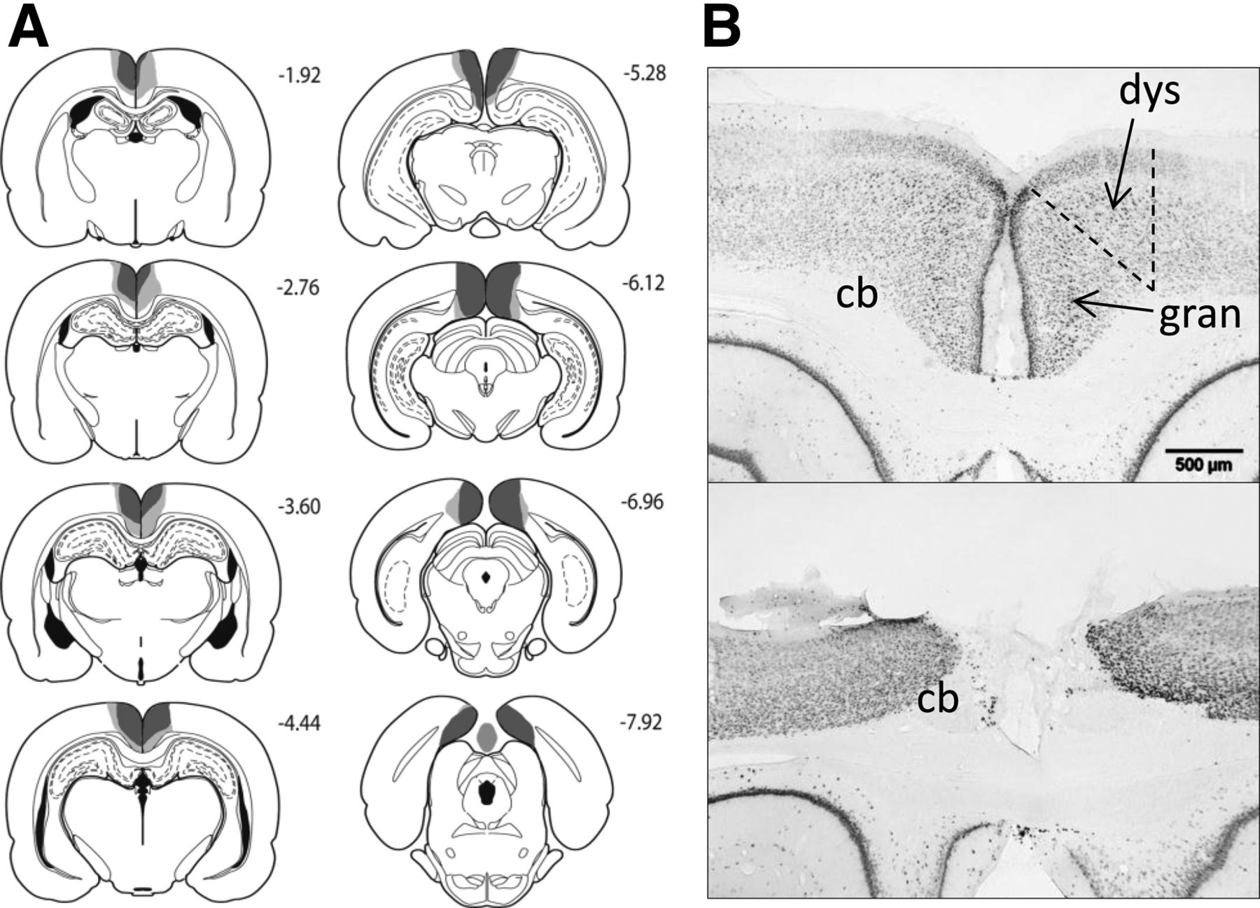
Figure 3.
(A) Series of coronal sections showing the cases with the largest (light gray) and smallest (dark gray) lesions in the combined dysgranular and granular retrosplenial (RScomb) lesion cohort. The numbers correspond to the distance behind bregma in millimeters (Paxinos and Watson 2007). (B) Coronal NeuN sections showing the retrosplenial cortex (both hemispheres) from a sham surgery control rat (top), and a representative rat from the RScomb lesion group (bottom). (cb) Cingulum bundle; (dys) dysgranular retrosplenial cortex; (gran) granular retrosplenial cortex.



