


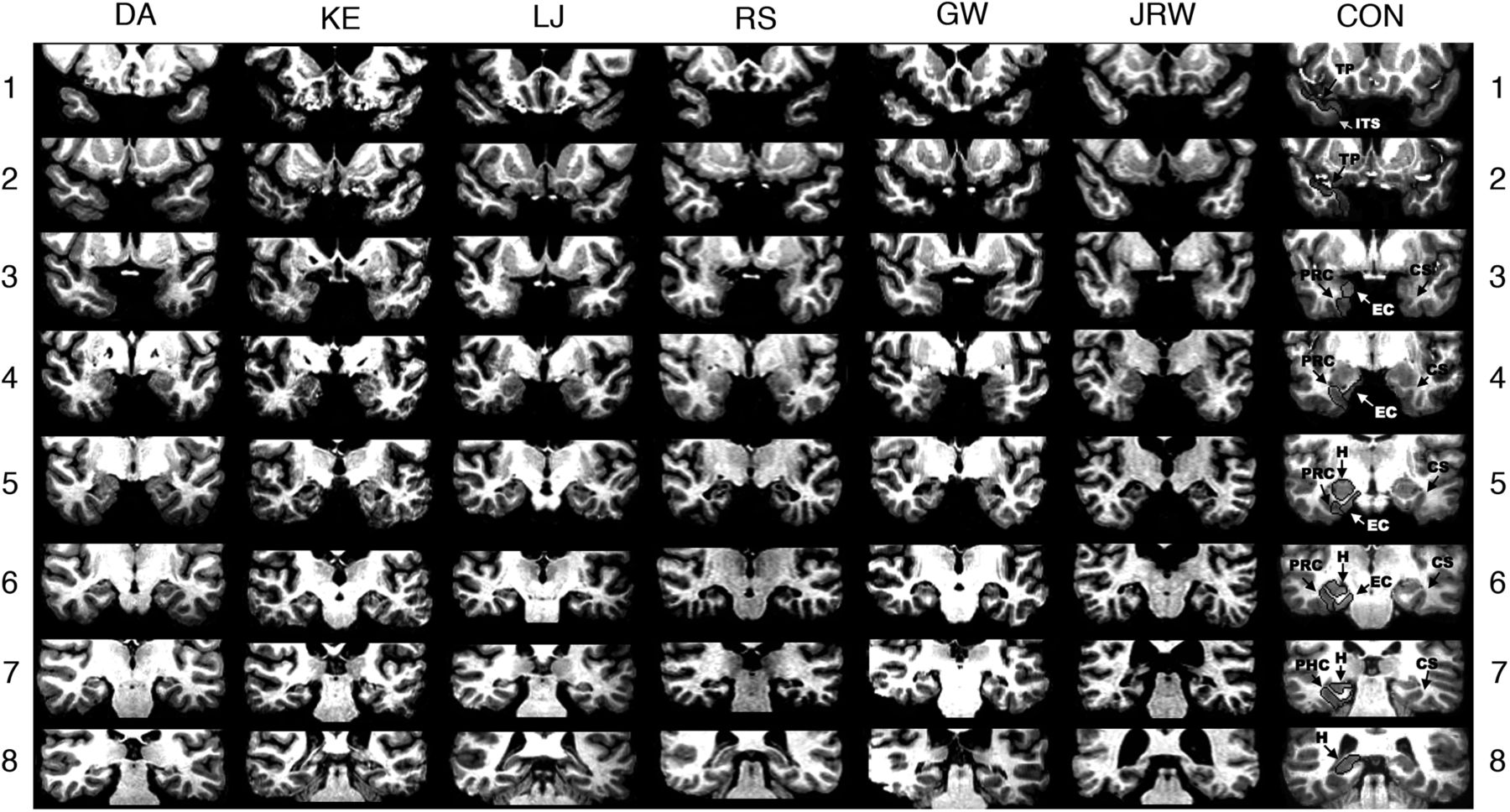
Series of eight T1-weighted coronal images of six patients with limited hippocampal lesions (DA, KE, LJ, RS, GW, and JRW) and one control (CON) are illustrated. The sections proceed posteriorly in 7-mm intervals from the temporopolar (TP) cortex in the top section. The left side of the brain is on the right side of each image. As described by Insausti et al. (1998), TP cortex extends medially from the inferotemporal sulcus (ITS) to the fundus of the TP sulcus. TP cortex extends rostrally from the tip of the temporal pole caudally to the limen insula (LI), which approximates the border between the TP cortex and perirhinal cortex (PRC). Caudal to TP cortex, the collateral sulcus (CS) is the most important structure for the identification of medial temporal lobe cortices. At its most rostral extent, the CS is surrounded entirely by PRC. Caudally, entorhinal cortex (EC) extends from the midpoint of the medial bank of the CS to the subiculum, while PRC extends laterally from the midpoint of the medial bank of the CS to the inferotemporal cortex. Two millimeters caudal to the disappearance of the gyrus intralimbicus of the hippocampus (H), the CS is surrounded by parahippocampal cortex (PHC). The caudal border of the posterior PHC is defined as lying 1.5 mm posterior to the crus of the fornix at the point where the fimbria turns upward to continue as the posterior pillars of the fornix and posterior to the pulvinar nucleus of the thalamus (Franko et al., 2012). The top section (1) shows the TP cortex and the ITS in the control brain. None of the patients has damage evident at this level. The ITS is visible bilaterally at this level for patients KE, RS, GW, and JRW. For DA, the ITS is not visible on either side at this level. For LJ, only the right ITS is visible. The second section (2) shows TP cortex and the ITS in the control brain. The ITS and TP cortex is evident in all patients at this level. None of the patients has damage evident at this level. The CS is visible, indicating the beginning of PRC, in patients KE and RS (right side only). The third section (3) shows the CS and surrounding PRC and EC in the control brain. None of the patients has damage evident at this level with the exception of KE, who has damage in the basal ganglia secondary to toxic shock syndrome (and to a lesser extent in section 4). For patient DA, the CS is not evident at this level and PRC is evident bilaterally. For patients KE, LJ, and GW, the PRC is evident on the left side, bounded by the LI and CS. On the right side, both EC and PRC are evident. For patients RS and JRW, both EC and PRC are evident bilaterally. The fourth section (4) shows the anterior hippocampus and the adjacent PRC and EC in the control brain. At this level hippocampal damage is evident in patient DA. The hippocampus is not yet visible at this level in any of the other patients. For DA, bilateral damage to the globus pallidus is evident at this level, presumably secondary to heroin overdose. No damage to the PRC or EC is evident for any of the patients at this level. The fifth section (5) shows the hippocampus and the adjacent PRC and EC in the control brain. The CS and the surrounding PRC and EC appear normal in all patients at this level. Damage to the hippocampus is evident in all patients at this level. The sixth section (6) shows the hippocampus and the adjacent PRC and EC in the control brain. Damage to the hippocampus is evident in all patients at this level. The surrounding PRC and EC appear normal in all patients. Both the PRC and EC are visible in all patients bilaterally, with the exception of JRW for whom only PRC is visible on the left side, indicated by the disappearance of the gyrus intralimbicus 2 mm rostral to the sixth section (not shown). The seventh section (7) shows the hippocampus and the CS, surrounded by PHC in the control brain. Damage to the hippocampus is evident in all patients at this level. The PHC is evident in all patients. For DA, the PRC is still visible on the right side. The eighth section (8) shows the hippocampus in the control brain. Bilateral hippocampal damage is evident in patients DA, KE, and GW at this level. Patient LJ shows hippocampal damage only on the left side, and no damage is evident in patient RS. At this level, the hippocampus is no longer evident in patient JRW. PHC is no longer evident at this level in patients DA, KE, LJ, RS, and JRW, and PHC appears normal in patient GW. The warping artifact in the right lateral temporal lobe of GW on this section did not interfere with the assessment of his damage. No damage is evident posterior to this level for all patients.



