


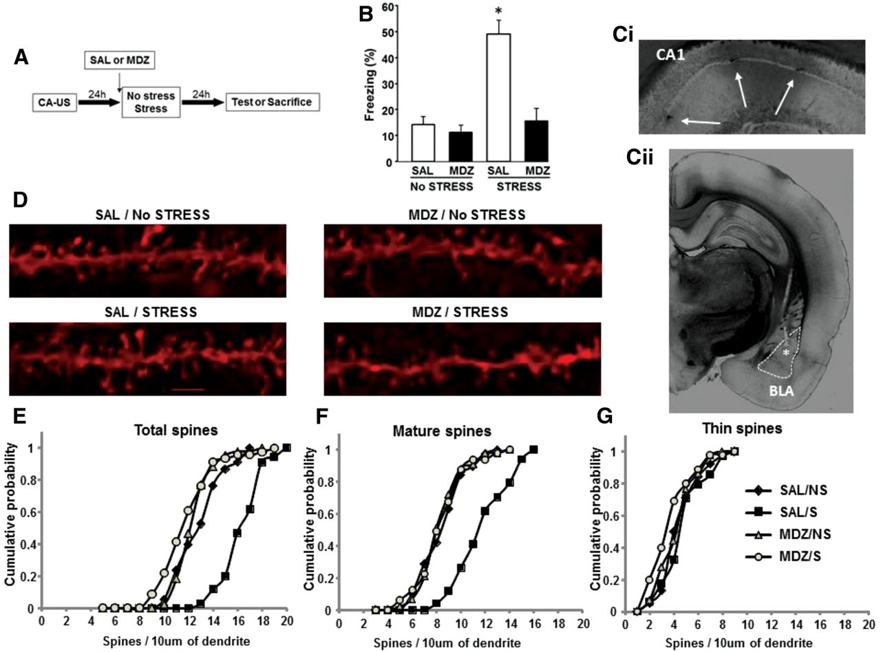
Intra-basolateral amygdala infusion of MDZ prior to stress attenuates the promoting influence of restraint on both fear retention and DH structural remodeling. (A) Schematic representation of the experimental design. (B) Freezing behavior response observed during testing in rats that received either intra-BLA MDZ or SAL administration 10 min prior to restraint. Data are expressed as mean ± SEM of the percentage of freezing spent during the test (n = 8 rats for all groups). (*) P < 0.05 compared to the rest of the experimental groups (two-way ANOVA, Bonferroni post-hoc test). (C) Bright field image of a representative coronal section showing the location of the cannula placement in BLA (Cii) (anterior-posterior, −3.12 mm from bregma [Paxinos and Watson 2007]; [*] injector site for SAL or MDZ infusions intra-BLA) and DH CA1 region (Ci), where arrows indicate the spots of DiI injection. (D) Representative examples of apical dendritic segments of DH CA1 pyramidal neurons which were selected for quantitative analysis of dendritic spines from animals: SAL/S (n = 3 rats), SAL/NS (n = 3), MDZ/S (n = 4), and MDZ/NS (n = 3). Bar, 2 μm. (E–G) Cumulative frequency of total (E), mature (F), and thin (G) spine density on apical dendrites of hippocampal pyramidal cells CA1 stratum radiatum (P < 0.05, by Kolmogorov–Smirnov test).



