


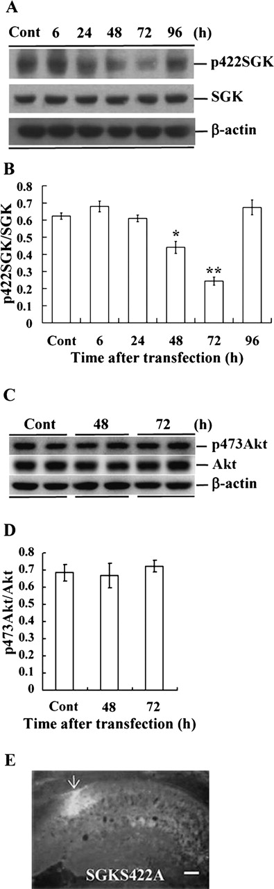
Time-course study of SGK phosphorylation after SGKS422A transfection. (A) Representative gel pattern showing p422SGK, SGK, and β-actin protein bands at different time points after SGKS422A transfection to the hippocampal CA1 area. (B) Statistical analysis revealed that SGKS422A transfection markedly decreased Ser422 SGK phosphorylation 48 h and 72 h later, but this effect is recovered at 96 h. *P < 0.05; **P < 0.01 (one-way ANOVA followed by Dunnett’s t-test). (C) Representative gel pattern showing p473Akt, Akt, and β-actin protein bands 48 h and 72 h after SGKS422A transfection to CA1 area. (D) The same SGKS422A transfection did not alter Ser473 Akt phosphorylation at 48-h and 72-h time intervals. Data are mean ±SEM. (E) Immunohistochemical staining showing fluorescence labeling in CA1 neurons upon HA-SGKS422A transfection to this area. An anti-HA primary antibody and a FITC-conjugated IgG secondary antibody were used. The arrow indicates the area of transfection. Bar, 220 μm.



