


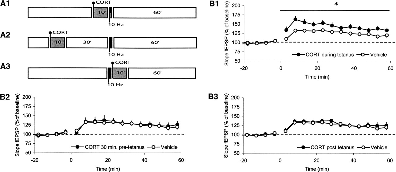
Corticosterone facilitates synaptic potentiation in a specific time window. The schematic representation of the experimental design (A) and the corresponding results (B) are shown. Corticosterone was applied for 10 min (gray area) at three time points: Starting 10 min before high-frequency stimulation (HFS, 900 pulses at 10 Hz; black area) and lasting until the end of the HFS (A1); starting 40 min before HFS (A2); or starting directly after the end of HFS (A3). In all cases, signals were recorded for 60 min after HFS. (B1) Application of 100 nM of corticosterone to hippocampal slices (filled circles) 10 min prior to and during repetitive stimulation (10 Hz, 900 pulses) significantly enhances synaptic potentiation up to 60 min after stimulation, compared with untreated slices (open circles). (B2) When the hormone was administered half an hour before repetitive stimulation (t = −40 to −30 min) corticosterone did not affect subsequent synaptic potentiation. (B3) Similarly, when corticosterone administration was started after the repetitive stimulation was completed, no effect was seen on the development and maintenance of potentiation. Statistical analysis was performed over t = 0–60 min with analysis of variance for repeated measures. *P < 0.05. For clarity, only average data for a selection of time points (once every 5 min) are shown in these pictures, starting at t = −18 min. The interval between the last value before and the first value after high-frequency stimulation was left open. Statistical analysis, though, was performed on all data (values for each minute).



