


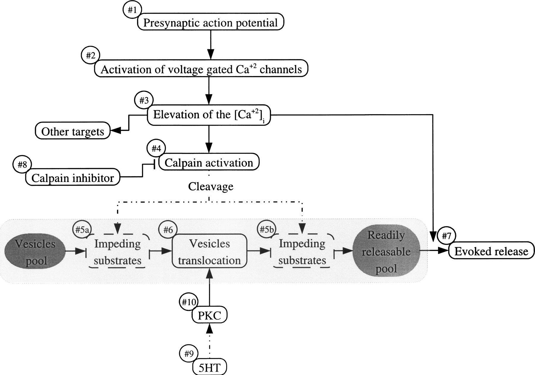
A model for the role of calpain in the sensory motor synapse. Presynaptic action potentials (#1) induce calcium influx through voltage-gated calcium channels (#2). The elevated [Ca2+]i (#3) activate calpain (#4), and in parallel, invokes neurotransmitter release (#7). The activated calpain cleaves substrates (#5a; #5b) that impede vesicle translocation from the reserve pool to a readily releasable pool of neurotransmitter (#6). The nature of the calpain substrate is not known. Nevertheless, based on the experimental results, the impeding substrates could be located upstream (#5a) or downstream (#5b) with respect to the vesicle-mobilization functions of PKC. When calpain activation is inhibited (#8), the impeding substrates are not cleaved, and as a consequence, the translocation of vesicles is slowed down, leading to an increased rate of homosynaptic depression. Furthermore, following homosynaptic depression, in the presence of calpeptin, 5HT application (#9) and PKC activation (#10) fail to mobilize vesicles, since vesicle translocation is impeded by the hypothetical uncleaved calpain substrate.



