


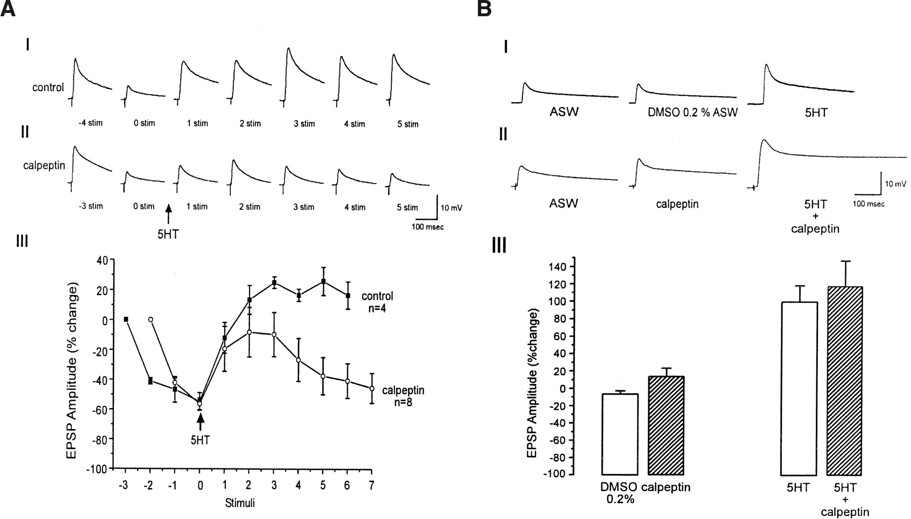
Effect of calpeptin on 5HT-induced synaptic facilitation of moderately depressed and nondepressed synapses. Examples of 5HT-induced facilitation of moderately depressed synapses in controls (0.2% DMSO ASW) (AI) and preparations bathed for 15 min in 100μM calpeptin (AII). The figure shows representative traces of EPSP in the course of the experiments. The number shown under the EPSPs in AI and AII corresponds to the order of the EPSP's generation. To moderately depress the synaptic potentials to about 45% of the initial level, four stimuli at 0.05 Hz were given to the sensory neuron in control experiments. Since the rate of depression is faster in calpeptin, only three stimuli at 0.05 Hz were given to depress the synapses to about 45% of the initial level in calpeptin. The arrow (time 0) indicates 5HT application. In the control experiments, 5HT induces facilitation of the depressed EPSPs. In the presence of calpeptin, 5HT induces partial recovery, which then rapidly declines. The kinetics of synaptic depression and 5HT-induced facilitation in control (AIII, ▪) and in the presence of calpeptin (AIII, ○) are shown. Significant difference between control and calpeptin experiments becomes apparent from the fourth stimulus after 5HT application, (P < 0.05; t-test). (B) In rested synapses, calpeptin has no effect on 5HT-induced facilitation. BI and BII are examples of 5HT-evoked facilitation of rested synapses in the controls and in the neurons incubated in 100 μM calpeptin for 15 min, respectively. (BII) EPSP before (ASW), 15 min after calpeptin application (calpeptin) and 3 min following 5HT application (5HT + calpeptin). In control experiments, DMSO 0.2% was added instead of calpeptin (BI). (BIII) Summary of the short-term facilitation of rested synapses in the presence and absence of calpeptin. Each bar represents the mean percent change ±SEM of the EPSP amplitude with respect to the first EPSP. Note that facilitation produced by 5HT in rested synapses is not affected by calpeptin (P > 0.05; t-test).



