


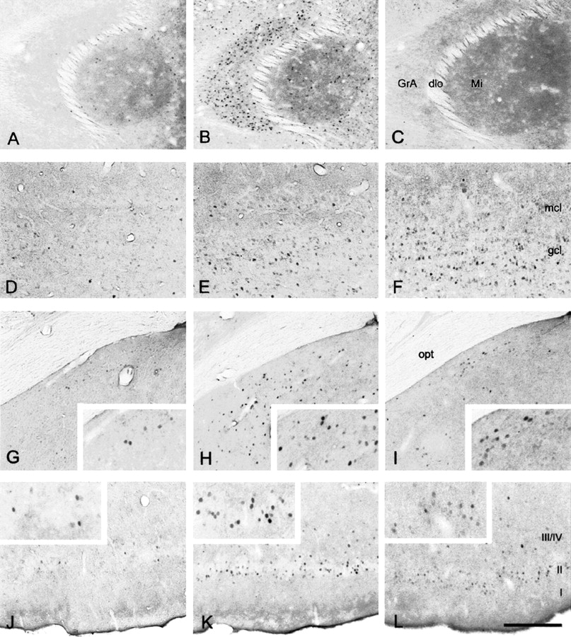
Representative microphotographs of Fos immunoreactivity in brain slices of adult male mice that were killed either unstimulated (left panel), 70 min after sampling-like juvenile exposure (middle panel), or 70 min after carvone exposure (right panel). The figure shows (A–C) the accessory olfactory bulb, (D–F) the main olfactory bulb, (G–I) the medial amygdala, and (J–L) the piriform cortex. GrA, granule cell layer of the accessory olfactory bulb; dlo, dorsolateral olfactory tract; Mi, mitral cell layer of the accessory olfactory bulb; mcl, mitral cell layer of the olfactory bulb; gcl, granule cell layer of the olfactory bulb; opt, optic tract; I, II, III/IV, layers of the piriform cortex. Scale: G–L, 300 μm; A–F and insets, 150 μm.



